INVESTIGATING THE ETIOLOGY OF THE SHORT ROOT ANOMALY
Ashley Teresa Hill
A thesis submitted to the faculty at the University of North Carolina at Chapel Hill in partial fulfillment of the requirements for the degree of Master of Science in the School of Dentistry (Orthodontics).
Chapel Hill 2017
Approved by:
ABSTRACT
Ashley Teresa Hill: Investigating the Etiology of the Short Root Anomaly (Under the direction of Sylvia Frazier-Bowers)
Objectives: Short Root Anomaly (SRA) is an emerging clinical problem but the clinical features and etiology are poorly understood. In order to test the hypothesis that one or more genes contribute to the SRA phenotype, we aimed to 1) characterize the SRA phenotype and its associated risk for
orthodontically induced external apical root resorption (OIEARR) and 2) complete mutational analysis.
Methods: The affected teeth and presence of other dental anomalies was assessed. PCR based mutational analysis was completed for 9 SRA samples for BMP4, BMP2, NFI-C and PTH1R. Pre- and
post-treatment panoramic radiographs for 7 patients were evaluated for incidence and severity of OIEARR of SRA affected maxillary central incisors. Results: We identified 3 broad types of SRA and 5 SNPs. OIEARR was observed in 64% of affected maxillary central incisors. Mild resorption was observed in 50%, moderate resorption was observed in 7% and severe resorption was observed in 7% of teeth. Conclusions: SRA can present as three distinct phenotypes. SRA affected maxillary central incisors are
ACKNOWLEDGMENTS
TABLE OF CONTENTS
LIST OF TABLES ... viii
LIST OF FIGURES ... ix
LIST OF ABBREVIATIONS ... x
LIST OF SYMBOLS ... xi
REVIEW OF THE LITERATURE ... 1
Developmental Disorders of Root Formation ………....1
The Short Root Anomaly ... 2
The Etiology of the Short Root Anomaly ……….…...3
Overview of Root Development ……….….4
Signaling Pathways Involved in Tooth Root Development……….…….5
Transforming Growth Factor Beta ………..………..6
Bone Morphogenetic Proteins (BMPs)………...6
Sonic Hedgehog……….6
Nuclear Factor 1-C (NFI-C) ……….….7
Wingless-type ……….………...7
Osterix ……….………..8
Parathyroid Hormone 1 Receptor (PTH1R) ……….………….8
Risk of Root Resorption with the Short Root Anomaly……….……….…….9
Summary ... 10
References ... 12
Introduction ... 16
Materials and Method……….………18
Phenotype Characterization ……….………..18
PCR-Based Mutational Analysis…...………19
Radiographic Analysis of OIEARR………...20
Statistical Analysis ... 21
Results ... 21
Phenotype Characterization ... 21
PCR-Based Mutational Analysis…...………22
Radiographic Analysis of OIEARR………...22
Discussion……….………..23
Conclusions ... 27
Tables ... 29
Figures ... 32
LIST OF TABLES
Table 1 - Descriptive characteristics of the SRA sample in the phenotype characterization ... 29
Table 2 -Descriptive characteristics of the sample in the mutational analysis ... 29
Table 3 - PTH1R, BMP4, BMP2, and NFI-C primers ………..……..30
Table 4 - Demographic and clinical characteristics of SRA sample evaluated for OIEARR…………..…31
Table 5 - Phenotype characterization of SRA sample………...31
Table 6 - Sequencing variations found in mutational analysis……….…...32
Table 7 - Mean age of patients based on incidence of OIEARR ……….……….….….32
LIST OF FIGURES
Figure 1 - Modification of Malmgren and Levander scale………..32
Figure 2.1 - Pretreatment panoramic radiograph of 18 yr. old Hispanic male with maxillary central incisors affected (Type 1 SRA)………...….………...33
Figure 2.2 - Periapical of SRA affected maxillary central incisors ... 33
Figure 2.3 - Post-treatment panoramic radiograph with no evidence of OIEARR…………...…………...33
Figure 3.1 - Panoramic radiograph of 9 year old Hispanic female with generalized SRA (Type 3)………….………...…34
Figure 3.2 - Panoramic radiograph of patient in Figure 3a at 12 years old with generalized SRA……….….34
Figure 4 - Panoramic radiograph of 13 yr. old Hispanic female with maxillary central incisors, mandibular and maxillary premolars affected (Type 2 SRA)…...…35
Figure 5 - Affection of premolars………....35
Figure 6 - Chromatogram of PTH1R c.1389T>C………..…..36
Figure 7 - Chromatogram of PTH1R c.178+28G>T ………..….…36
Figure 8 - Chromatogram of PTH1R c.1116+58T>C………...……...36
Figure 9 - Chromatogram of BMP2 c.570A>T………...………….37
Figure 10 - Chromatogram of NFIC c.562+33C>T………...………..37
Figure 11 - Incidence of OIEARR of SRA affected maxillary central incisors after orthodontic treatment………..…...……..37
LIST OF ABBREVIATIONS
A Adenine
BMP Bone Morphogenetic protein C Celsius
C Cytosine
CBCT Cone Beam Computed Tomography G Guanine
HERS Hertwig’s Epithelial Root Sheath IEE Inner Enamel Epithelial
MAF Minor Allele Frequency NFI-C Nuclear Factor 1 C OEE Outer Enamel Epithelium Osx Osterix
PTH Parathyroid Hormone PCR Polymerase Chain Reaction SHH Sonic Hedgehog
Smad Small Mother Against Decapentaplegic SNP Single nucleotide polymorphism SRA Short Root Anomaly
T Thymine
TGF- β Transforming Growth Factor Beta US United States
LIST OF SYMBOLS
° Degrees
A REVIEW OF THE LITERATURE
Developmental Disorders of Root Formation
Odontogenesis is a complex, yet eloquent process that begins in the developing human at six weeks in utero. The tooth is derived from the cells of the ectoderm and the underlying ectomesenchyme of the neural crest. Each tooth develops through the initiation, bud, cap, bell, apposition and maturation stages. After crown formation, root development begins. The root is vital to the existence of the tooth in the alveolar bone as it allows for 1) anchorage of the tooth in the alveolar process by the fibers of the PDL, 2) attachment of gingival fibers, and 3) provides a foramen for passage of vessels and nerves.1
Developmental defects such as dentinogenesis imperfecta, dentin dysplasia and the Short Root Anomaly (SRA) all result in abnormally short roots.3 Providing orthodontic treatment to these patients given the unavoidable outcome of external apical root resorption provides a challenge to clinicians and more importantly a concern for the outcome of the affected teeth. Most studies that have investigated orthodontic treatment in patients with these disorders are case studies and lack the large cohorts needed to provide evidence of proper management of these affected individuals.
Outside of their orthodontic implications, extensive molecular research related to dentinogenesis imperfecta and dentin dysplasia is available in the literature. Mutational analyses have revealed mutations in one key gene, dentin sialophosphoprotein as the cause.3 SRA on the other hand has an etiology that has not been investigated and remains poorly understood in the dental community. SRA has received less attention because it is less common and often misdiagnosed as root resorption.4 However, as minority populations in the United States continue to grow there is a concomitant increase in the prevalence of SRA patients in dental practices that will likely continue to increase. Taken together this trend presents a challenge to dental professionals, particularly orthodontists.
The Short Root Anomaly
The prevalence of SRA is low and varies for different populations.4-6 The prevalence has been reported in Japanese populations as 10% and 1- 3% in European populations.4-6 Puranik et al. investigated a cohort of primarily Hispanic patients with SRA7 representing the first known report documenting SRA in a Hispanic sample. Although the prevalence rate among this population still has not been published, Puranik et al. have seen an increase in prevalence of SRA that correlates well with the increase in the US Hispanic population.8 The relatively recent increase in the Hispanic population is responsible for a large portion of the population growth in this country.9 As of 2015, the Hispanic population had grown to be the largest minority group in the United States.8 Although the Pew Research Center found that in the last ten years the Hispanic growth rates have fallen to record lows of 2% due to the recession, lower
entire US population by 2060.8,9 With the expected growth of this minority group, we can expect
continued rise in the prevalence of SRA, making it vitally important to investigate this anomaly.
To date, only radiographic signs indicate that the condition is present which contributes to the low reported prevalence.4 In 1972, Volmer Lind was the first to describe SRA and described it as “abnormally short roots of characteristic plump shape mainly affected maxillary central incisors.”4 SRA most often presents as bilaterally short maxillary central incisor roots but can also affect lateral incisors, bicuspids, mandibular incisors or the entire dentition.4,7 Mandibular second premolars are most commonly affected behind maxillary central incisors.7 No attempt has been made to determine if different types or variations of SRA exist given that different teeth can be involved; establishing a pattern of affection can aid in ease of SRA diagnosis. Affected teeth also have a distinctive appearance.7 Maxillary central incisor roots are often plump with rounded apices while mandibular second premolars often have blunted apices.7 Teeth are always affected bilaterally.7 The crown length: root length ratios are approximately 1:1 with normal crown size and shape.4,7 The tissues surrounding the teeth are normal and the hard tissues of the tooth are also normal which distinguishes the abnormally short roots from dentinogenesis imperfect and dentin dysplasia.7
Root resorption will most often be included in the differential diagnosis for SRA. Root resorption, like SRA, can be idiopathic which makes the distinction between the two difficult.
The Etiology of the Short Root Anomaly
Series of patient radiographs prior to the establishment of the permanent dentition with evidence of lack of continued root formation as well as family history have provided evidence of a distinction of SRA from root resorption caused by trauma or occlusal load. 4,5,7 Familial cases of SRA indicate a potential genetic etiology that has yet to be explored. Identification of the specific genetic etiology provides an additional mode for diagnosis of SRA in affected individuals but will also contribute to the growing body of literature on root development.
Although the specific etiology of SRA remains unknown, an autosomal dominant inheritance pattern for SRA has been documented.4,5,7 Lind was the first to hypothesize a possible genetic etiology.4 Lind evaluated 112 SRA cases and found that siblings and parents of SRA affected individuals also presented with the anomaly, although he did not report the percentage of sporadic versus familial cases.4Apajalahati investigated 8 families of probands with SRA.5 In 3/8 families there was evidence of autosomal dominant inheritance and 5/8 families showed affection in only one generation.5 Apajalahati speculated that the cases lacking an autosomal dominant inheritance pattern may be the result of
spontaneous mutations.5 Puranik et al. also provided evidence of familial cases with 7 affected individuals in two generations of a single family.7 The evidence of SRA as a heritable condition in the literature warrants investigation of the specific genetic etiology. Given that the crown remains unaffected in SRA and only the root is malformed, the likely cause of reduced root length in SRA patients has been hypothesized to lie in the mechanisms of root formation. A review of the cellular and molecular mechanisms involved in root development below provide a basis for understanding the processes of abnormal root development in the SRA.
Overview of Root Development
once the crown is developed.1 Hertwig’s epithelial root sheath (HERS) is formed by the bi-layered epithelium of the cervical loop.1 HERS has been considered the region controlling root development just as the enamel knot controls crown formation. While continuing to elongate apically, HERS moves laterally at a forty five degree angle to form the epithelial diaphragm.1 The epithelial diaphragm
eventually creates the narrow opening of the apical foramen.1 Multirooted teeth form by extensions of the epithelial diaphragm that separate the initially single apical foramen into two or three.1
The epithelial cells of HERS play a major role in dentinogenesis and cementogenesis, which lead to the formation of the hard tissues of the root.1 The inner cell layer of HERS, the IEE, stimulates the outer cells of the dental papilla to differentiate into preodontoblast and subsequently odontoblasts.1 The odontoblasts secrete the predentin layer.1 As calcification centers form in the predentin, odontoblasts recede and continue secreting predentin.1,7,11 Contact of dental follicle cells with the surface dentin after disintegration of HERS has traditionally been considered the impetus for cementoblast differentiation and secretion of the cementum matrix.10,11 Mineralization occurs after cementum matrix deposition. Areas lacking embedded cementocytes are called acellular cementum, which covers the coronal two-thirds of the root while cellular cementum covers the remainder of the root.1 HERS proliferation slows once the tooth enters the oral cavity.2,12
Signaling Pathways Involved in Root Formation
Signaling between the dental epithelium and ectomesenchyme mediate the cellular processes of root formation discussed above. Signaling molecules bind to receptors on cell membranes and trigger intracellular gene expression for transcription factors that control odontogenesis.13 The pathways of signaling molecules are well documented in crown formation and are suggested to also control root formation but less evidence exists to support their role in root formation. Studies of transgenic rodents have allowed for continued increase in knowledge of the molecular pathways controlling root
into the potential etiology of SRA.13-28 In this manuscript we consider the individual contributions of selected genes to the root developmental process and the potential role they may play in the pathogenesis of SRA.
Transforming Growth Factor Beta (TGF-β)
TGF-β signaling with Smad4 is required for root development. TGF-β and BMPs are members of the transforming growth factor family.14 TGF-β and BMP signaling involve Small Mother Against Decapentaplegic (Smad) proteins. TGF-β binds its receptor and causes phosphorylation of the Smad proteins, which then form a complex with Smad4 and leads to targeted gene expression in the cell. 14 Inactivation of Smad4 inhibits root formation by inhibiting HERS development and elongation.14 Therefore, regulation of HERS by TGF-β signaling may impact root size, shape and number. Lack of elongation of HERS can lead to reduction of root length and may contribute to the SRA phenotype.
Bone Morphogenetic Proteins (BMPs)
BMP signaling plays a major role in all stages of odontogenesis. BMP2, BMP4 and BMP7 have all been detected locally during root formation.15 Evidence suggests that BMP2 and BMP4 are necessary for normal root development. One major role of BMP4 in root development is its influence on HERS.16 BMP4 is expressed by the dental mesenchyme adjacent to HERS where the epithelial cells have BMP4 receptors.16 Hosoya et al. showed that this local BMP4 expression regulates HERS elongation.16
Inhibition of BMP4 signaling by the antagonist Noggin leads to lengthening of HERS, while increase in BMP4 leads to decreased HERS elongation.16 BMP4 like TGF-β may influence root length through HERS regulation and disruption of these signaling pathways may lead to abnormal root length as seen in SRA.
BMP2 participates in odontoblast and cementoblast differentiation.17, 18 BMP2 stimulates
differentiation of preodontoblasts to mature odontoblasts and then stimulates dentinogenesis.19 BMP2 also induces expression of Osterix, an important transcription factor in root formation, and dentin
root dentin formation and lack of cellular cementum.17, 19 Mutations present in BMP2 could potentially contribute to reduction in root length in SRA.
Sonic Hedgehog
Sonic hedgehog signaling determines normal root lengthening and may be involved in SRA phenotype expression. Sonic hedgehog (Shh) is the only hedgehog protein that participates in
odontogenesis.20 Smoothened (Smo), Patched1 (Ptc1) and Gli1are transcription factors involved inShh signaling. 21 Shh localizes in the IEE and epithelial diaphragm while Ptc1 and Glil are also expressed in the epithelium and adjacent to HERs in the mesenchyme during root formation.21 Nakatomi inhibited Shh signaling by alteration of PATCHED, the Shh receptor, and revealed a reduced cell proliferation in the mesenchyme and reduction of root length.21 Shh expression by HERS acts through Gli1 in the dental mesenchyme to induce the expression of NFI-C.13,14 Shh’s regulation of NFI-C is likely what contributes to its control of root development.
Nuclear Factor 1-C (NFI-C)
NFI-C is expressed by odontoblast and preodontoblast and is necessary for odontoblast differentiation during root development.22,23 Without NFI-Cgene expression, odontoblasts are dysmorphic and fail to differentiate properly which leads to decreased expression of proteins such as dentin sialophosphoprotein and consequently a decrease in dentin formation.13, 23, 24 Additionally, NFI-C deficiency leads to suppression of cell proliferation and increase in apoptosis in the cervical loop and mesenchmye.24
sialophosphoprotein expression.23 The NFI-C knock-out phenotype shares similarities with the human SRA phenotype. NFI-C mutational analysis in humans may provide evidence of the cause of SRA.
Wingless-type
Wnts (wingless-type) are glycoproteins whose receptor binding leads to β-cantenin accumulation in the cell cytoplasm and eventual targeted gene expression.25,26 Wnts are expressed by HERS and odontoblasts and induce odontoblast and cementoblast differentiation.15, 25 Inhibition of Wnt/ β-cantenin signaling results in short or absent roots and reduced dentin formation, while the prolonged signaling activation can accelerate odontoblast differentiation and excessive dentin and cementoblast formation.15, 25, 26 BMP2 and Wnt/ β-cantenin signaling both induce odontoblast differentiation and dentinogenesis and
interact in the process.19 BMP2 can intermingle with Wnt signaling by increasing β-cantenin expression by the p38 mitogen-activated protein kinase pathway.19
Osterix
Osx’s role in dentinogenesis and cementogenesis warrants its investigation in the SRA
phenotype. The transcription factor Osterix (Osx) is a well-known regulator of osteoblast differentiation during bone formation but the literature also suggests its regulation of odontoblast differentiation and cementoblast differentiation during root development.25,27 Kim et al. inactivated Osx in mice
odontoblasts to evaluate the impact on dentinogenesis.27 They found short molar roots and thin interradicular dentin with normal crowns due to disrupted odontoblast maturation.27
Parathyroid Hormone 1 Receptor (PTH1R)
Abnormal PTH1R may not only influence failure of eruption but evidence suggests is role in abnormal root development.29 PTH1R is a transmembrane G-protein-coupled receptor that is activated after binding of parathyroid hormone (PTH) and parathyroid hormone–related peptide (PTHrP).29 PTHrP is expressed by dental follicle cells, cementoblasts and PDL cells while its receptor PTH1R is expressed in odontoblasts and dental follicle cells during root formation.28 Ono et al. recently investigated
parathyroid hormone receptor signaling in mice and found that lack of the PTH1R receptor in
and leads to truncated roots.28 PTH1R signaling appears to be an important part of normal root formation and should be investigated for contribution to SRA.
Evidence of TGF- β and BMP4 as regulators of HERs and mice phenotypes lacking normal root length due to abnormal signaling involving BMP2, SHH, NFI-C, WnT, Osterix and PTH1R provide a starting point for investigation of the etiology of SRA.
Risk of Root Resorption with the Short Root Anomaly
As stated previously remodeling of the root surface normally occurs during orthodontic treatment. When a portion of the root is not replaced, resorption has occurred. Root resorption during orthodontic treatment is most often unavoidable but usually not clinically significant.30 The incidence of
orthodontically induced external apical root resorption (OIEARR) as observed radiographically has been reported to be between 66-73%.30, 31, 32 When OIEARR does occur it is often minor or less than 2.5 mm.30 Severe OIEARR where at least a third of the root has resorbed occurs only in 1-5% of teeth.30,32 The risk for OIEARR has been found to be multifactorial and dependent on both biologic and environment or clinical factors.30,33 Interleukin 1 receptor antagonist gene for example, has been linked to a predisposition for OIEARR.33As the literature suggests, SRA may be another factor that contributes to the risk for OIEARR.
Lind found the prevalence of root resorption among an SRA sample to be 41%, much higher than the normal sample that he evaluated.4 The observed root resorption was did not include OIEARR but resorption due to other factors such as impacted teeth. Because of the high prevalence or resorption, Lind hypothesized that SRA affected teeth may have a low resistance to resorption.4 This theory has remained associated with SRA although little evidence supports a true elevated risk of resorption in SRA patients. No studies have evaluated orthodontic treatment in an SRA sample; only case reports have been
presented.
30.8% of these teeth had moderate or severe OIEARR and concluded that these patients tend to have more severe resorption than the normal population.37 Because Newman did not specifically include SRA patients in his study the relevance of his conclusion is unclear. It can be concluded that there is lack of sufficient evidence regarding the risk of OIEARR in SRA patients or teeth. This is likely due to the low prevalence of SRA and difficulty in establishing a large cohort. Without evidence of the outcome after orthodontic treatment for SRA patients, it is not possible to provide adequate informed consent to
patients. Additionally, whether these patients are more likely to experience OIEARR and potentially more severe resorption will impact an orthodontist’s’ treatment plan and mechanics. The decision to extract or not in a case could be influenced by the patient’s risk and therefore investigation of the risk of resorption in these patients is necessary.
Summary
Many gaps remain in the complete understanding of SRA because it has received little attention by the dental community due to its rarity. Hence, it is often misdiagnosed and presents with no clinical signs until a radiograph is taken. No efforts have been made to identify the specific genetic etiology and no studies have attempted to determine the risk of OIEARR in an SRA sample. Establishing a better understanding of SRA and determining its etiology aids in distinguishing the anomaly from other
conditions such as root resorption and contributes to our understanding of root development. Investigating the outcome of SRA affected teeth after orthodontic treatment directly impacts the patient’s treatment plan and long-term health of their dentition.
formation and must be further researched. Understanding the molecular mechanisms of root odontogenesis is important first because it provides a basis for investigating etiologies of root
developmental disorders such as SRA and secondly because it lays the foundation for tissue engineering and regeneration research.
REFERENCES
1.Bath-Balogh M, Fehrenbach M. Dental Embryology, Histology and Anatomy. St. Louis, MO: Elsevier; 2006.
2. Profitt W, Fields H, Sarver D. Contemporary Orthodontics. St. Louis, MO: Elsevier; 2013. 3. Neville B, Damm D, Allen C, Bouqut J. Oral and Maxillofacial Pathology. St. Louis, MO:
Saunders;2009.
4. Lind V. Short root anomaly. Scand J Dent Res. 1972; 80(2): 85-93.
5. Apajalahti S, Arte S, Pirinen S. Short root anomaly in families and its association with other dental anomalies. Eur J Oral Sci. 1999; 107: 97 –101.
6. Apajalahti S, Holtta P, Turtola L, Pirinen S. Prevalence of short-root anomaly in healthy young adults. Acta Odontol Scand 2002;60:56–59.
7. Puranik CP, Hill A, Henderson Jeffries K, Harrell SN, Taylor RW, Frazier-Bowers SA. Characterization of short root anomaly in a Mexican cohort--hereditary idiopathic root malformation. Orthod Craniofac Res. 2015 Suppl 1:62-70.
8. United States Census Bureau. FFF: Hispanic Heritage Month.
https://www.census.gov/newsroom/facts-for-features/2016/cb16-ff16.html. Published October 12, 2016. Accessed February 25, 2017.
9. Gomez A.Hispanic growth rate in U.S. lowest on record. USA Today. September 8, 2016.
http://www.usatoday.com/story/news/2016/09/08/hispanic-growth-rate-united-states-lowest-decades/89975372/. Access February 24, 2016.
10. Selvig KA. Electron microscopy of hertwig's epithelial sheath and of early dentin and cementum formation in the mouse incisor. Acta Odontol Scand.1963;21(2): 175-186. doi:
10.3109/00016356308993957.
11. Ten Cate AR. A fine structural study of coronal and root dentinogenesis in the mouse: observations on the so-called 'von Korff fibres' and their contribution to mantle dentine. J Anat .1978;125(1):183-197.
12. Thomas, HF. Root formation. Int J Dev Biol.1995;39:231-237.
13. Jernvall J, Thesleff I. Reiterative signaling and patterning during mammalian tooth morphogenesis. Mech Devel.2000;92:19-29.
14. Huang X, Xu X, Bringas P, Hung YP, Chai Y. Smad4-Shh-Nfic signaling cascade–mediated
epithelial-mesenchymal interaction is crucial in regulating tooth root development. J Bone Miner Res. 2010;25(5):1167–1178.
16. Hosoya A, Kim J, Cho S. BMP4 signaling regulates formation for hertwig’s epithelial root sheath during tooth root development. Cell Tissue Res.2008; 333:503–509.
17. Rakian A, Yang W, Gluhak-Heinrich, et al. Bone morphogenetic protein-2 gene controls tooth development in coordination with formation of the periodontium. Int J Oral Sci.2013;5:75–84. 18. Yang J, Ye L, Hui T, et al. Bone morphogenetic protein 2-induced human dental pulp cell
differentiation involves p38 mitogen-activated protein kinase-activated canonical WNT pathway. Int J Oral Sci.2015; 7: 95–102. doi:10.1038/ijos.2015.7
19. Yang W, Harris MA, Cui Y, Mishina Y, Harris SE, Gluhak-Heinrich J. Bmp2 Is required for odontoblast differentiation and pulp vasculogenesis. J Dent Res.2012; 91(1):58-64.
20. Zhang YD, Chen A, Song, YQ, Liu C, Chen YP. Making a tooth: growth factors, transcription factors and stem cells. Cell Res.2005; 15(5):301-316.
21. Nakatomi M, Morita I, Eto K, Ota MS. Sonic hedgehog signaling is important in tooth root development. J Dent Res.2006; 85(5):427-431. doi: 10.1177/154405910608500506.
22. Steele-Perkins G, Butz KG, Lyons GE, et al. Essential roles for NFI-C/CTF transcription-replication factor in tooth root development. Mol Cell Biol.2003;23(3):1075–1084.
23. Park J, Herr Y, Kim H, Gronostajski RM, Cho M. Nfic gene disruption inhibits differentiation of Odontoblasts Responsible for Root Formation and Results in Formation of Short and Abnormal Roots in Mice. J Periodontol.2007;78(9):1795-1802.
24. Lee D, Park J, Kim H, et al. Nuclear factor I-C is essential for odontogenic cell proliferation and odontoblast differentiation during tooth root development. J Biol Chem. 2009;284(25):17293– 17303.
25. Nemoto E, Sakisaka Y, Tsuchiya M, et al. Wnt3a signaling induces murine dental follicle cells to differentiate into cementoblastic/osteoblastic cells via an osterix-dependent pathway. J Periodont Res.2016; 51:164–174.
26. Kim TH, Bae CH, Lee JC, et al.β-catenin is required in odontoblasts for tooth root formation. J Dent Res.2013;92(3):215-221.
27. Kim TH, Bae CH, Lee JC, et al. Osterix regulates tooth root formation in a site-specific manner. J Dent Res.2015;94(3):430–438.
28. Ono W, Sakagami N, Nishimori S, Ono N, Kronenberg H. Parathyroid hormone receptor signaling in osterix-expressing mesenchymal progenitors is essential for tooth root formation. Nat.
Commun.2016; 7:11277. doi: 10.1038/ncomms11277
29.Frazier-Bowers S, Hendricks HM, Wright JT, et al. Novel Mutations in PTH1R Associated with Primary Failure of Eruption and Osteoarthritis. J Dent Res. 2014;93(2):134-139.
bone loss in orthodontically treated adults. Am J Orthod Dentofacial Orthop.1996;109(1):28-37. 32. Levander E, Malmgren O. Evaluation of the risk of root resorption during orthodontic treatment: a
study of upper incisors. Eur J Orthod. 1988;10(1):30-8.
33. Iglesias-Linares A, Sonnenberg B, Solano B et al. Orthodontically induced external apical root resorption in patients treated with fixed appliances vs removable aligners. Angle Orthod. 2017;87(1):3-10. doi: 10.2319/02016-101.1.
34. Farret MM, Farret MM. Retreatment of a Class II Patient with Short-Root Anomaly.Clin Orthod. 2015;49(10):659-65.
35. Marques LS, Generoso R, Armond MC,and Pazzini CA. Short-root anomaly in an orthodontic patient.Am J Orthod Dentofacial Orthop. 2010;138:346-8.
36. Valladares Neto J, Rino Neto J, Paiva JB. Orthodontic movement of teeth with short root anomaly: Should it be avoided, faced or ignored? Dental Press J Orthod. 2013;18(6):72-85
INVESTIGATING THE ETIOLOGY OF THE SHORT ROOT ANOMALY
Introduction
The Short Root Anomaly (SRA) is a developmental dental condition that results in idiopathic short roots of permanent teeth. The etiology and clinical sequela remain poorly understood in the dental community and the subsequent outcomes associated with orthodontic treatment are unknown. Overall, there is a paucity of research data and information available to clinicians that allow better diagnosis and management. Improving the orthodontists’ understanding of SRA will ultimately aid in distinguishing the anomaly from other disorders or pathologic conditions such as root resorption but it will also enhance our understanding of root development. Determining the outcomes of orthodontic tooth movement on SRA affected teeth, specifically regarding apical root resorption, is important for 1) allowing orthodontists to develop treatment plans and mechanics that provide the most favorable outcome and 2) providing patients with the necessary informed consent prior to treatment.
SRA was first described by Volmer Lind in 1972.1 Lind defined SRA as idiopathic short
permanent tooth roots with crown: root length ratios approximately 1:1 or less and normal crown size and shape.1 Lack of clinical signs or symptoms and the need for radiographic evidence provide challenges in diagnosis. The anomaly always affects maxillary central incisors bilaterally but may also affect other teeth, particularly mandibular premolars or the entire dentition.1,2 Variations exist in the teeth that may be affected and variation may exist in different populations. Determination of whether a pattern of affection can be categorized into types of SRA has not been established but would aid in refinement of SRA diagnosis.
general population.2 Puranik et al. reported an increase in the prevalence of SRA that correlates well with the growth of the Hispanic population in the United States.4 With the expected continued growth of this minority group, we also anticipate a concomitant rise in the prevalence of SRA, making it vitally
important to investigate this anomaly.
It is of significance that SRA appears heritable and therefore likely due to a specific genetic etiology.2,3 Investigating the genetic etiology of SRA may not only provides an additional means for diagnosis through genetic testing but also provides a model to study root development. The molecular basis of root development is not completely understood and determination of the molecular genetic etiology of SRA will contribute to the growing literature. Some aspects of root development, however, are known and provide the rationale for investigating the four candidate genes, PTH1R, BMP4, BMP2 and NFI-C, in the pathogenesis of SRA.
Ono et al. recently investigated parathyroid hormone receptor signaling in mice and found that lack of PTH1R/PPR in mesenchymal progenitor cells impairs proliferation of mesenchymal cells in the dental papilla and follicle and leads to truncated roots.5 Therefore, abnormal PTH1R may not only influence failure of eruption but also abnormal root development.BMP4 is expressed adjacent to HERS and regulates its elongation, indicating a possible role in control of root length.6 BMP2 on the other hand, stimulates differentiation of preodontoblasts to mature odontoblasts and then stimulates dentinogenesis.6 The phenotype that results from BMP2 deletion (by deletion of exon 3) in mice is lack of root formation by reduction in root dentin formation and lack of cellular cementum.7,8 Finally, NFI-C is also necessary for odontoblast differentiation. NFI-C deficiency in mice by removal of exon 2 leads to abnormal odontoblast differentiation and results in normal crowns but short roots.9,10 Given that PTH1R, BMP4, BMP2 and NFI-C are key factors in root formation and influence root length, they may be related to expression of the SRA phenotype in humans.
treatment. We speculate that individuals with short roots initially, as seen in SRA, would have a more guarded prognosis of affected teeth subsequent to root resorption. The literature suggests that affection with SRA confers an increased risk for external apical root resorption.1,11 However, few studies in the literature have evaluated orthodontic treatment in SRA patients and no studies quantify the amount of apical root resorption in individuals with SRA as a result of orthodontic treatment. Based on the literature we cannot definitively say whether individuals with SRA are more likely to have apical root resorption during orthodontic treatment or have severe resorption. Assessing the amount of apical root resorption in SRA patients treated orthodontically in our study will allow orthodontists to modify treatment and treatment mechanics prior to initiating orthodontic treatment to minimize root resorption and provide informed consent to patients with SRA.
In this report, we aimed to improve our understanding of SRA through an established SRA patient database to 1) characterize the SRA phenotype 2) conduct mutational analyses to determine whether one or more of four candidate genes, PTH1R, BMP4, BMP2 or NFI-C, contribute to the SRA phenotype and 3) determine the incidence and severity of OIEARR in individuals with SRA.
Materials and Methods
Phenotype Characterization
A dataset of 28 patients with a history of SRA was collected from 2011-2016. The sample consisted of patients from the University of North Carolina School of Dentistry (UNC SOD), Wake County Health Department (Raleigh, NC) and various private practices (sent by referral). SRA diagnosis was based on the presence of maxillary central incisors with a root length:crown length ratio of
teeth, chemotherapy or radiation or disorders associated with root resorption (scleroderma, Stevens Johnson syndrome, Down syndrome, dwarfism, etc). Affected teeth and presence of dilacerations and presence of other dental anomalies (congenitally missing teeth, peg teeth or supernumerary teeth) were recorded for each patient. Missing third molars were excluded due to lack of information on history of extraction. The radiographs for the sample included 25 panoramic radiographs and 15 periapical
radiographs. Three patient’s SRA diagnosis was based solely on periapical radiographs. The mean age of the sample was 14.8 ± 5.7 years (range 9-30). The sample included 24 Hispanic patients and 4 patients without known ethnicity. Additional demographic information for the sample is in Table 1.
PCR-Based Mutational Analysis
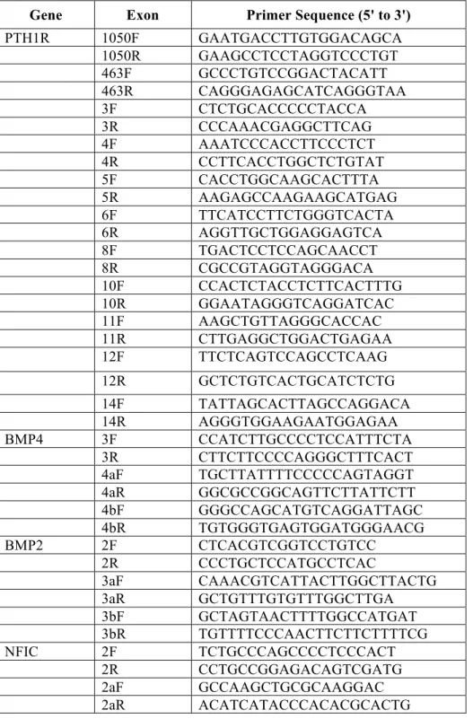
Mutational analysis was carried for 4 candidate genes (below) on 9 patients recruited from the SRA dataset of 28 patients between July 2016 and November 2016 (Table 2). All 9 patients were sporadic cases and unrelated. Assent and consent forms were obtained from the patients and their legal guardians. Extraction and purification of DNA was carried out following collection of a saliva sample from each affected patient (Oragene kits; DNA Genotek, Toronto, Canada). DNA samples were subsequently amplified by polymerase chain reactions (PCR) using primers specific for all coding exons of PTH1R and for exons 3 and 4 of BMP4, for exon 2 and 3 of BMP2 and for exon 2 of NFI-C. Exons were selected based on the exons targeted in knockout mice studies that revealed abnormal root development.The conditions for PCR were as follows: 10 minutes 95°C activation/premelt, followed by 35 cycles of 30 seconds at 95°C melt, 1 minute at 60°C anneal, and 3 min of 72°C extension. Purification of PCR products was completed with Exosapit (USB, Cleveland OH) and sequenced by Eton Biosciences (Durham, NC). The primer sequences used for the four genes are listed in Table 3.
program in Lasergene (DNAstar Inc., Madison, WI, USA) was used for alignments.
Radiographic Analysis of OIEARR
Seven patients included in the SRA database had completed orthodontic treatment or were under treatment. Digitized pretreatment and posttreatment or progress panoramic radiographs were collected for all 7 patients. The median age for the sample was 15 years (IQR 12-16). Four females and 3 males were included and all patients were Hispanic. Six patients had completed orthodontic treatment and their digitized posttreatment panoramic radiographs were collected and only 1 patient was still in treatment and their digitized progress panoramic radiograph was obtained. All patients were treated by orthodontic residents at either the UNC SOD Graduate Orthodontic Clinic or the Wake County Dental Clinic. All subjects were treated comprehensively with fixed edgewise appliances. Five patients were treated with an .022 slot appliance and two were treated in an .018 slot appliances. Demographic and remaining clinical characteristics for the sample are presented in Table 4.
For the clinical portion of the study, the following inclusion criteria were applied: subject age of 9 years or older with pretreatment and posttreatment or progress treatment panoramic radiographs of adequate diagnostic quality. Exclusion criteria included endodontically treated teeth, evidence of
resorption on the pretreatment panoramic radiographs, contributing history of trauma, dilacerations of the maxillary central incisor roots, impacted canines, radiographs with poor diagnostic quality and history of orthodontic treatment prior to available pretreatment radiographs. Panoramic radiographs with obvious errors in head positioning with respect to tilting were also excluded.
resorbed (minor resorption); 2 indicates moderate blunting from 1/4 -1/3 of the root resorbed (moderate resorption); and 3 indicates more than 1/3 of the root has resorbed (severe resorption) (Figure 1). One clinician, a third year orthodontic resident performed all assessments.
Statistical Analysis
Intra-observer reliability was examined by evaluating the maxillary right central incisor for five individuals after 2 weeks of the first assessment and scoring the severity. The weighted kappa statistic was 0.706 indicating substantial reliability. Association between age, treatment duration and sex with incidence of OIEARR was evaluated. An unpaired t-test was used to determine whether age was
associated with incidence of OIEARR. The Wilcoxon Rank Sum test was used to evaluate association of treatment duration with OIEARR and a Fisher’s Exact test was used to determine association of sex. Statistical significance was set a P<.05. Severity of OIEARR was not evaluated for association with age, sex or treatment duration due to lack of sufficient sample size in each category of severity.
Results
Phenotype Characterization
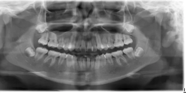
The SRA affected teeth in the sample were evaluated (Table 5). Maxillary central incisors with no other teeth affected was evident in 64.3% of individuals (Figure 2a-c). Three generalized cases were present where all teeth were affected accounting for 10.7% of the sample (Figure 3a-b). In 25% of subjects at least one set of premolars (bilateral) was affected along with the maxillary central incisors (Figure 4). The mandibular second premolars were affected most often followed by maxillary first premolars, maxillary second premolars and mandibular first premolars (Figure 5). In two individuals all premolars were affected. Two individuals had only mandibular second premolars affected. One individual had only maxillary first premolars affected. One individual had only maxillary second premolars affected. One individual had all maxillary premolars affected and mandibular second premolars affected.
Another individual with affected maxillary central incisors and mandibular second premolars had peg-shaped maxillary lateral incisors. No supernumerary teeth were present among the sample. One individual had a dilacerated mandibular lateral incisor.
PCR-Based Mutational Analysis
Among the 9 individuals included in the mutational analysis, five single nucleotide
polymorphisms (SNPs) were found that were previously documented in Ensembl and NCBI: PTH1R c.1389T>C, PTH1R c.178+28G>T, PTH1R c.1116+58T>C, BMP2 c.570A>T, and NFIC c.562+33C>T (Table 6).
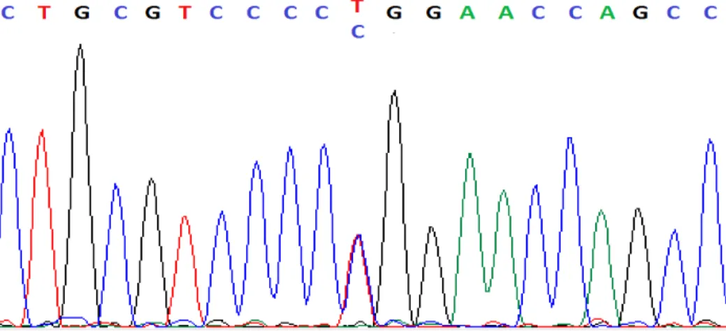
PTH1R c.1389T>C was present in 3 (33%) individuals (Figure 6). The CT genotype was present in 22% of the individuals and the CC genotype was in 11%. The minor allele frequency (MAF) of T is 0.37 in Hispanics and C is the more common allele with a frequency of 0.63.13 PTH1R c.1389T>C is present in exon 14 and results in a synonymous variation with the amino acid remaining asparagine.13 PTH1R c.178+28G>T was present in 1 (11%) individual (Figure 7). The GT genotype was present in this one individual. The MAF of T is 0.29.13 This SNP occurs in the intron region of PTH1R. PTH1R
c.1116+58T>C was present in 1 individual (11%) (Figure 8). The CT genotype was present in this one individual. The MAF of T is 0.36.13 This SNP occurs in the intron region of PTH1R. BMP2 c.570A>T was present in 7 (78%) individuals (Figure 9). The TT genotype was present in 4 (44%) individuals and the AT genotype was present in 3 (33%) individuals. The MAF for A is 0.23.13 This SNP results in a missense mutation and the amino acid change is arginine to serine.13 NFI-C c.562+33C>T was present in 4 (44%) individuals (Figure 10). Three (33%) individuals had the TT genotype and 1 individual had the CT genotype. The MAF of T is 0.37.13 NFI-C c.562+33C>T occurs in the intron region of NFI-C.
Radiographic Analysis of OIEARR
Type 2 SRA. Of the seven individuals, five had resorption in at least one maxillary central incisor and two did not. The variables age, treatment duration and sex were evaluated for association with incidence of OIEARR in the individuals and there was no difference in the incidence of OIEARR based on these variables (Table 7 and Table 8).
There was no difference in incidence of OIEARR based on SRA type. The sample was not large enough to determine if there was a difference in severity based on SRA type.
Discussion
In this study we aimed first to characterize the SRA phenotype based on an established SRA patient database to aid in distinguishing the anomaly from other disorders or pathologic conditions. The phenotype characterization revealed a pattern of affection among the SRA sample. Individuals either had only maxillary central incisors affected, maxillary central incisors and at least one set of premolars affected or all teeth affected. Based on these patterns, three types of SRA can be distinguished- Type 1, Type 2 or Type 3. This study represents the first to clinically categorize SRA patients. Type 1 SRA is localized and characterized by maxillary central incisors with a root length:crown length ratio of 1:1 or less as noted by Volmer Lind.1 In comparison, normal maxillary central incisors have a root length: crown length ratio of greater than 1.6:1.1,14 Type 2 SRA is also localized and based on our sample is
distinguished from SRA by obliteration of the pulp chambers.15 Dentinogenesis imperfecta on the other hand usually presents with tooth discoloration and obliterated pulps as well.15
Lind found that teeth besides maxillary central incisors can be affected with SRA as well but less often including premolars and canines.1 Premolars were the only other teeth affected in our sample unless all teeth were affected in the individual. Lind, however, evaluated a Swedish population while we looked at primarily a sample of Hispanic patients.1 Other studies have found SRA among teeth besides premolars and maxillary central incisors.1,16,17 Marques presented a case report with a Brazilian female with
mandibular central incisors affected along with maxillary premolars and mandibular second premolars.17 Apaghalati et al. noted SRA involving maxillary lateral incisors, which was not seen in our sample.16 Their sample however included a Finnish population.16 It appears that differences may exist based on ethnicity. Pattern of affection should be evaluated in other populations to determine if other types exist. It is possible that Type 2 SRA should include any localized affection of teeth besides the maxillary central incisors including premolars, canines, maxillary lateral incisors and mandibular incisors. Type 1 SRA appears to be the most common and distinct from Type 2 or Type 3 and should remain a separate category.
There was a low prevalence of other dental anomalies among the SRA sample. An association of SRA with other dental anomalies has not been established. Apaghalati et al. reported a 12% prevalence of hypodontia among a Finnish SRA sample as compared to 3.6% in our sample.16 Similarly peg lateral incisors had a low prevalence in Aphaghalti’s sample as in ours.16 Dental anomalies such as hypodontia have a genetic predisposition and don’t appear to be related to the SRA phenotype. The lack of
supernumerary teeth among the sample could also be due to extraction at an early age.
5 SNPs detected in our sample the MAF was at least 0.23 or greater.13 Due to the small sample size in our study and lack of a control group the five polymorphisms found in this study cannot be directly associated with SRA.
We focused on specific exons for BMP2, BMP4 and NFI-C and evaluation of all coding exons in these genes could yield additional SNPs. The regions that yielded no novel mutations or common SNPs can be used to narrow the search for the genetic etiology of SRA. Other genes involved in root
development such as sonic hedgehog, transforming growth factor β and wingless-type should also be investigated in the future. Whole genome or whole exome sequencing are alternate routes for detecting the specific genes that are associated with SRA but are costly and more time consuming.
A limitation of our mutational analysis was our small sample size. A larger sample with all SRA types would allow genotype and phenotype associations. Additional studies should also include families with SRA affected individuals; our sample was limited to sporadic cases. We collected saliva samples for our DNA extraction. Some of the sequencing results had evidence of contaminants. Blood or buccal cell samples would be ideal for future studies as to limit the contaminants in the sequencing results.
Severity of OIEARR was evaluated in SRA affected maxillary central incisors to determine whether these teeth tend to experience severe resorption during comprehensive treatment. Our results are similar to what has been published for normal teeth for the severity of resorption. Resorption of greater than one-third of the root occurs in 1- 5% of teeth during orthodontic treatment.18 Only one incisor (7%) experienced severe OIEARR equivalent to greater than one-third of the root. Levander and Malmgren’s evaluation of 390 maxillary incisors, had comparable results.12 They found no resorption in 34% of teeth, minor resorption in 48%, moderate resorption in 17% of incisors and severe resorption in 1% of
incisors.12 Most teeth in our study had none or minor resorption.12 Our study also agrees with Levander and Malmgren’s investigation of root resorption based on root form. They found that short roots do not have a higher degree of root resorption but that blunt or pipette shaped roots do, however they did not document whether any of the short rooted teeth had SRA.12 Although SRA affected incisors are not more susceptible than normal incisors to experience OIEARR and are not more likely to have severe resorption, because the roots are significantly shorter than normal incisors at the start of treatment any presence of resorption can be more detrimental compared to the same amount of resorption in a normal incisor. This is important to explain to SRA patients at the start of orthodontic treatment.
One potential bias that may have affected the outcome of the incidence and severity of OIEARR is that all providers were aware of the short roots and may have adjusted treatment mechanics to minimize resorption including lighter forces and avoiding extraction treatments. Two patients did have premolar extractions and one of these patients was the only patient that experienced greater than minor OIEARR. One of his incisors had moderate resorption and the other had severe resorption. This patient also had the longest treatment duration, which is known to contribute to increased risk for resorption. Another reason for the severity of resorption in this patient may be a genetic predisposition, which was not tested for.
The limitations of the radiographic evaluation of OIEARR in our study include the small sample size. Due to the low prevalence of SRA, establishing a large cohort of SRA patients treated
research related to these patients some clinicians may be reluctant to treatment them comprehensively. The results of this portion of the study provide reassurance to clinicians that SRA affected incisors have a similar risk compared to normal incisors.
For this study, panoramic radiographs were used to qualitatively assess OIEARR. Although other techniques such as CBCT have been shown to allow better visualization of root resorption, Stramotas provided evidence that measuring root lengths on panoramic films are relatively accurate when there is not excessive tilt of the occlusal plane. 18,20 In our study, any radiograph showing evidence of significant positioning errors were excluded. Additionally, this was a retrospective study where we were limited to the available radiographs for the orthodontic patients. 3-D imagining is not the standard of care for orthodontic diagnostic records and in most cases panoramic radiographs are taken prior to treatment, for progress and final records. Also, our radiographic evaluation was meant to mimic a clinical environment where in most orthodontic practices there is rarely an indication for 3-D imaging to assess root resorption after treatment.
We opted for qualitative evaluation over quantitative because some form of calibration would have been necessary on the panoramic films for measurement of root length. Given that the study was done in a university setting and a retrospective study, calibration for example with an object of known length in the panoramic image, would have required a more controlled setting for the imaging.
Conclusions
The following conclusions can be drawn from this investigation.
1. SRA can present as three distinct phenotypes. Variations may exist based on race.
2. Mutational analysis of PTH1R, and select exons in BMP2, BMP4 and NFI-C did not reveal novel mutations but 5 common SNPs. Further investigation is needed to determine the genetic etiology of SRA.
3. Most SRA affected maxillary central incisors have none or minor OIEARR; they are not more susceptible to OIEARR or severe root resorption during orthodontic treatment.
Tables
Table 1: Descriptive characteristics of the SRA sample in the phenotype characterization (n=28)
Characteristics n (%)
Mean age (y) 14.8 ± 5.7
Sex
Female 17 (60.7)
Male 7 (25.0)
Unknown 4 (14.3)
Ethnicity
Hispanic 24 (85.7)
Unknown 4 (14.3)
Source
UNC SOD 13 (46.4)
Wake County Dental 4 (14.3)
Private Practice 11 (39.3)
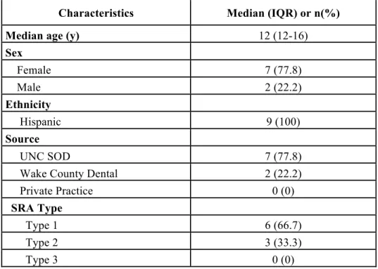
Table 2: Descriptive characteristics of the sample in the mutational analysis (n=9)
Characteristics Median (IQR) or n(%)
Median age (y) 12 (12-16)
Sex
Female 7 (77.8)
Male 2 (22.2)
Ethnicity
Hispanic 9 (100)
Source
UNC SOD 7 (77.8)
Wake County Dental 2 (22.2)
Private Practice 0 (0)
SRA Type
Type 1 6 (66.7)
Type 2 3 (33.3)
Table 3: PTH1R, BMP4, BMP2, and NFI-C primers used for PCR amplification and sequencing
Gene Exon Primer Sequence (5' to 3')
PTH1R 1050F GAATGACCTTGTGGACAGCA
1050R GAAGCCTCCTAGGTCCCTGT
463F GCCCTGTCCGGACTACATT
463R CAGGGAGAGCATCAGGGTAA
3F CTCTGCACCCCCTACCA
3R CCCAAACGAGGCTTCAG
4F AAATCCCACCTTCCCTCT
4R CCTTCACCTGGCTCTGTAT
5F CACCTGGCAAGCACTTTA
5R AAGAGCCAAGAAGCATGAG
6F TTCATCCTTCTGGGTCACTA
6R AGGTTGCTGGAGGAGTCA
8F TGACTCCTCCAGCAACCT
8R CGCCGTAGGTAGGGACA
10F CCACTCTACCTCTTCACTTTG
10R GGAATAGGGTCAGGATCAC
11F AAGCTGTTAGGGCACCAC
11R CTTGAGGCTGGACTGAGAA
12F TTCTCAGTCCAGCCTCAAG
12R GCTCTGTCACTGCATCTCTG
14F TATTAGCACTTAGCCAGGACA
14R AGGGTGGAAGAATGGAGAA
BMP4 3F CCATCTTGCCCCTCCATTTCTA
3R CTTCTTCCCCAGGGCTTTCACT
4aF TGCTTATTTTCCCCCAGTAGGT
4aR GGCGCCGGCAGTTCTTATTCTT
4bF GGGCCAGCATGTCAGGATTAGC
4bR TGTGGGTGAGTGGATGGGAACG
BMP2 2F CTCACGTCGGTCCTGTCC
2R CCCTGCTCCATGCCTCAC
3aF CAAACGTCATTACTTGGCTTACTG
3aR GCTGTTTGTGTTTGGCTTGA
3bF GCTAGTAACTTTTGGCCATGAT
3bR TGTTTTCCCAACTTCTTCTTTTCG
NFIC 2F TCTGCCCAGCCCCTCCCACT
2R CCTGCCGGAGACAGTCGATG
2aF GCCAAGCTGCGCAAGGAC
Table 4: Demographic and clinical characteristics of SRA sample evaluated for OIEARR
Characteristics Median (IQR) or n (%)
Demographics
Age (y) 15 (13-17)
Sex
Female 4 (57.1)
Male 3 (42.9)
Ethnicity
Hispanic 7 (100)
SRA Type
Type 1 4 (57.1)
Type 2 2 (28.6)
Type 3 1 (14.3)
Clinical
Treatment Duration (mo) 24.2 (21.4-29.5) Treatment Complete
Yes 6 (85.7)
No 1 (14.3)
Extraction
Yes 2 (28.6)
No 5 (71.4)
Table 5: Phenotype characterization of SRA sample (n=28)
Variable n (%)
Teeth Affected
Maxillary central incisors only 18 (64.3)
Max centrals and at least one premolar set 7 (25.0)
All teeth 3 (10.7)
Presence of other Dental Anomalies
Dilacerated Roots 1 (3.6)
Congenitally missing at least one tooth
(except 3rd molars) 1 (3.6)
Peg laterals 1 (3.6)
Table 6: Sequencing variations found in mutational analysis (n=9)
Gene Exon Sequence Variation Functional Consequence N (%) Minor Allele Frequency
PTH1R 4 c.178+28G>T Intron variant 1 (11%) T= 0.29
12 c.1116+58T>C Intron variant 1 (11%) C=0.46
14 c.1389T>C Synonomous variant 3 (33%) T=0.36
BMP2 3 c.570A>T Nonsynonomous variant 7 (78%) A=0.23
NFI-C 2 c.562+33C>T Intron variant 4 (44%) T=0.37
Table 7: Mean age of patients based on incidence of OIEARR
Incidence of OIEARR n(%) Mean age (SD) CI 95% P-value
No 2 (29) 18.0 (1.41) 5.29-30.7 0.0681
Yes 5 (71) 14.4 (1.95) 11.9- 16.8
Table 8: Median treatment time of patients based on incidence of OIEARR
Incidence of OIERARR n (%) Median treatment time (IQR) CI 95% P-value
No 2 (29) 16.95 (9.89-24.02) 9.89-24.02 0.1905
Yes 5 (71) 28.09 (24.21-30.82) 18.86-39.98
Figures
Figure 2.1: Pretreatment panoramic radiograph of 18 yr. old Hispanic male with maxillary central incisors affected (Type 1 SRA). Patient was included in the phenotype characterization, mutational analysis and radiographic analysis of OIEARR.
Figure 2.2: Periapical of SRA affected maxillary central incisors.
Figure 3.1: Panoramic radiograph of 9 year old Hispanic female with generalized SRA (Type 3). The image reveals short roots at the time of eruption of second premolars and maxillary canines.
Figure 4: Panoramic radiograph of 13 yr. old Hispanic female with maxillary central incisors, mandibular and maxillary premolars affected (Type 2 SRA). Patient was included in the phenotype characterization.
Figure 5: Affection of premolars among 7 individuals with at least one set of premolars affected in the phenotype characterization.Mandibular second premolars were affected in 6 of the 7 individuals.
0 1 2 3 4 5 6 7
Maxillary First Premolars
Maxillary Second Premolars
Mandibular First Premolars
Mandibular Second Premolars
Number of
Individuals
Figure 6: Chromatogram of PTH1R c.1389T>C variation present in exon 14 of patient with Type 1 SRA.
Figure 7: Chromatogram of PTH1R c.178+28G>T variation present in intron region in patient with Type 1 SRA.
Figure 9: Chromatogram of BMP2 c.570A>T variation present in exon 3 of patient with Type 2 SRA.
Figure 10: Chromatogram of NFIC c.562+33C>T variation in present in exon 2 of patient with Type 1 SRA.
Figure 11: Incidence of OIEARR of SRA affected maxillary central incisors after orthodontic treatment.
0
20
40
60
80
Yes
No
Percentage
of Maxillary
Central
Incisors
Figure 12: Severity of OIEARR of SRA affected maxillary central incisors after orthodontic treatment.
0
10
20
30
40
50
60
0
1
2
3
Percentage
of Maxillary
Central
Incisors
REFERENCES
1. Lind V. Short root anomaly. Scand J Dent Res. 1972; 80(2): 85-93.
2. Puranik CP, Hill A, Henderson Jeffries K, Harrell SN, Taylor RW, Frazier-Bowers SA. Characterization of short root anomaly in a Mexican cohort--hereditary idiopathic root malformation. Orthod Craniofac Res. 2015 Suppl 1:62-70.
3. Apajalahti S, Arte S, Pirinen S. Short root anomaly in families and its association with other dental anomalies. Eur J Oral Sci 1999; 107: 97 –101.
4. United States Census Bureau. FFF: Hispanic Heritage Month.
https://www.census.gov/newsroom/facts-for-features/2016/cb16-ff16.html. Published October 12, 2016. Accessed February 25, 2017.
5. Ono W, Sakagami N, Nishimori S, Ono N, Kronenberg H. Parathyroid hormone receptor signaling in osterix-expressing mesenchymal progenitors is essential for tooth root formation. Nat.
Commun.2016; 7:11277. doi: 10.1038/ncomms11277
6. Hosoya A, Kim J, Cho S. BMP4 signaling regulates formation of Hertwig’s epithelial root sheath during tooth root development. Cell Tissue Res. 2008; 333: 503-509.
7. Rakian A, Yang W, Gluhak-Heinrich, et al. Bone morphogenetic protein-2 gene controls tooth development in coordination with formation of the periodontium. Int J Oral Sci.2013;5:75–84. 8. Yang W, Harris MA, Cui Y, Mishina Y, Harris SE, Gluhak-Heinrich J. Bmp2 Is required for
odontoblast differentiation and pulp vasculogenesis. J Dent Res.2012; 91(1):58-64.
9. Steele-Perkins G, Butz KG, Lyons GE, et al. Essential roles for NFI-C/CTF transcription-replication factor in tooth root development. Mol Cell Biol.2003;23(3):1075–1084.
10. Park J, Herr Y, Kim H, Gronostajski RM, Cho M. Nfic gene disruption inhibits differentiation of Odontoblasts Responsible for Root Formation and Results in Formation of Short and Abnormal Roots in Mice. J Periodontol.2007;78(9):1795-1802.
11. Newman WG. Possible etiologic factors in external root resorption. Am J Orthod 1975; 67: 522 –39. 12. Levander E, Malmgren O. Evaluation of the risk of root resorption during orthodontic treatment: a
study of upper incisors. Eur J Orthod. 1988;10(1):30-8. 13.Variant Displays. Ensembl.
http://useast.ensembl.org/Homo_sapiens/Variation/Population?r=20:6777968-6778968;v=rs235768;vdb=variation;vf=124762. Published December 2016. Accessed March 11, 2017.
14. Haghanifar S, Moudi E, Abbasi S, Bijani A, Bejeh MirAP, Ghasemi N. Root-Crown Ratio in Permanent Dentition Using Panoramic Radiography in a Selected Iranian Population. J Dent Shiraz Univ Med Sci., December 2014; 15(4): 173-179.
Saunders;2009.
16. Apajalahti S, Holtta P, Turtola L, Pirinen S. Prevalence of short-root anomaly in healthy young adults. Acta Odontol Scand. 2002;60:56–59.
17. Marques LS, Generoso R, Armond MC, and Pazzini CA. Short-root anomaly in an orthodontic patient. Am J Orthod Dentofacial Orthop. 2010;138:346-8.
18. Weltman B, Vig K, Fields HW, Shanke, S, Kaizar E. Root resorption associated with orthodontic tooth movement: A systematic review. Am J Orthod Dentofacial Orthop. 2010;137:462-76. 19. Lupi J, Handleman CS, Sadosky C. Prevalence and severity of apical root resorption and alveolar
bone loss in orthodontically treated adults. Am J Orthod Dentofacial Orthop.1996;109(1):28-37. 20. Stramotas S, Geenty JP, Petocz P, Darendeliler MA. Accuracy of linear and angular measurements on