1 Abstract
The ability to store iron through the use of iron storage proteins, ferritins, has evolved as
an advantageous strategy for numerous diatom species. Iron is a micronutrient in the ocean that
typically exists at low concentrations throughout much of the marine system. Therefore, iron
deficiency often plays a central role in controlling primary productivity, and thus, diatom
populations within the ocean (Martin, 1990). This project investigates the expression of an iron
storage gene, ferritin, in three pennate diatoms and four centric diatoms. The strains were
cultured at Bigelow Laboratory for Ocean Sciences and the University of North Carolina at
Chapel Hill (UNC) and grown as triplicate cultures under various iron conditions. This
experiment allowed for the comparison of ferritin gene expression along with iron storage
capabilities, in contrasting iron environments among phylogenetically distinct diatoms. This was
achieved by utilizing the ΔCt method. Our results suggest that pennates may upregulate ferritin
levels in high iron environments. Marchetti et al (2009) similarly demonstrated that pennates
such as Pseudo-nitzschia granii store iron during temporary pulses, which allows them to
continue to proliferate once iron limited conditions return. Two of the examined centrics,
however, demonstrated greatest ferritin gene expression under low iron conditions. This result is
counterintuitive, as ferritin is a storage gene and high expression during low iron levels appears
inefficient. However, this finding supports the theory that centrics may be using their ferritin as
an iron buffer and thus eludes to physiological differences in the way these two diatom lineages
are storing iron and utilizing their ferritin genes. There is also evidence of a biogeographical
distinction among ferritin use in certain species. Diatoms in open ocean regions, where iron is
often limited, may be utilizing their ferritin differently than those persisting in high- iron coastal
2 Introduction
Iron is a critical micronutrient to all living organisms. However, iron exists in low
concentrations in surface waters (<1 nM) throughout much of the world’s oceans and is the most
growth limiting micronutrient to ecosystem-level primary productivity in 30-40% of the marine
environment (Armbrust, 2009). Because of this, iron plays a key role in controlling diatom
populations and abundance. Open ocean regions often experience sporadic pulses of iron from
aeolian dust blown from continental deserts and periodic upwelling events. During intervals
between such inputs, these regions must depend on tightly recycled iron to fuel primary
productivity. Coastal regions, however, often experience constant influxes of iron from river
runoff, sediment inputs, coastal upwelling, and other continental sources. Thus, coastal regions
are not often classified as iron-limited regions (though iron limitation has been found in some
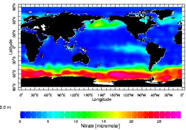
coastal regions) (Moore et al, 2002). Figure 1 displays nitrate concentrations in the ocean
system. The warmer colors depict areas with higher concentrations of nitrate. These regions are
often classified as High Nitrate, Low Chlorophyll (HNLC) regions and cover approximately 20%
of the ocean system. These locations contain a substantial amount of nitrate, another essential
nutrient for phytoplankton, however are often low in primary productivity (chlorophyll) due to a
deficit of iron. The well-characterized regions of the oceans known to be iron limited are the
3
Iron has an essential role in cellular and molecular functions in the phytoplankton
community. This micronutrient is particularly utilized in the electron transport chain due to iron
demanding proteins and enzymes that are effectively able to transfer electrons. Iron is also
involved in synthesis of chlorophyll and reduction of important elements such as carbon dioxide
(Street and Paytan, 2005). Phytoplankton have developed mechanisms to cope with low iron
environments. These include the ability to lower iron requirements via molecular mechanisms
and also increasing iron uptake by using iron storage proteins. In iron-limited environments,
many diatoms down-regulate their use of iron-demanding proteins and switch to a reliance on
less efficient, iron-independent proteins (Marchetti et al, 2012). When iron concentrations are
low, certain diatoms convert to the use of flavodoxin, an iron-free protein that is less efficient
than the iron-demanding protein, ferredoxin (McKay, Geider, & LaRoche, 1997).
Iron storage is an advantageous ability possessed by a plethora of organisms including,
bacteria, protists and plants (Theil, 1987). This adaptation allows certain phytoplankton to store
iron and thus persist during low iron intervals. Iron can be stored via iron storage proteins called Figure 1. Annual average nitrate concentrations in the
4
ferritins. Ferritins allow for the rapid uptake and storage of iron (Marchetti et al, 2009). Because
free iron within the cell can be toxic, it is advantageous for organisms to store iron in these
specialized proteins.
Ferritin is a spherical protein with 24 subunits that form the nanocage and are arranged as
a hollow shell called an apoferritin. Iron is stored in the ferritin iron core located in the center of
the sphere as Fe (III), called ferrihydrite. The iron core commonly possesses 2,000 Fe (III) iron
atoms, although the molecule can contain up to 4,500 iron atoms. Gated pores present around the
nanocage allow iron atoms to enter and exit the core, permitting the cell to regulate intracellular
iron (Harrison, 2010). The control of ferrous iron through the gated pores is regulated by contact
between ferrihydrite in the iron core and biological reductants outside the nanocage (Theil, Liu,
& Tosha, 2008).
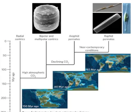
There are two major phylogenetic groups of diatoms: pennates and centrics. Centric
diatoms evolved approximately 150 million years ago (Mya) and possess radial symmetry.
Centrics can be classified as either bipolar or radial. Pennate diatoms diverged from centrics
between 50-90 Mya and these cells have bilateral symmetry and appear needle-like (Figure 2).
This group consists of raphids, pennates with a raphe, and araphids, pennates lacking a raphe. A
raphe is a longitudinal slit which allows the diatoms to glide on a substrate. The evolution of the
raphe is significant to diatom diversification, as it expanded ecological niche availability.
However, the particular benefit of a raphe to open ocean pennate diatoms is yet to be fully
understood. As is depicted in the image below collected from Armbrust (2009), centric diatoms
evolved earlier in Earth’s history, during a more anoxic environment in which iron was more
readily available in the ocean. Pennates diverged millions of years later, during a more oxic
5
Pennate and centric diatoms only share 57% of their genes and hence can differ
tremendously in their life processes and abilities to cope with fluctuating levels of iron in their
environments (Bowler et al, 2008). Iron storage has been observed largely in pennates. It has
been shown in pennate species, such as Pseudo-nitzschia granii, that ferritins allow the diatom to
rapidly uptake iron, during iron pulses into the open ocean, and store it. This iron storage
capability permits pennate diatoms to continue to proliferate even once iron-limited conditions
have returned (Marchetti et al, 2009). Because of their ability to use stored iron in times of iron
limitation, it is speculated that pennates may have an ecological advantage over centrics in open
ocean environments where iron inputs can be ephemeral. The presence of the ferritin gene has
more recently been observed in certain centric diatoms as well. The function of the ferritin gene
in centric diatoms, and how centrics utilize their stored iron, is not as well understood as in Figure 2. Approximate centric and pennate evolutionary divergence
6
pennate species. The objective of this project is to investigate if there are physiological
differences in the way pennates and centrics use their ferritin gene to store iron and regulate
ferritin gene expression under varying iron conditions.
Materials and Methods
This experiment, which is part of a greater project investigating the ecological
importance of iron storage in diatoms, is a collaboration between Benjamin Twining’s research
group at Bigelow Laboratory for Ocean Sciences (East Boothbay, ME) and the Marchetti Lab at
UNC. This particular aspect of the project investigated ferritin gene expression of three pennate
and four centric diatoms when grown under varying iron concentrations (Table 1).
Isolate Phylogeny Biogeographical Isolation
Location
Pseudo-nitzschia granii
UNC 1102
Raphid pennate North Pacific (open ocean)
Thalassionema frauenfeldii
CCMP 1798
Araphid pennate Caribbean Sea (coastal)
Amphora coffeaeformis
CCMP 127
Raphid pennate Nantucket Sound MA (coastal)
Minutocellus polymorphus
CCMP 3303
Bipolar centric South Atlantic (coastal)
Thalassiosira sp. strain
NH16
Bipolar centric North Pacific (coastal)
Thalassiosira rotula
CCMP 3096
7
Marine Microbial Eukaryote Transcriptome Sequencing Project (MMETSP) is a
publically available database that contains the transcriptomes of many centric and pennate
diatoms. A known reference gene of ferritin from Pseudo-nitzschia granii was searched via
BLAST across this database to find both pennate and centric diatoms that possess a ferritin gene
homolog. Using the MMETSP database, the sequences of ferritin (FTN), the target gene, and
actin (ACT), which served as the housekeeping gene, were queried in order to identify the
transcriptomes of the species selected for this study.
To measure the relative gene expression of ferritin, the ΔCt method was utilized. This
method relies on the threshold cycle of the target gene and the housekeeping gene, actin, when
amplified in a quantitative PCR (qPCR). We also compared the results for M. polymorphus using
the ΔCt method to those of creating qPCR standards to ensure the accuracy of the ΔCt method.
A housekeeping gene is that which is required for basic cell maintenance and is constitutively
expressed in both normal and sub-optimal conditions, therefore, the gene can serve as a reference
when assessing the expression levels of the target gene. The expression of one gene relative to
another can be quantified using the ΔCt method equation displayed in Eq. 1. This equation uses
the threshold cycle differences between the target and housekeeping gene (Pfaffl, 2001).
Eq. 1. R = 2-[Ct sample-Ct housekeeping]
The particular diatom strains analyzed were originally purchased from the National
Center for Marine Algae and Microbiota (NCMA) located in East Boothbay, Maine. These
strains were then grown in 30mL polycarbonate tubes at Bigelow Laboratory, excluding
Amphora coffeaeformis which was cultured at UNC. The strains were grown in high iron (Total Table 1. Diatom species investigated for ferritin gene expression
8
Fe = 8.41 µM), medium iron (Total Fe = 1.37 µM Fe) and low iron (various concentrations to
achieve iron-limited growth) conditions, each in the presence of 100 µM EDTA, with the
exception of Thalassionema frauenfeldii and Amphora coffeaeformis which were grown in only
medium and low iron. The goal in the lowest iron treatment was to slightly iron limit the diatoms
in order to assess how iron limitation would affect the expression of ferritin in each diatom
isolate. We did strive to avoid severe iron limitation that would induce other molecular
mechanisms for coping with low iron availability. This iron limited concentration was
determined for each species by growing each strain under various low iron conditions and
targeting for ~10% reduction in growth rate compared to the higher iron cultures. Growth rates
were determined by fluorometric measurements (Brand, Sunda, & Guillard, 1981).
Once the respective strains were successfully iron limited, displaying a consistently
reduced growth rate in the low iron medium, the cultures were grown in triplicate 1 liter
polycarbonate bottles under each iron concentration. After reaching the respective high biomass
determined by fluorometer measurements, the 1 liter cultures were then filtered through a 0.2
micron filter. The filters were then put in labeled cryotubes and flash frozen in liquid nitrogen.
The frozen filters were then sent from the Twining Laboratory to the Marchetti Laboratory. This
was done for all strains except Amphora coffeaeformis, currently being cultured at UNC to
achieve iron limitation.
The RNA was extracted from the diatom filters using the RNAqueous 4-PCR kit
(Ambion). Once the RNA was successfully extracted, the solution could be quantified using
absorbance measurements via spectrophotometry with the Nanodrop. An absorbance peak at 230
nm revealed contaminants such as phenols and carbohydrates, which absorb at that wavelength.
9
(Qiagen). Once the samples were free of contamination they were DNased, a process in which
the samples were treated with an enzyme, DNase (Ambion), which served to catalyze a reaction
to degrade any contaminating DNA to ensure the RNA sample was pure. Following DNasing, a
qPCR run was performed with each RNA sample and eukaryotic 18S-specific primers to check
for remaining DNA contamination. These primers are very sensitive and in the qPCR will
amplify any DNA that is in the sample, therefore, the qPCR had to be prepared under a
laboratory hood in a highly sterile environment. If the qPCR revealed DNA contamination then
it was necessary to re-DNase the contaminated samples. DNasing results in a small loss of the
RNA sample and therefore it was not ideal to have to perform this step more than once. After
each of the RNA samples were free from contamination the next step of reverse transcription
was performed. Reverse transcription (RT) is the process of creating a complementary DNA
(cDNA) template from the RNA. General RT primers were used, which reversed transcribed all
mRNA to create the total cDNA template.
For each species, ferritin and actin OligoDT primers were designed by searching via
BLAST a known Pseudo-nitzschia granii ferritin and actin reference gene sequence of each
examined diatom within the MMETSP database. Thalassionema frauenfeldii and Amphora
coffeaeformis both possess two ferritin genes. This is likely a result of horizontal gene transfer
(Groussman et al, 2015). Appropriate OligoDT primers were designed for both ferritin genes.
Following RT, a PCR was conducted using the cDNA to test the ability of the ferritin and actin
primers to amplify their target sequences. After completing the PCR, a gel electrophoresis using
the ferritin and actin PCR product was performed to confirm the correct size of DNA fragment
10
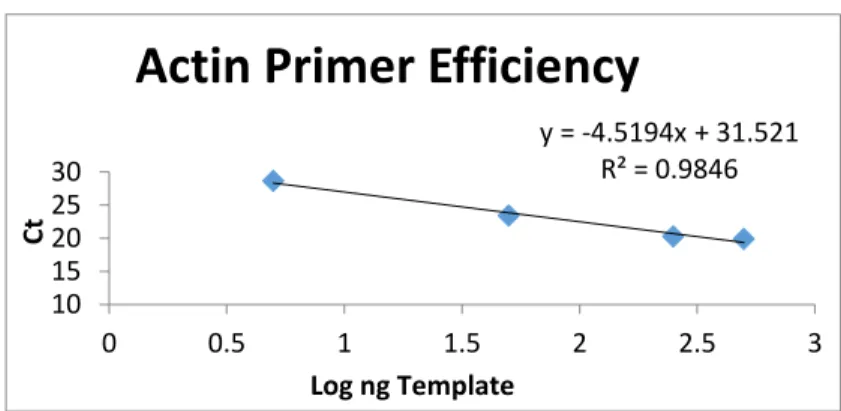
Primer efficiency testing was also necessary and conducted using qPCR to ensure the
primers yielded the correct value of 2 from the efficiency equation (Eq. 2). Bias or inefficient
primers could have produced results that did not appropriately reflect the relationship between
the housekeeping gene and the target gene. A value of E (efficiency) equal to 2 is anticipated to
ensure the primers were competent and could be used in the ΔCt method. The reason for the
primer efficiency to yield E=2 is because for each cycle the amount of product should double.
Eq. 2. E=10(-1/slope)
y = -4.5194x + 31.521 R² = 0.9846
10 15 20 25 30
0 0.5 1 1.5 2 2.5 3
Ct
Log ng Template
Actin Primer Efficiency
Figure 3a.Actin primer efficiency
Figure 3b. Ferritin primer efficiency
y = -1.6128x + 29.197 R² = 0.8041
10 15 20 25 30
0 0.5 1 1.5 2 2.5 3
Ct
Log ng Template
11
Eq. 2 represents the calculation necessary to find the efficiency of the primers. To do this,
the log ng template of both the actin and ferritin primers was compared to the Ct. The slope
derived from the plot of the log ng template versus Ct for both primers was used to calculate the
primer efficiency (Figures 3a and 3b). It was found that most of the primers were acceptable,
except for those designed for the ferritin gene of M. polymorphus. Because of this, it was
necessary to create gene standards to compare those results to the ΔCt method results.
The standard method for gene expression analysis is based on quantified copies per
microliter. Standards were created by incorporating both the ferritin and actin gene into a vector
that contained kanamycin resistance. That vector was then transformed into the competent
bacterial cells of Escherichia coli (E. coli) that were grown-up on an agar plate containing
kanamycin. This ensured that only the E. coli cells which had taken up the vector (with our target
gene fragment) could grow on the plate. Using a toothpick, certain bacterial colonies were
removed and underwent PCR using M13 forward and reverse primers designed for the specific
vector. The PCR product was then run on a gel and the bands which showed positive for the
ferritin and actin genes were grown up overnight in liquid colonies. The following day the
plasmids were extracted from the removed gel bands via a plasmid extraction kit (Qiagen). The
plasmid sample then underwent restriction enzyme digestion to linearize the desired gene insert.
Dilutions of 101-106 of the DNA were created to determine how much transcript was in each
sample in copies per microliter. A qPCR was run including both the standard dilutions and the
cDNA template to generate a standard curve. The graph of Ct cycles versus number of amplicon
copies showed a standard curve in which the number of copies for both actin and ferritin
12
When comparing the results of M. polymorphus ΔCt method to those derived from
creating standards, it was found that both methods portrayed relatively the same trend, with a
significant difference in ferritin gene expression between high and low treatments (Figures 4a
and 4b). Because of this result, it was accepted that the DNA was likely very diluted during the
standard curve creation process, however, that does not necessarily mean the ΔCt method is not
reflecting the appropriate relationships when the concentrations are normal.
Once the primers were deemed efficient, a full plate qPCR was performed. Each cDNA
sample was loaded, in triplicate, into the plate wells for samples with ferritin primers and
separate samples containing actin primers. Water was used as a negative control. Running a full
plate allowed the assessment of ferritin gene expression compared to actin gene expression
within the various iron treatments. This was done by comparing the average threshold cycle
numbers of the triplicates for each iron condition with the ferritin primers to that of its respective
triplicate treatment with actin primers. This relative comparison of gene expression allowed for Figure 4a.M. polymorphus relative
ferritin gene expression derived using Standard method assessment.
13
us to evaluate under which iron conditions certain species were increasing their target gene
expression.
Results
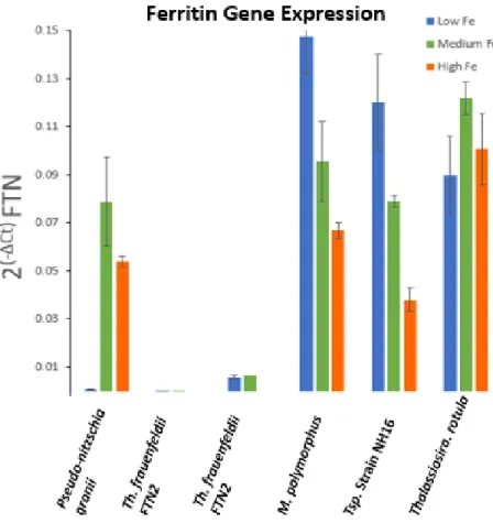
Using the ΔCt method, ferritin gene expression of diatoms in high, medium, and low iron
conditions was observed. There is a significant difference in ferritin gene expression between the
open ocean pennate diatom, P. granii, and the coastal centric diatoms. The open ocean pennate,
P. granii, showed a 72-fold increase in ferritin gene expression from the low to high culture, the
greatest ferritin gene expression being under high iron conditions. In contrast, the centric
diatoms, M. polymorphus and Thalassiosira sp., upregulated their ferritin gene in low iron
conditions (Figure 5). There was an approximate 2-fold and 3-fold decrease in ferritin expression
from the low to high iron cultures for both M. polymorphus and Thalassiosira sp. respectively. T.
rotula displayed very little variation in ferritin expression in all three iron treatments, however, it
expressed ferritin at a relatively large amount in all treatments. Th. frauenfeldii did not appear to
produce as many transcripts and perhaps was not using its ferritin to the same degree as the other
diatoms (Figure 5). This anomalous result could also be a consequence of a high actin levels in
Th. frauenfeldii, therefore, relative to actin, ferritin is being expressed at low levels. Although
there does not appear to be differences in ferritin gene expression between the middle and low
14
Figure 5. The use of the ΔCt method to show ferritin gene expression by pennate and centric diatoms in various iron conditions.
The results of the centric diatoms appear counterintuitive for an iron storage protein;
portraying lowest gene expression within a high iron environment. It is puzzling and intriguing
that the centrics would not express the iron storage gene in an environment that would be optimal
for storing iron. These results suggest ferritin is functioning differently in centric diatoms than in
pennates.
Discussion
This study successfully reveals that the role and expression of ferritin can vary among
15
previous observations made by Marchetti et al (2009). Our findings correspondingly displayed
that P.granii had high ferritin gene expression in high iron conditions, allowing them to store
iron when available, and had low ferritin gene expression when iron conditions were limited and
storing iron was not ideal. The differing iron conditions in which pennate and centrics express
ferritin begs the question of how the centrics are using their iron storage genes? This data
supports the idea that centrics are using their ferritins differently than pennates. As the centrics
express their iron storage gene the most in low iron conditions, these diatoms may not be
utilizing their ferritins purely as a means for storing iron during times in which iron is in surplus
supply in the environment.
Similar results to those observed by the examined centric diatoms have been reported
with Chlamydomonas reinhardtii, a green microalgae. C. reinhardtii possess Fer1 and Fer2, two
unlinked genes that encode for ferritins. In a study conducted by Long et al (2008) involving iron
assimilation in C. reinhardtii, it was found that the concentration of Fer1 was at high levels in
iron-limited environments. These results are similar to those observed in M. polymorphus and
Thalassiosira sp. Long et al (2008) suggest that ferritin is playing a role in buffering iron or
holding on to iron when it is released during photosynthesis breakdown. Thus, the increased
expression of Fer1 in low iron environments allows for iron binding within the chloroplast of
cells that are iron limited (Long et al, 2008). The comparable results between this green alga and
these centric diatoms suggest that centrics may be using their ferritins in a similar way to C.
reinhardtii. Centrics could be using the ferritin geneas a means to increase the binding capacity
of free iron in iron-limited environments, utilizing their ferritin proteins to buffer iron and
16
An alternative reason for the behavior of the centrics under low iron conditions was
proposed by Dr. William Sunda. He suggested that ferritin protein levels are increasing in the
centrics under low iron conditions as a precaution against iron toxicity in iron-limited cells in the
event of an ephemeral iron input. In this case, ferritin is upregulated in preparation to capture and
store iron during a sudden high influx of iron, as without ferritins, iron levels could quickly
become toxic to the cell.
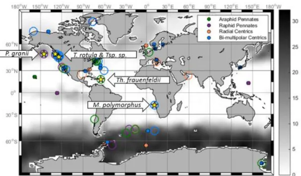
An important distinction that can be concluded from these results is the likelihood of
biogeography playing a key role in determining the use of the ferritin gene in diatom species.
Although there was only a single open ocean species, the pennate P. granii appeared to be using
its ferritins to store iron when it was readily available in its environment. The P. granii species
used in this experiment was isolated from the Northeast Pacific, where iron concentrations are
often sub-nanomolar and iron inputs are short-lived, coming from dust and periodic upwelling
events (Figure 1 and 6). Thus, it is beneficial for an open ocean species living in this
environment to have the capability to store iron during the sporadic iron events in order to
continue to proliferate during iron limited intervals. All other species were coastal diatoms
(Table 1 and Figure 6). The coastal species do not appear to be using their ferritins to store iron
when it is readily available in the environment. Coastal regions of the ocean commonly (but not
always) have a more constant influx of iron, which is often not the limiting nutrient in these
areas. Therefore, it would be probable that coastal species utilize their ferritin gene for
alternative purposes other than strategies developed by open ocean strains to survive in low-iron
environments. This biogeography distinction is likely a factor controlling how diatom species
17 Future Directions
To expand the depth of understanding of the factors controlling ferritin expression in
these two distinct diatom lineages, it would be important to analyze gene expression in a greater
number of open ocean centrics and pennates. Our study was limited to a single open ocean
species, however including both another open ocean pennate and at least two open ocean
centrics, which contain ferritin, would permit a more comprehensive understanding of the factors
controlling how and for what purpose these species are using ferritin. Expanding this study
would provide a stronger argument for a biogeographical distinction and the influence of
environmental pressures determining how diatoms utilize their ferritins.
Further investigation into the role of ferritin as an iron buffer in diatoms could provide a
more solid foundation for the explanation of centric diatoms usingferritins to buffer iron in the
18
centrics possessing ferritin could strengthen or weaken this claim depending on gene expression
results.
It has been shown that ferritin proteins can be used to store iron in high iron
environments and to serve as an iron buffer in the cell. However, the function of the ferritin gene
is not definitively limited to these two purposes. Further exploration into other functions of
ferritin proteinswithin the phytoplankton community, and diatoms specifically, would expand
the comprehensive understanding of the ecological importance of iron storage in marine
19 REFERENCES
Bai L., et al (2015) Genome-wide comparison of ferritin family from Archaea, Bacteria,
Eukarya, and Viruses: its distribution, characteristic motif, and phylogenetic relationship. The Science of Nature 102:64.
Bowler C., et al. (2008) The Phaeodactylum genome reveals the evolutionary history of diatom genomes. Nature 456:239-244.
Brand LE, Sunda W, Guillard RRL (1981) A method for the rapid and precise determination of acclimated phytoplankton reproductive rates. Journal of Plankton Research 3:193-201.
Groussman R.D., et al (2015) Diversity and evolutionary history of iron metabolism genes in diatoms. PloS One 10(6). doi:10.1371/journal.pone.0129081.
Harrison, Pauline (2010) The Structure and Function of Ferritin. Biochemical Education 14(4): 154-162.
Long J.C., et al. (2008) FER1 and FER2 encoding two ferritin complexes in Chlamydomonas reinhardtii chloroplasts are regulated by iron. Genetics 179:137–147.
Marchetti A, et al. (2009) Ferritin is used for iron storage in bloom-forming pennate diatoms. Nature 457:467-470.
Martin, J.H. (1990), Glacial-interglacial CO2 change: The Iron Hypothesis. Paleoceanography 5(1): 1-13, doi: 10.1029/PA005i001p00001.
McKay R.M.L, Geider R.J, LaRoche J (1997) Physiological and biochemical response of the photosynthetic apparatus of two marine diatoms to Fe stress. Plant Physiology 114: 615– 622.
Moore JK et al. (2002) Iron cycling and nutrient-limitation patterns in surface waters of the World Ocean. Deep-Sea Research II 49:463-507.
Pfaffl M.W. (2001) A new mathematical model for relative quantification in real-time RT-PCR. Nucleic Acids Res 29(9): e45.
Spokes, L. (2003) Iron in the oceans. Retrieved from
ESPERE:http://www.xplora.org/downloads/Knoppix/ESPERE/ESPEREdez05/ESPEREd
e/www.atmosphere.mpg.de/enid/0,55a304092d09/2__Oceanic_nutrients/-_Iron_in_the_oceans_og.html.
Street J.H. and Paytan A. (2005) Iron, phytoplankton growth, and the carbon cycle. Metal Ions in Biological Systems 43:153-93.
20