GENETIC AND PHARMACOGENETIC ASSOCIATIONS WITH HEART FAILURE PATIENT SURVIVAL
Jasmine A. Talameh
A dissertation submitted to the faculty of the University of North Carolina at Chapel Hill in partial fulfillment of the requirements for the degree of Doctor of Philosophy in Pharmaceutical Sciences in the Eshelman School of Pharmacy (Pharmacotherapy and
Experimental Therapeutics).
Chapel Hill 2013
Approved by
iii ABSTRACT
JASMINE A. TALAMEH: Genetic and Pharmacogenetic Associations with Heart Failure Patient Survival
(Under the direction of J. Herbert Patterson)
iv
v
To Matt, family, friends, and faculty who have supported me.
For those living with heart failure, those we have lost to heart failure,
vi
ACKNOWLEDGEMENTS
First and foremost, I would like to thank my advisor, Dr. Herb Patterson. I will always be grateful for the countless opportunities, guidance, and motivation that Dr. Patterson provided for me to succeed in my career. Without Dr. Patterson, none of my accomplishments in graduate school would have been possible. Next, I would like to thank the chair of my dissertation committee, Dr. Howard McLeod. Dr. McLeod’s advice was integral in all steps of my dissertation research and the search for the next phase in my career. Dr. McLeod welcomed me into his laboratory, and I could not have been in a better environment to learn about pharmacogenetics. I would like to thank Dr. Kirkwood Adams, Jr. for the opportunity to work with his heart failure research group. Dr. Adams knows everything there is to know about heart failure, and I'm grateful that he shared some of his knowledge with me. I would like to thank Dr. Craig Lee for always holding me and my work to a high standard. Dr. Lee is a role model for any young scientist, and I know that I have become the best scientist I could be because he was on my committee. I would like to thank Dr. Jo Ellen Rodgers for being my clinical preceptor and providing invaluable clinical context for this dissertation. Dr. Rodgers is also an inspirational role model for young women striving to be faculty members.
In the rest of Dr. Adams’ and Dr. Patterson’s heart failure research group, I would
vii
management, Dr. Nurjahan Begum for the DNA, and Dr. Amanda Pruett and Jana Glotzer for clinical instruction. I would like to thank the UNITE-HF investigators that contributed patients and DNA for this research. In the rest of Dr. McLeod’s laboratory, I
would like to thank Dr. Janelle Hoskins and Anne Misher for genotyping instruction, Dr. Daniel Hertz for our discussions during lab meetings and input on my defense presentation, Tammy Havener for laboratory management, Kevin Long and Walter Scheper for the genotype database management and IT troubleshooting, Dr. Michael Wagner for always challenging me during laboratory meeting and teaching me about genetics, and Dr. Mary Roederer for her feedback and clinical insight during laboratory meetings. I would like to thank Dr. Alison Motsinger-Reif and her graduate student Ronglin Che for their guidance with the genetic risk scores and gene-gene interaction analysis. I would like to thank Dr. Robert Gibbs for introducing me to research, and I’m grateful that I can still call on Dr. Gibbs for advice. I would like to thank Dr. Leslie Lerea for listening and advice, and Dr. Boka Hadzija for her award and inspiration.
I would like to thank Amber Allen, Kathy Maboll, Arlo Brown, Chrissy Crockett, and Rochelle Hurt for their administrative support. I would also like to thank everyone in UNC ESOP IT for IT troubleshooting. I would like to acknowledge Dr. Julie Johnson’s and Dr. Mias Pretorius’s laboratories for providing DNA samples for genotype assay
viii
ix
TABLE OF CONTENTS
LIST OF TABLES ...xv
LIST OF FIGURES ... xvii
LIST OF ABBREVIATIONS ... xviii
Chapter Page I. INTRODUCTION ...1
Summary ...1
The Problem of Heart Failure (HF) ...1
The Sympathetic Nervous System (SNS) ...3
SNS Hyperactivity in HF ...4
Consequences of SNS Hyperactivity in HF ...5
The Renin-Angiotensin-Aldosterone System (RAAS) ...5
RAAS Hyperactivity in HF ...6
Consequences of RAAS Hyperactivity in HF ...6
Interaction with SNS ...7
Beta-blockers (BB) in HF ...7
Genetic Variation within the SNS and RAAS ...9
Perspective ...13
Specific Aims ...14
x
Figures...16
References ...19
II. IDENTIFICATION OF CANDIDATE GENETIC VARIANTS ...25
Summary ...25
Introduction ...25
Methods...28
Selection criteria ...28
Search strategy ...28
Results ...29
Discussion ...29
Tables ...31
References ...35
III. CHARACTERIZATION OF CANDIDATE GENETIC VARIANTS ...38
Summary ...38
Introduction ...38
Methods...41
DNA samples ...41
Quality control ...42
DNA sample preparation ...42
TaqMan® fluorescent allele discrimination. ...42
QIAxcel® capillary electrophoresis for BDKRB2 9-bp indel ...43
xi
QIAxcel® capillary electrophoresis for ACE 287-bp
indel...44
Sequenom® MALDI-TOF mass spectrometry ...46
Linkage disequilibrium ...47
Results ...47
Discussion ...48
Tables ...52
References ...58
IV. CLINICAL FACTORS ASSOCIATED WITH HF PATIENT SURVIVAL AND BETA-BLOCKER RESPONSE ...60
Summary ...60
Introduction ...60
Methods...63
Registry ...63
Baseline analysis ...64
Survival analysis ...64
BB response analysis ...65
Results ...66
Baseline characteristics and follow-up ...66
Survival analysis ...67
BB response analysis ...67
Discussion ...68
xii
Objective #2: Determine the independent clinical
factors associated with survival in UNITE-DNA ...70
Objective #3: Determine the independent association of BB treatment with survival in UNITE-DNA...72
Objective #4: Determine clinical factors associated with BB response in UNITE-DNA ...73
Tables ...75
Figure legends ...85
Figures...86
References ...89
V. INDIVIDUAL GENETIC VARIANTS ASSOCIATED WITH HF PATIENT SURVIVAL AND BETA-BLOCKER RESPONSE ...93
Summary ...93
Introduction ...93
Methods...97
Statistical analysis ...97
Results ...99
Survival ...99
Beta-blocker response ...100
Discussion ...101
Survival ...101
ADRB1 Ser49Gly and BB response. ...102
ADRB1 Arg389Gly and BB response. ...106
ADRB2 Gly16Arg and BB response. ...110
xiii
ADRA2C indel and BB response...111
GRK5 Gln41Leu and BB response. ...113
RAAS variants and BB response. ...115
Tables ...119
Figure legends ...126
Figures...127
References ...129
VI. GENETIC RISK SCORES ASSOCIATED WITH HF PATIENT SURVIVAL AND BETA-BLOCKER RESPONSE...137
Summary ...137
Introduction ...137
Methods...144
Genetic risk score calculation ...144
Statistical analysis ...145
Results ...145
Discussion ...147
Tables ...151
Figure legends ...156
Figures...157
References ...160
VII. GENE-GENE INTERACTIONS ASSOCIATED WITH HF PATIENT SURVIVAL AND BETA-BLOCKER RESPONSE...165
Summary ...165
xiv
Methods...169
Recursive partitioning ...169
Results ...171
Discussion ...172
Table ...180
Figure legends ...181
Figures...183
References ...188
VIII. DISCUSSION AND PERSPECTIVE ...191
Summary ...191
Discussion ...191
Perspective ...197
References ...200
APPENDICES ...207
I. Genetic tailoring of pharmacotherapy in heart failure: optimize the old, while we wait for something new. Journal of Cardiac Failure ...205
II. Pharmacogenetics in chronic heart failure: new developments and current challenges. Current Heart Failure Reports ...243
III.Beta-1 adrenergic receptor genotype Ser49Gly is associated with beta-blocker survival benefit in patients with heart failure. Journal of the American College of Cardiology ...272
xv
LIST OF TABLES
Table Page
1. Identification and location information of candidate genetic variants...31
2. Minor allele frequencies of candidate genetic variants...32
3. Gene function and molecular and clinical phenotypes of candidate SNS genetic variants ...33
4. Gene function and molecular and clinical phenotypes of candidate RAAS genetic variants ...34
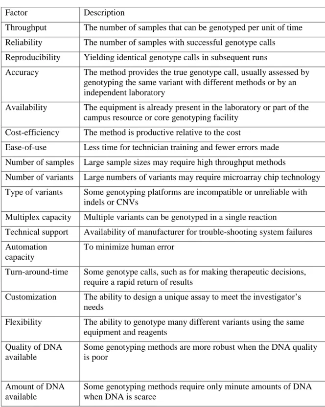
5. Factors to consider when selecting genotyping methods. ...52
6. Primers used in Sequenom® genotyping assay ...53
7. Concordance and call rates for 11 candidate variants ...54
8. Allele frequencies in the literature and UNITE-DNA and HWE p-value by race for 11 candidate variants ...55
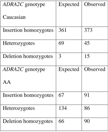
9. Expected and observed genotype distributions for the ADRA2C indel by race ...56
10. Linkage disequilibrium, diplotypes identified, and frequencies by race for the ADRB1 and ADRB2 genetic variants ...57
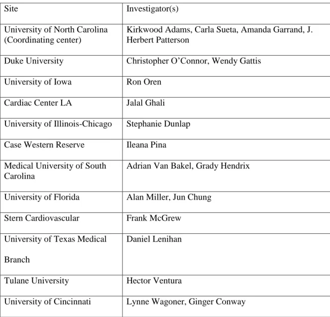
11. UNITE-HF study sites and investigators that contributed subjects for this research ...75
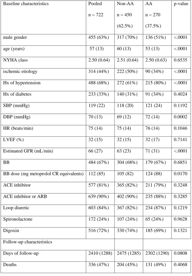
12. Baseline characteristics in pooled UNITE-DNA and by race ...76
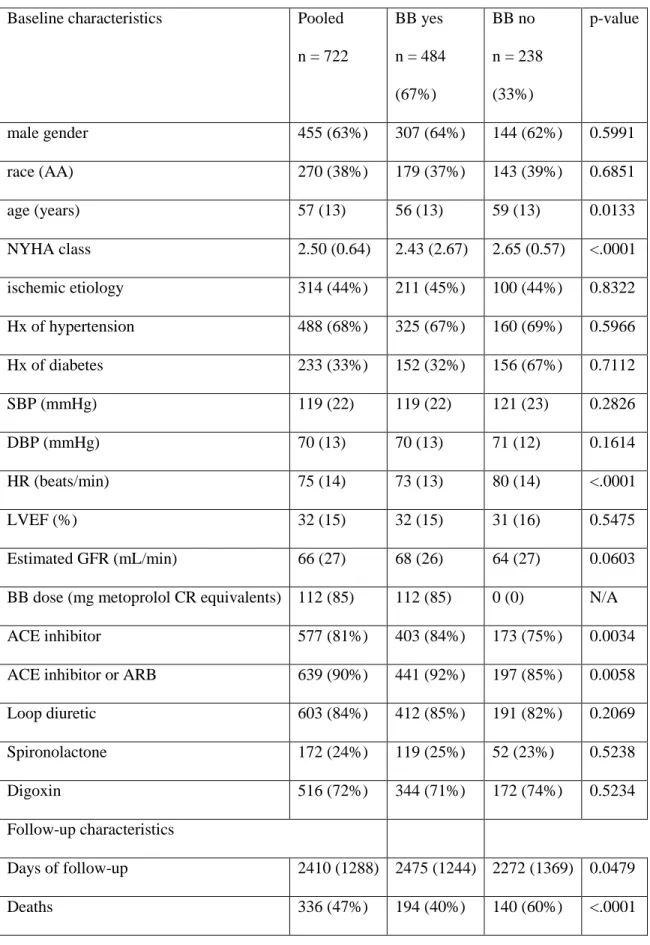
13. Baseline characteristics in pooled UNITE-DNA and by BB treatment status ...77
14. Baseline characteristics in pooled UNITE-DNA and by etiology ...78
15. Baseline characteristics in pooled UNITE-DNA and by vital status ...79
16. Baseline characteristics of propensity-matched dataset ...80
17. Baseline characteristics of propensity-matched patients and unmatched ...81
xvi
19. Final multivariable clinical model ...83
20. Adjusted interaction between 11 candidate clinical variables and BB response ...84
21. Independent association of 11 candidate genetic variants and 2 haplotypes with survival...119
22. Ten lowest p-values in the exploratory analyses for an association with survival and beta-blocker response prior to and after correction for 5% FDR modified for dependency ...120
23. Adjusted interaction between 11 candidate genetic variants and 2 haplotypes and BB survival benefit ...122
24. Baseline characteristics by Ser49Gly genotype ...123
25. Propensity-matched and propensity-adjusted Ser49Gly-stratified BB response ...124
26. Stratified and contrast-derived HR for BB in Ser49Gly genotypes adjusted with reduced clinical model ...125
27. Designation of risk alleles for 11 variant panel used in GRS calculation ...151
28. Mean ± sd, median, minimum, and maximum GRS in all patients and sub-groups ...152
29. Baseline clinical characteristics, drug utilization, and vital status by GRS quintiles ...153
30. Adjusted survival by GRS quintiles ...154
31. Adjusted interaction between the GRS and BB in all patients and sub-groups ...155
xvii
LIST OF FIGURES
Figure Page
1. Comparison of healthy SNS activation and in heart failure ...16
2. Interaction between SNS and RAAS ...17
3. Hypothetical scenarios demonstrating need for untreated patients in pharmacogenetic studies ...18
4. Kaplan-Meier survival curve for UNITE-DNA ...86
5. Adjusted survival curves by BB treatment in UNITE-DNA ...87
6. Adjusted BB dose associated survival benefit in all UNITE-DNA patients. ...88
7. Adjusted survival curves stratified by Ser49Gly genotype and BB treatment ...127
8. Adjusted BB dose-associated survival benefit by Ser49Gly genotype. ...128
9. Distribution of GRS in alive and deceased patients...157
10. Adjusted HR for GRS in all UNITE-DNA patients and sub-groups. ...158
11. ROC curves for clinical risk factors ± GRS ...159
12. Decision tree for all UNITE-DNA patients. ...183
13. Complete decision tree for Non-AA UNITE-DNA patients...184
14. Branches of Non-AA decision tree leading to gene-gene interaction. ...185
15. Complete decision tree for Non-AA UNITE-DNA patients...186
xviii
LIST OF ABBREVIATIONS
AA African-American
Arg arginine
ACE angiotensin-converting enzyme
ACE gene for the angiotensin-converting enzyme
ADRA2C gene for the alpha-2C adrenergic receptor
ADRB1 gene for the beta-1 adrenergic receptor
ADRB2 gene for the beta-2 adrenergic receptor
ADRB3 gene for the beta-3 adrenergic receptor
AGT gene for angiotensinogen
AGTR1 gene for the angiotensin II receptor type 1 ARB angiotensin receptor blocker
AUC area under the curve BB beta-blocker
BDKRB2 bradykinin receptor type B2
bp base pair
BP blood pressure bpm beats per minute
C cytosine
CART classification and regression tree CI confidence interval
xix
dbSNP database of single nucleotide polymorphisms
dl deciliters
DM diabetes mellitus DNA deoxyribonucleic acid
dNTP deoxynucleotide triphosphate EPI epinephrine
FDR false discovery rate
G guanine
GFR glomerular filtration rate
Gly glycine
GRK5 gene for G protein-coupled receptor kinase 5 GRS genetic risk score
GWAS genome wide association study HF heart failure
HR hazard ratio HTN hypertension
HWE Hardy-Weinberg equilibrium
Hx history
indel insertion/deletion polymorphism IDCM idiopathic dilated cardiomyopathy LDL low-density lipoprotein
xx LVEF left ventricular ejection fraction
M methionine
MAF minor allele frequency
MALDI-TOF matrix assisted laser desorption and ionization time-of-flight
Met methionine
MI myocardial infarction
min minute(s)
mg milligram(s) ml milliliter(s)
mmHg millimeters of mercury MS mass spectrometry
n sample size
NCBI National Center for Biotechnology Information NE norepinephrine
NOS gene for nitric oxide synthase NYHA New York Heart Association
RAAS renin-angiotensin-aldosterone system
REN gene for renin
RFLP restriction fragment length polymorphism ROC receiver operating characteristic
xxi sd standard deviation
Ser serine
SLC6A2 gene for solute carrier family 6 member 2 SNP single nucleotide polymorphism
SNS sympathetic nervous system
Thr Threonine
UNITE Unified Investigators to Evaluate Heart Failure UTR untranslated region
CHAPTER I: INTRODUCTION
Summary
Heart failure (HF) is an enormous public health problem. Hyperactivity of the sympathetic nervous system (SNS) and renin-angiotensin-aldosterone system (RAAS) plays a major role in HF pathophysiology. Direct pharmacologic inhibition of the SNS and indirect inhibition of the RAAS with beta-blockers significantly improves survival in patients with HF, on average, but the individual patient responses to beta-blockade widely vary. Genetic variation, affecting the functional activity of the SNS and RAAS, may be a possible explanation for variation in HF patient survival and beta-blocker response. The aims of this dissertation research were to determine if genetic variants in the SNS and RAAS, individually, additively, or with interactions between, are associated with survival and beta-blocker survival benefit in patients with HF.
The Problem of Heart Failure
2
have HF (2). This burden of HF is expected to increase as the population ages, acute mortality from myocardial infarction (MI) declines, and the survival of patients with HF is prolonged. HF continues to have high morbidity and mortality rates. One out of four HF patients admitted to the hospital will be readmitted within 30 days (4). Overall, 20% of HF patients will die within one year, and 50% will die within five years (2). For end-stage HF, the one-year mortality rate with optimal medical management is 75%, which is higher than many types of cancer (5). Heart transplant is the only cure for HF, which improves end-stage HF one-year survival to approximately 88% and five-year survival to approximately 75% (6). However in 2010, there were only 2,406 hearts donated (6), and the low donation rate has remained constant with no substantive increase anticipated in the near future (6).
3
predominately caused by infection, bleeding, neurologic dysfunction, and device malfunction (10).
The current therapies for HF do not distinguish among the complex types of HF, in which there are many potential etiologies, diverse clinical features, and numerous clinical subsets. For example, patients with HF caused by MI, chemotherapy, or no known identifiable cause (i.e. idiopathic dilated cardiomyopathy which could possibly be viral or inherited) are generally treated the same (7), even though the source and manifestation of myocardial damage are grossly different. Also, there is not unequivocal evidence for pharmacotherapies in patients with HF and preserved left ventricular ejection fraction (LVEF). Although the syndromes under the moniker of HF are diverse, a final common pathway among the heterogeneous HF patient population is neurohormonal activation. The major neurohormonal systems that are activated in HF are the sympathetic nervous system (SNS) and renin-angiotensin-aldosterone (RAAS) system.
The Sympathetic Nervous System
4
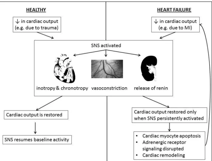
release at the sinus and atrioventricular nodes and left ventricle; 2) epinephrine (EPI) released in the circulation by the adrenal cortex; 3) local release of EPI and NE in the peripheral vessels; and 4) circulating NE which can act in multiple locations (12). Both NE and EPI exert their biological actions via activation of nine different adrenergic receptor subtypes: alpha-1 (1A, 1B, and 1D), alpha-2 (2A, 2B, and 2C), and beta (1, 2, and 3). Adrenergic receptors are members of the super-family of seven transmembrane receptors that signal primarily via interaction with heterotrimeric G proteins. These healthy SNS responses may have evolved to compensate for non-specific, short-term loss of blood volume and/or pressure. However in HF, cardiac output is persistently affected, for example due to myocardial infarction, ventricular hypertrophy, or idiopathic dilated cardiomyopathy. Thus, HF results in long-term activation of the SNS, which in turn leads to abnormalities in SNS function and adverse consequences (Figure 1).
5
cardiovascular reflexes, such as the arterial baroreceptor reflex, are significantly suppressed (17), whereas the sympatho-excitatory reflexes, such as the cardiac sympathetic afferent reflex (18), are augmented. Angiotensin II levels are also increased in HF, which can facilitate sympathetic neurotransmission via several mechanisms described in detail below (See: Interaction with SNS).
Consequences of SNS hyperactivity in HF. SNS hyperactivity in HF has adverse consequences at molecular, physiologic, and clinical levels. There are multiple alterations in the beta-adrenergic receptor signaling pathway, including down-regulation of receptors (decreased beta receptor density) (19), desensitization via uncoupling from Gs (stimulatory G protein) (20), and an increase in Gi (inhibitory G protein) (21). Notably, receptor down-regulation in HF is specific to beta-1 receptors, and beta-2 receptor density remains unchanged (22). Down-regulation and desensitization of beta-receptors is thought to be a protective adaptation in HF because persistent sympathetic stimulation is toxic to the cardiac myocyte (23). Persistent SNS activation also leads to the pathophysiologic cardiac remodeling process, in which the heart goes through maladaptive changes in size, shape, and function after an injury (24). The degree of SNS hyperactivity is also correlated with hemodynamic abnormalities (25), symptoms (26), and survival (27) in patients with HF (25).
The Renin-Angiotensin-Aldosterone System
6
into the circulation, which converts angiotensinogen (synthesized in the liver) to angiotensin I. Angiotensin I is converted to angiotensin II by the angiotensin-converting enzyme. Angiotensin II elicits the majority of its actions via the angiotensin II type 1 receptor, which includes vasoconstriction, sodium and water retention, and aldosterone secretion. Notably, the release of renin is the rate-limiting step in the RAAS activation (28).
RAAS hyperactivity in HF. Like the SNS, the RAAS is hyperactive in HF to compensate for the persistent decrease in cardiac output, which has been demonstrated in experimental HF (29) and HF patients (30,31) by increased plasma levels of renin, angiotensin II, and aldosterone. Also like the SNS, there are several mechanisms that can account for the loss of the counter-regulatory balance in the RAAS. Baroreceptor dysfunction is a common mechanism for RAAS and SNS hyperactivity. In a healthy person, under- and over-filling of the vasculature initiates afferent signals from various sensory receptors (e.g. atrial and arterial baroreceptors), aiming ultimately to restore perfusion pressures with sodium and water retention or induce natriuresis to relieve circulatory congestion. However in HF, there are disturbances in the afferent signaling from volume-sensing sites resulting in blunted natriuresis in the face of venous congestion and elevated cardiac filling pressures (32). In addition, the suppression of renin release and renal excretion responses to natriuretic peptide are attenuated in HF (33).
7
several mechanisms: increasing DNA, protein, and collagen synthesis in cardiac fibroblasts (34,35); mediating stretch-induced hypertrophy (36); and cardiac myocyte necrosis (37). Therefore it is not surprising that RAAS activity is associated with left ventricular dysfunction (13). Other physiologic consequences of RAAS hyperactivity in HF include peripheral vasoconstriction, sodium retention, and hence, circulatory congestion. RAAS activity is associated with the progression to HF from asymptomatic left ventricular dysfunction (13) and HF morbidity and mortality (38-40).
Interaction with SNS. Although discussed separately, the SNS and RAAS mutually facilitate each other’s hyperactivity (Figure 2). Specifically, angiotensin II can
facilitate sympathetic neurotransmission via several mechanisms: stimulatory action on sympathetic ganglia (41); increasing neurotransmitter release at sympathetic nerve endings (42); preventing NE uptake at sympathetic nerve terminals (43); centrally stimulating angiotensin II type 1 receptors in the brain (44); increasing central sympathetic nerve activity (45); the release of catecholamines from the adrenal medulla (46); facilitation of NE release from sympathetic nerve terminals (47); and modulation of baroreflex control of heart rate (48). In turn, SNS stimulation of beta-1 adrenergic receptors in the kidney results in renin release (49).
Beta-Blockers in Heart Failure
8
significant reductions in morbidity and mortality with the use of beta-blockers in HF (50,51). The long-term benefits of inhibition of the adverse effects of SNS hyperactivity greatly outweigh the negative inotropic effects of beta-blockers. Metoprolol CR/XL, carvedilol, and bisoprolol are the three beta-blockers that significantly reduced mortality in large HF trials, and hence, are recommended for HF in the treatment guidelines (7). Despite the success of these three beta-blockers, a class effect cannot be assumed because bucindolol failed to significantly reduce mortality in a large clinical trial (52). Bucindolol is a non-selective β-1and β-2 adrenergic receptor antagonist, but bucindolol also has the unique pharmacologic property of marked sympatholysis. Metoprolol and bisoprolol are selective β-1 receptor antagonists, whereas carvedilol also blocks β-2 and α-1. Although,
the pharmacologic differences between metoprolol, bisoprolol, and carvedilol have not translated into differences in efficacy.
Importantly, these results from large beta-blocker trials demonstrate an average benefit, but the individual patient responses to beta-blockers vary. For example, long-term optimal dosing of beta-blockers fails to improve LVEF over 5% in as many as 43% of HF patients (53). In a randomized, double-blind trial of metoprolol versus carvedilol in 150 HF patients, the 95% CI for change in LVEF was -8.2% to +22.6% for metoprolol and -11.1% to +32.9% for carvedilol (54). In a study of 171 chronic HF patients treated with metoprolol or carvedilol for 9 to 12 months, only 22% of patients had an increase in LVEF ≥ 15% (55). Controversy exists over whether there is racial and regional variation
9
0.63), but the estimate was not statistically significant in 545 AAs (56). Less than half of the patients enrolled in the large beta-blocker trials were from the United States, and the relative risk reduction for each beta-blocker was of smaller magnitude and not statistically significant in Americans compared to the rest of the world (57).
Genetic Variation within the SNS and RAAS
Genetic variation is differences in DNA sequences between individuals and populations, and it can take on a variety of forms, frequencies, and functions. The most common form of genetic variation is the single nucleotide polymorphism (SNP), which occurs every 100 to 300 base-pairs along the 3 billion base-pair human genome. Other common forms of genetic variation are insertion/deletions (indels) and copy number variations (CNVs). Genetic variation can be common, i.e. occurring in greater than 5% of the general population, or an extremely rare mutation. Most genetic variation is believed to be random mutation and have neutral effect, but some genetic variation could have profound effect on phenotype, in which a single genetic variant is sufficient to cause disease. The functional effect of genetic variation on phenotype can be easily seen for some human phenotypic traits, such as eye, hair, and skin color, but genetic variation also affects traits that are not readily visible, such as the SNS and RAAS (58,59).
10
variants reported within the angiotensin-converting enzyme gene (ACE). There are both uncommon and common genetic variants identified within the SNS and RAAS. For example, an indel in the alpha-2C adrenergic receptor gene (ADRA2C) occurs in only 4% of Caucasians, while an indel in ACE occurs in 44% of Caucasians. Importantly, the frequencies of genetic variants in the SNS and RAAS can differ between races. For comparison, the same ADRA2C indel that is 4% frequent in Caucasians is ten times more common in AAs (frequency 43%), but the ACE indel has nearly identical frequency in Caucasians (44%) and AAs (43%). This genetic variation in the SNS and RAAS is not merely random mutation; these genetic variants can have profound effects on protein function or expression. For example, the ACE indel mentioned above explains 50% of the variation in serum ACE levels (61). Another example is that the beta-1 adrenergic receptor has agonist-stimulated activity that is three times higher with an arginine at amino acid 389 compared to glycine (62).
11
was associated with decreased survival (adjusted risk ratio 2.03) (66). This same genetic variant was also associated with survival benefit of beta-blockers in HF patients (67).
The relationship between SNS and RAAS genetic variation and HF clinical outcomes, such as survival and beta-blocker response, has not been fully characterized. The genetic and pharmacogenetic association literature for HF is still in very early stages and subject to several limitations. For example, many of the previous HF genetic and pharmacogenetic studies were low power due to short follow-up (most < 5 years) and/or small sample size (most n < 400), which could lead to falsely negative results. Positive associations from small, single-center HF genetic and pharmacogenetic studies could be false due to selection bias or chance, and those associations have not yet been replicated in larger, multicenter HF patient cohorts to rule out those possibilities. Most of the previous studies only tested one to three variants; therefore they are unable to determine the association of multiple variant combinations or interactions. Previous studies are also limited by retrospective design, the exclusion of HF patients with preserved ejection fraction, and poor or no representation of AAs. Retrospective studies were often not initially designed for genetic and pharmacogenetic research, and hence they often lack power. The inclusion of HF patients with preserved ejection fraction in pharmacogenetic studies is important because of differences in the HF phenotype and drug response compared to HF patients with reduced ejection fraction. Including AAs in genetic and pharmacogenetic HF studies is important because, as exemplified above, there are racial discrepancies in allele frequencies.
12
dose analysis, lack of specific beta-blocker analysis, and the analysis of the non-FDA approved beta-blocker bucindolol. Falsely negative or the reverse pharmacogenetic associations could stem from 100% beta-blocker treatment rates present in some of the previous HF pharmacogenetic literature. Hypothetical scenarios demonstrating the need for an untreated portion of patients in pharmacogenetic studies are shown in Figure 3. Many previous HF pharmacogenetic studies only evaluated intermediate phenotypes such as ventricular remodeling and not clinical outcomes such as survival, and ventricular remodeling is not a perfect surrogate for beta-blocker survival benefit. Many previous HF pharmacogenetic studies also lacked beta-blocker dose, which is important because genetic effects could vary by dose. For example, ADRB1 Ser49-homozygous patients treated with a high dose of beta-blockade had a greater survival benefit as compared with a low dose. Whereas ADRB1 Gly49-carriers had a similar survival rate regardless of blocker dose (67). Many previous HF pharmacogenetic studies lacked specific beta-blocker data or did not test for beta-beta-blocker specific interactions. However in vitro data suggests that pharmacogenetic interactions may be beta-blocker specific (68), which may translate into clinical differences. The most robust HF pharmacogenetic data comes from sub-studies of the Beta-Blocker Evaluation of Survival Trial (BEST) (52). However the beta-blocker tested in BEST is bucindolol, which has unique pharmacologic properties including marked sympatholysis (69), and bucindolol did not significantly reduce mortality like other FDA-approved beta-blockers (52). Therefore the pharmacogenetic data on bucindolol may not be applicable to metoprolol CR/XL, carvedilol, or bisoprolol.
13
relationship between SNS and RAAS genetic variants and HF survival and beta-blocker survival benefit. This dissertation research addresses the limitations described above by using a well-powered, HF patient cohort and the novel application of advanced analytical methods to determine the individual (Specific Aim I), additive (Specific Aim II), and interactive (Specific Aim III) association of multiple SNS and RAAS variants with HF patient survival and beta-blocker survival benefit.
Perspective
14 Specific Aims
I. Determine if functionally annotated genetic variants within the SNS & RAAS are
independently associated with survival and beta-blocker response in patients with
HF. Hypothesis: Genetic variants causing increased activity of the SNS or RAAS in vitro
and in vivo will be independently associated with higher risk of all-cause mortality in patients with HF, but patients with the higher activity genotypes will have a greater reduction in mortality with beta-blockers.
II. Determine if a genetic risk score, composed of a panel of functionally annotated
SNS & RAAS genetic variants, is associated with survival and beta-blocker response
in patients with HF. Hypothesis: The SNS & RAAS genetic variants individually will have a modest association with survival and beta-blocker response, but patients possessing a combination of high activity variants will have additive risk for all-cause mortality and beta-blocker response.
III. Determine if gene-gene interactions among functionally annotated SNS & RAAS
genetic variants are associated with survival and beta-blocker response in HF
15 Figure legends
Figure 1. A comparison of healthy SNS activation (left half of figure) and in HF (right half of figure) is shown. When a decrease in cardiac output occurs in an otherwise healthy person, the SNS is activated, resulting in increased inotropy, chronotropy, vasoconstriction, and the release of renin. Once cardiac output is restored, the SNS resumes baseline activity. However in a person with HF, cardiac output is only maintained with continuous SNS activation, which leads to adverse consequences such as cardiac myocyte apoptosis, disrupted adrenergic receptor signaling, and cardiac remodeling.
Figure 2. The interaction between the SNS and RAAS is shown. Sites labeled AII are sites in which angiotensin II facilitates the SNS, and those labeled SNS are sites in which the SNS facilitates the RAAS.
Figure 3. Hypothetical scenarios demonstrating the need for an untreated portion of patients in pharmacogenetic studies. Values are hypothetical mortality rates in ADRB1
16
18
19 REFERENCES
(1) Writing group members, Lloyd-Jones D, Adams RJ, Brown TM, Carnethon M, Dai S, et al. Heart disease and stroke statistics--2010 update: a report from the American Heart Association. Circulation 2010 Feb 23;121(7):e46-e215.
(2) Roger VL. Heart Disease and Stroke Statistics--2012 Update: A Report From the American Heart Association. Circulation (New York, N.Y.) 2012 -01-03;125(1):e2-e220.
(3) Page RL, Hogan C, Strongin K, Mills R, Lindenfeld J. Medicare Beneficiaries with Mild to Severe Heart Failure See 15–23 Different Providers Annually. Circulation 2008;118:S_1030.
(4) Centers for Medicare & Medicaid Services. Medicare Hospital Quality Chartbook. 2011; Available at: https://www.cms.gov/HospitalQualityInits/Downloads/HospitalChartBook2011.pdf. Accessed January 26, 2012.
(5) Rose EA, Gelijns AC, Moskowitz AJ, Heitjan DF, Stevenson LW, Dembitsky W, et al. Long-term use of a left ventricular assist device for end-stage heart failure. N Engl J Med 2001 Nov 15;345(20):1435-1443.
(6) Organ Procurement and Transplantation Network. 2009 Annual Report. Available at:
http://optn.transplant.hrsa.gov/.
(7) Heart Failure Society of America, Lindenfeld J, Albert NM, Boehmer JP, Collins SP, Ezekowitz JA, et al. HFSA 2010 Comprehensive Heart Failure Practice Guideline. J Card Fail 2010 Jun;16(6):e1-194.
(8) Flesch M, Erdmann E. The problem of polypharmacy in heart failure. Curr Cardiol Rep 2006 May;8(3):217-225.
(9) Kirklin JK, Naftel DC, Kormos RL, Stevenson LW, Pagani FD, Miller MA, et al. Second INTERMACS annual report: more than 1,000 primary left ventricular assist device implants. J Heart Lung Transplant 2010 Jan;29(1):1-10.
(10) Rose EA. Long-term use of a left ventricular assist device for end-stage heart failure. N Engl J Med 2001 -11-15;345(20):1435-43.
(11) Malliani A, Pagani M, Pizzinelli P, Furlan R, Guzzetti S. Cardiovascular reflexes mediated by sympathetic afferent fibers. J Auton Nerv Syst 1983 Mar-Apr;7(3-4):295-301.
(12) Triposkiadis F, Karayannis G, Giamouzis G, Skoularigis J, Louridas G, Butler J. The sympathetic nervous system in heart failure physiology, pathophysiology, and clinical implications. J Am Coll Cardiol 2009 Nov 3;54(19):1747-1762.
20
(14) Hasking GJ, Esler MD, Jennings GL, Burton D, Johns JA, Korner PI. Norepinephrine spillover to plasma in patients with congestive heart failure: evidence of increased overall and cardiorenal sympathetic nervous activity. Circulation 1986 Apr;73(4):615-621.
(15) Morris MJ, Cox HS, Lambert GW, Kaye DM, Jennings GL, Meredith IT, et al. Region-specific neuropeptide Y overflows at rest and during sympathetic activation in humans. Hypertension 1997 Jan;29(1 Pt 1):137-143.
(16) Leimbach WN,Jr, Wallin BG, Victor RG, Aylward PE, Sundlof G, Mark AL. Direct evidence from intraneural recordings for increased central sympathetic outflow in patients with heart failure. Circulation 1986 May;73(5):913-919.
(17) Ferguson DW, Berg WJ, Roach PJ, Oren RM, Mark AL. Effects of heart failure on baroreflex control of sympathetic neural activity. Am J Cardiol 1992 Feb 15;69(5):523-531.
(18) Wang W, Zucker IH. Cardiac sympathetic afferent reflex in dogs with congestive heart failure. Am J Physiol 1996 Sep;271(3 Pt 2):R751-6.
(19) Bristow MR, Ginsburg R, Minobe W, Cubicciotti RS, Sageman WS, Lurie K, et al. Decreased catecholamine sensitivity and beta-adrenergic-receptor density in failing human hearts. N Engl J Med 1982 Jul 22;307(4):205-211.
(20) Sibley DR, Benovic JL, Caron MG, Lefkowitz RJ. Regulation of transmembrane signaling by receptor phosphorylation. Cell 1987 Mar 27;48(6):913-922.
(21) Feldman AM, Cates AE, Veazey WB, Hershberger RE, Bristow MR, Baughman KL, et al. Increase of the 40,000-mol wt pertussis toxin substrate (G protein) in the failing human heart. J Clin Invest 1988 Jul;82(1):189-197.
(22) Bristow MR, Ginsburg R, Umans V, Fowler M, Minobe W, Rasmussen R, et al. Beta 1- and beta 2-adrenergic-receptor subpopulations in nonfailing and failing human ventricular myocardium: coupling of both receptor subtypes to muscle contraction and selective beta 1-receptor down-regulation in heart failure. Circ Res 1986 Sep;59(3):297-309.
(23) Engelhardt S, Hein L, Wiesmann F, Lohse MJ. Progressive hypertrophy and heart failure in beta1-adrenergic receptor transgenic mice. Proc Natl Acad Sci U S A 1999 Jun 8;96(12):7059-7064.
(24) Kudej RK. Effects of chronic beta-adrenergic receptor stimulation in mice. J Mol Cell Cardiol 1997 -10;29(10):2735-46.
(25) Levine TB, Francis GS, Goldsmith SR, Simon AB, Cohn JN. Activity of the sympathetic nervous system and renin-angiotensin system assessed by plasma hormone levels and their relation to hemodynamic abnormalities in congestive heart failure. Am J Cardiol 1982 May;49(7):1659-1666.
21
(27) Cohn JN, Levine TB, Olivari MT, Garberg V, Lura D, Francis GS, et al. Plasma norepinephrine as a guide to prognosis in patients with chronic congestive heart failure. N Engl J Med 1984 Sep 27;311(13):819-823.
(28) Skeggs LT,Jr, Kahn JR, Lentz K, Shumway NP. The preparation, purification, and amino acid sequence of a polypeptide renin substrate. J Exp Med 1957 Sep 1;106(3):439-453.
(29) Davis JO. Adrenocortical and renal hormonal function in experimental cardiac failure. Circulation 1962 Jun;25:1002-1014.
(30) Merrill AJ, Morrison JL, Branno ES. Concentration of renin in renal venous blood in patients with chronic heart failure. Am J Med 1946 Nov;1(5):468.
(31) Dzau VJ, Colucci WS, Hollenberg NK, Williams GH. Relation of the renin-angiotensin-aldosterone system to clinical state in congestive heart failure. Circulation 1981 Mar;63(3):645-651.
(32) Greenberg TT, Richmond WH, Stocking RA, Gupta PD, Meehan JP, Henry JP. Impaired atrial receptor responses in dogs with heart failure due to tricuspid insufficiency and pulmonary artery stenosis. Circ Res 1973 Apr;32(4):424-433.
(33) Cody RJ, Atlas SA, Laragh JH, Kubo SH, Covit AB, Ryman KS, et al. Atrial natriuretic factor in normal subjects and heart failure patients. Plasma levels and renal, hormonal, and hemodynamic responses to peptide infusion. J Clin Invest 1986 Nov;78(5):1362-1374.
(34) Brilla CG. Collagen metabolism in cultured adult rat cardiac fibroblasts: response to angiotensin II and aldosterone. J Mol Cell Cardiol 1994 -07;26(7):809-20.
(35) Sadoshima J. Molecular characterization of angiotensin II--induced hypertrophy of cardiac myocytes and hyperplasia of cardiac fibroblasts. Critical role of the AT1 receptor subtype. Circ Res 1993 -09;73(3):413-23.
(36) Sadoshima J, Xu Y, Slayter HS, Izumo S. Autocrine release of angiotensin II mediates stretch-induced hypertrophy of cardiac myocytes in vitro. Cell 1993 Dec 3;75(5):977-984.
(37) Kabour A, Henegar JR, Janicki JS. Angiotensin II (AII)-induced myocyte necrosis: role of the AII receptor. J Cardiovasc Pharmacol 1994 Apr;23(4):547-553.
(38) Szymanski MK. Prognostic value of renin and prorenin in heart failure patients with decreased kidney function. The American heart journal 2011 -09;162(3):487-93.
(39) Tsutamoto T. Comparison of active renin concentration and plasma renin activity as a prognostic predictor in patients with heart failure. Circulation journal : official journal of the Japanese Circulation Society 2007 -06;71(6):915-21.
22
(41) Lewis GP. The action of angiotensin and bradykinin on the superior cervical ganglion of the cat. J Physiol (Lond ) 1965 -08;179(3):538-53.
(42) Lokhandwala MF, Amelang E, Buckley JP. Facilitation of cardiac sympathetic function by angiotensin II: role of presynaptic angiotensin receptors. Eur J Pharmacol 1978 Dec 1;52(3-4):405-409.
(43) Peach MJ, Bumpus FM, Khairallah PA. Inhibition of norepinephrine uptake in hearts by angiotensin II and analogs. J Pharmacol Exp Ther 1969 Jun;167(2):291-299.
(44) Watson AM, Mogulkoc R, McAllen RM, May CN. Stimulation of cardiac sympathetic nerve activity by central angiotensinergic mechanisms in conscious sheep. Am J Physiol Regul Integr Comp Physiol 2004 Jun;286(6):R1051-6.
(45) Keim KL, Sigg EB. Activation of central sympathetic neurons by angiotensin II. Life Sci I 1971 May 15;10(10):565-574.
(46) Peach MJ, Cline WH,Jr, Watts DT. Release of adrenal catecholamines by angiotensin. II. Circ Res 1966 Sep;19(3):571-575.
(47) Boke T, Malik KU. Enhancement by locally generated angiotensin II of release of the adrenergic transmitter in the isolated rat kidney. J Pharmacol Exp Ther 1983 Sep;226(3):900-907.
(48) Guo GB, Abboud FM. Angiotensin II attenuates baroreflex control of heart rate and sympathetic activity. Am J Physiol 1984 Jan;246(1 Pt 2):H80-9.
(49) Osborn JL, DiBona GF, Thames MD. Beta-1 receptor mediation of renin secretion elicited by low-frequency renal nerve stimulation. J Pharmacol Exp Ther 1981 Feb;216(2):265-269.
(50) MERIT-HF Investigators. Effect of metoprolol CR/XL in chronic heart failure: Metoprolol CR/XL Randomised Intervention Trial in Congestive Heart Failure (MERIT-HF). Lancet 1999 Jun 12;353(9169):2001-2007.
(51) Packer M, Bristow MR, Cohn JN, Colucci WS, Fowler MB, Gilbert EM, et al. The effect of carvedilol on morbidity and mortality in patients with chronic heart failure. U.S. Carvedilol Heart Failure Study Group. N Engl J Med 1996 May 23;334(21):1349-1355.
(52) Beta-Blocker Evaluation of Survival Trial Investigators. A trial of the beta-blocker bucindolol in patients with advanced chronic heart failure. N Engl J Med 2001 May 31;344(22):1659-1667.
(53) Chen L, Meyers D, Javorsky G, Burstow D, Lolekha P, Lucas M, et al. Arg389Gly-beta1-adrenergic receptors determine improvement in left ventricular systolic function in nonischemic cardiomyopathy patients with heart failure after chronic treatment with carvedilol. Pharmacogenet Genomics 2007 Nov;17(11):941-949.
23
(55) Metra M, Nodari S, Parrinello G, Giubbini R, Manca C, Dei Cas L. Marked improvement in left ventricular ejection fraction during long-term beta-blockade in patients with chronic heart failure: clinical correlates and prognostic significance. Am Heart J 2003 Feb;145(2):292-299.
(56) Shekelle PG, Rich MW, Morton SC, Atkinson CS, Tu W, Maglione M, et al. Efficacy of angiotensin-converting enzyme inhibitors and beta-blockers in the management of left ventricular systolic dysfunction according to race, gender, and diabetic status: a meta-analysis of major clinical trials. J Am Coll Cardiol 2003 May 7;41(9):1529-1538.
(57) O'Connor CM, Fiuzat M, Swedberg K, Caron M, Koch B, Carson PE, et al. Influence of global region on outcomes in heart failure beta-blocker trials. J Am Coll Cardiol 2011 Aug 23;58(9):915-922.
(58) Williams PD, Puddey IB, Beilin LJ, Vandongen R. Genetic influences on plasma catecholamines in human twins. J Clin Endocrinol Metab 1993 Sep;77(3):794-799.
(59) Kennedy BP, Rao F, Botiglieri T, Sharma S, Lillie EO, Ziegler MG, et al. Contributions of the sympathetic nervous system, glutathione, body mass and gender to blood pressure increase with normal aging: influence of heredity. J Hum Hypertens 2005 Dec;19(12):951-969.
(60) National Center for Biotechnology Information and National Library of Medicine. Database of Single Nucleotide Polymorphisms. Available at: http://www.ncbi.nlm.nih.gov/snp/, 2012.
(61) Rigat B, Hubert C, Alhenc-Gelas F, Cambien F, Corvol P, Soubrier F. An insertion/deletion polymorphism in the angiotensin I-converting enzyme gene accounting for half the variance of serum enzyme levels. J Clin Invest 1990 Oct;86(4):1343-1346.
(62) Mason DA, Moore JD, Green SA, Liggett SB. A gain-of-function polymorphism in a G-protein coupling domain of the human beta1-adrenergic receptor. J Biol Chem 1999 Apr 30;274(18):12670-12674.
(63) Talameh JA, Lanfear DE. Pharmacogenetics in chronic heart failure: new developments and current challenges. Curr Heart Fail Rep 2012 Mar;9(1):23-32.
(64) Talameh JA, McLeod HL, Adams KF,Jr, Patterson JH. Genetic tailoring of pharmacotherapy in heart failure: optimize the old, while we wait for something new. J Card Fail 2012 Apr;18(4):338-349.
(65) Podlowski S, Wenzel K, Luther HP, Muller J, Bramlage P, Baumann G, et al. Beta1-adrenoceptor gene variations: a role in idiopathic dilated cardiomyopathy? J Mol Med (Berl) 2000;78(2):87-93.
(66) Borjesson M, Magnusson Y, Hjalmarson A, Andersson B. A novel polymorphism in the gene coding for the beta(1)-adrenergic receptor associated with survival in patients with heart failure. Eur Heart J 2000 Nov;21(22):1853-1858.
24
(68) Liggett SB, Mialet-Perez J, Thaneemit-Chen S, Weber SA, Greene SM, Hodne D, et al. A polymorphism within a conserved beta(1)-adrenergic receptor motif alters cardiac function and beta-blocker response in human heart failure. Proc Natl Acad Sci U S A 2006 Jul 25;103(30):11288-11293.
CHAPTER II:
IDENTIFICATION OF CANDIDATE GENETIC VARIANTS
Summary
Chapter I introduced the severity of the HF public health problem and the roles that the SNS and RAAS play in HF pathophysiology and clinical outcomes. The goal of this chapter was to identify candidate genetic variants within the SNS and RAAS that have in vitro and in vivo evidence to support an association with HF survival and beta-blocker response. A final list of eleven candidate genetic variants was identified in the literature and will be used for analyses. A comparison of the candidate gene approach to other genetic association approaches was introduced, and the limitations of the candidate gene approach used herein were discussed.
Introduction
Because there are approximately three million differences in DNA sequences between individuals, deciding which genetic variants to test and how many to test is an extreme challenge for investigators. Genetic association study designs fall on an enormous spectrum. On one extreme, an investigator may test a single genetic variant. On the other extreme, an investigator may scan the entire genome. When an investigator decides to only test one or a few variants, this is referred to as a “candidate gene approach” because the investigator has a strong a priori hypothesis for the best
26
variants is ideally based on substantial pre-existing evidence for an association with the trait (e.g. molecular, physiologic, pre-clinical, or clinical evidence). When pre-existing evidence for particular candidate genes is lacking, an investigator may test all of the common variation in the genome. This is referred to as a genome-wide association study (GWAS), which has been referred to as “hypothesis-free,” or the broad hypothesis that
there are variants somewhere within the genome that will be associated with the trait. Many genetic association study designs fall somewhere in the middle of the two extremes, with intermediate numbers of genetic variants tested for a general hypothesis. For example, if the trait is drug response, the hypothesis could be that any genetic variation within pharmacokinetic genes (e.g. drug metabolizing enzymes and transporters) will be associated with drug response. This hypothesis is narrower than testing the entire genome, but it also does not make the hypothesis for specific variants. Instead of 1 variant or 3 million variants, this hypothesis falls in the middle range with a few thousand variants.
27
is the potential for false positives. For example, if testing 1 million variants, and using an alpha = 0.05, the investigator may find 50,000 variants associated with the trait by chance alone. Therefore to control for the potential false positives, stringent levels for statistical significance must be set, and therefore very large sample sizes are required to have the power to detect true associations. In this sense, candidate gene studies have the advantage of being flexible and having power to test few variants. Another advantage of candidate gene studies is coverage of uncommon or rare variants. GWAS typically only cover common genetic variation (minor allele frequency [MAF] > 5%). Cost is also a significant factor when deciding on the number of variants to test, as genotyping costs and sample sizes must increase with the number of variants. GWAS are typically hypothesis generating and therefore require validation samples as well. In addition, the data generated in GWAS are computationally intensive and often require more elaborate IT infrastructure and statistical support than a candidate gene study.
28
The rationale for candidate variant selection is as follows: Because the SNS and RAAS are so closely associated with HF pathophysiology and pharmacology (See Chapter I: Introduction), variants affecting those systems were chosen. Variants also known to affect gene expression or function were chosen because they are more likely to translate into clinically relevant outcomes. Much variation in the genome is thought to be random mutation and have a neutral effect; this variation would not be expected to translate into clinically meaningful outcomes. Because the best candidates for an association with HF clinical outcomes (survival and beta-blocker response) wanted to be chosen, variants that had effects further translating from molecular effects to physiologic or clinical outcomes (e.g. ventricular remodeling or beta-blocker response) were chosen. Only common variants (e.g. MAF >5% in Caucasian-Americans or AAs) were chosen due to statistical power and generalizability of the results to large numbers of patients. Herein the methods for finding candidate genetic variants that meet these criteria were described and the literature supporting the candidate variant list was summarized.
Methods
Selection criteria. Candidate genetic variants must be part of the SNS or RAAS, have a MAF of greater than 5% in Caucasian-Americans or AAs, affect gene expression or function, and are associated with HF patient clinical outcomes or relevant physiologic processes (i.e. ventricular remodeling). When linkage disequilibrium exists, the true functional variant was chosen if known.
29
sympathetic adrenergic system, renin-angiotensin-aldosterone system, genetic, polymorphism, beta-blocker, pharmacogenetic, survival, and left ventricular ejection fraction. Once variants were identified to have an association with HF relevant outcomes such as drug response, ventricular remodeling, or survival, then functional and pre-clinical data was searched for using the specific variant name or rsID (reference sequence identifier). Variant population frequencies were found in published literature, the HapMap database (2), or NCBI (3). Studies were limited to those published in English.
Results
Twelve candidate variants met the selection criteria. However one candidate variant, M235T in the gene for angiotensinogen (AGT), was excluded because it was found to be in complete linkage disequilibrium with G-6A in AGT, which was later determined to be the causal functional variant (4). Table 1 summarizes the identification and location information for the candidate genetic variants. Table 2 summarizes the MAF of the variants in populations of Caucasian and African descent. Tables 3 & 4 summarize the gene function within SNS or RAAS, respectively, and the molecular and clinical HF phenotypes of the variants.
Discussion
30
31
Table 1. Identification and location information of candidate genetic variants Gene rsID Nucleotide
substitution
Amino acid Substitution
Chromosome Chromosome position (GRCh37.p5 assembly)
Variant type
ADRB1 rs1801252 A1231G Ser49Gly 10 115804036 missense SNP
ADRB1 rs1801253 C1251G Arg389Gly 10 115805056 missense SNP
ADRB2 rs1042713 A285G Arg16Gly 5 148206440 missense
SNP
ADRB2 rs1042714 C318G Gln27Glu 5 148206473 missense
SNP
ACE rs1799752 287-bp deletion
n/a 17 61565890 intronic
indel
ADRA2C rs61767072 12-bp deletion
322GlyAla-GlyPro325
4 3769297 frameshift
indel
GRK5 rs17098707 A355T Gln41Leu 10 121086097 missense SNP
AGT rs5051 G-6A n/a 1 230849872 5’ UTR
SNP
AGTR1 rs5186 A1166C n/a 3 148459988 3’ UTR
SNP
CYP11B2 rs1799998 C-344T n/a 8 143999600 5’ UTR
SNP
BDKRB2 n/a 9-bp deletion
n/a 14 n/a
32
Table 2. Minor allele frequencies (MAF) of candidate genetic variants
Gene rsID Minor allele MAF Caucasian MAF African
ADRB1 rs1801252 G 0.17 0.25
ADRB1 rs1801253 G 0.27 0.38
ADRB2 rs1042713 A 0.40 0.50
ADRB2 rs1042714 G 0.42 0.20
ACE rs1799752 Ins 0.44 0.43
ADRA2C rs61767072 Del 0.04 0.43
GRK5 rs17098707 T 0.02 0.24
AGT rs5051 A 0.42 0.82
AGTR1 rs5186 C 0.25 0.05
CYP11B2 rs1799998 C 0.43 0.29
33
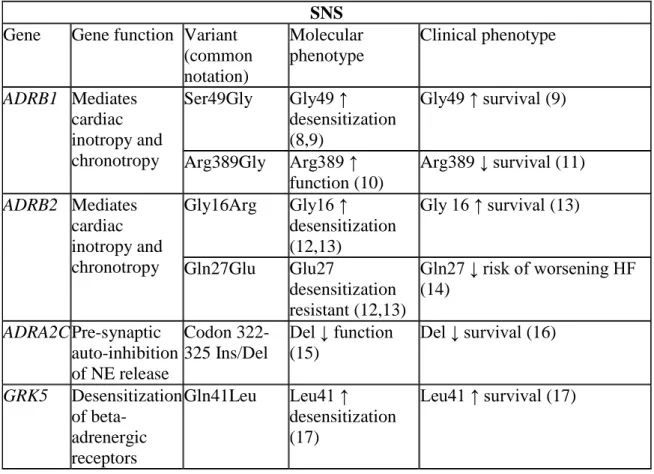
Table 3. Gene function and molecular and clinical phenotypes of candidate SNS genetic variants
SNS Gene Gene function Variant
(common notation)
Molecular phenotype
Clinical phenotype
ADRB1 Mediates cardiac inotropy and chronotropy
Ser49Gly Gly49 ↑ desensitization (8,9)
Gly49 ↑ survival (9)
Arg389Gly Arg389 ↑ function (10)
Arg389 ↓ survival (11)
ADRB2 Mediates cardiac inotropy and chronotropy
Gly16Arg Gly16 ↑ desensitization (12,13)
Gly 16 ↑ survival (13)
Gln27Glu Glu27
desensitization resistant (12,13)
Gln27 ↓ risk of worsening HF (14)
ADRA2C Pre-synaptic auto-inhibition of NE release
Codon 322-325 Ins/Del
Del ↓ function (15)
Del ↓ survival (16)
GRK5 Desensitization of
beta-adrenergic receptors
Gln41Leu Leu41 ↑ desensitization (17)
34
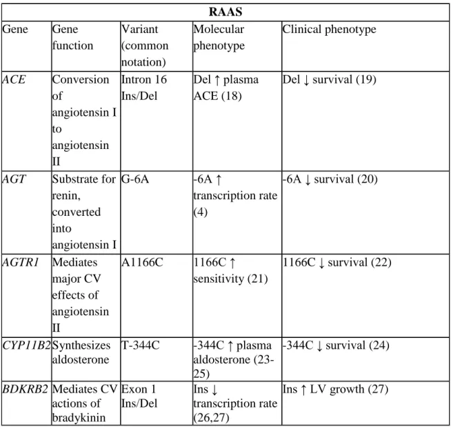
Table 4. Gene function and molecular and clinical phenotypes of candidate RAAS genetic variants
RAAS Gene Gene
function Variant (common notation) Molecular phenotype Clinical phenotype ACE Conversion of angiotensin I to angiotensin II Intron 16 Ins/Del
Del ↑ plasma ACE (18)
Del ↓ survival (19)
AGT Substrate for renin,
converted into
angiotensin I
G-6A -6A ↑
transcription rate (4)
-6A ↓ survival (20)
AGTR1 Mediates major CV effects of angiotensin II
A1166C 1166C ↑ sensitivity (21)
1166C ↓ survival (22)
CYP11B2 Synthesizes aldosterone
T-344C -344C ↑ plasma aldosterone (23-25)
-344C ↓ survival (24)
BDKRB2 Mediates CV actions of bradykinin Exon 1 Ins/Del Ins ↓ transcription rate (26,27)
35 REFERENCES
(1) Klein RJ, Zeiss C, Chew EY, Tsai JY, Sackler RS, Haynes C, et al. Complement factor H polymorphism in age-related macular degeneration. Science 2005 Apr 15;308(5720):385-389.
(2) International HapMap Consortium. A haplotype map of the human genome. Nature 2005 Oct 27;437(7063):1299-1320.
(3) National Center for Biotechnology Information and National Library of Medicine. Database of Single Nucleotide Polymorphisms. Available at: http://www.ncbi.nlm.nih.gov/snp/, 2012.
(4) Inoue I, Nakajima T, Williams CS, Quackenbush J, Puryear R, Powers M, et al. A nucleotide substitution in the promoter of human angiotensinogen is associated with essential hypertension and affects basal transcription in vitro. J Clin Invest 1997 Apr 1;99(7):1786-1797.
(5) Sehrt D, Meineke I, Tzvetkov M, Gultepe S, Brockmoller J. Carvedilol pharmacokinetics and pharmacodynamics in relation to CYP2D6 and ADRB pharmacogenetics. Pharmacogenomics 2011 May 23.
(6) Baudhuin LM, Miller WL, Train L, Bryant S, Hartman KA, Phelps M, et al. Relation of ADRB1, CYP2D6, and UGT1A1 polymorphisms with dose of, and response to, carvedilol or metoprolol therapy in patients with chronic heart failure. Am J Cardiol 2010 Aug 1;106(3):402-408.
(7) Colhoun HM, McKeigue PM, Davey Smith G. Problems of reporting genetic associations with complex outcomes. Lancet 2003 Mar 8;361(9360):865-872.
(8) Levin MC, Marullo S, Muntaner O, Andersson B, Magnusson Y. The myocardium-protective Gly-49 variant of the beta 1-adrenergic receptor exhibits constitutive activity and increased desensitization and down-regulation. J Biol Chem 2002 Aug 23;277(34):30429-30435.
(9) Borjesson M, Magnusson Y, Hjalmarson A, Andersson B. A novel polymorphism in the gene coding for the beta(1)-adrenergic receptor associated with survival in patients with heart failure. Eur Heart J 2000 Nov;21(22):1853-1858.
(10) Mason DA, Moore JD, Green SA, Liggett SB. A gain-of-function polymorphism in a G-protein coupling domain of the human beta1-adrenergic receptor. J Biol Chem 1999 Apr 30;274(18):12670-12674.
36
arrhythmogenesis, prognosis, and response to beta-blocker therapy. Am J Cardiol 2008 Sep 15;102(6):726-732.
(12) Green SA, Turki J, Innis M, Liggett SB. Amino-terminal polymorphisms of the human beta 2-adrenergic receptor impart distinct agonist-promoted regulatory properties. Biochemistry 1994 Aug 16;33(32):9414-9419.
(13) Shin J, Lobmeyer MT, Gong Y, Zineh I, Langaee TY, Yarandi H, et al. Relation of beta(2)-adrenoceptor haplotype to risk of death and heart transplantation in patients with heart failure. Am J Cardiol 2007 Jan 15;99(2):250-255.
(14) Forleo C, Resta N, Sorrentino S, Guida P, Manghisi A, De Luca V, et al. Association of beta-adrenergic receptor polymorphisms and progression to heart failure in patients with idiopathic dilated cardiomyopathy. Am J Med 2004 Oct 1;117(7):451-458.
(15) Small KM, Forbes SL, Rahman FF, Bridges KM, Liggett SB. A four amino acid deletion polymorphism in the third intracellular loop of the human alpha 2C-adrenergic receptor confers impaired coupling to multiple effectors. J Biol Chem 2000 Jul 28;275(30):23059-23064.
(16) Kardia SL, Kelly RJ, Keddache MA, Aronow BJ, Grabowski GA, Hahn HS, et al. Multiple interactions between the alpha 2C- and beta1-adrenergic receptors influence heart failure survival. BMC Med Genet 2008 Oct 23;9:93.
(17) Liggett SB, Cresci S, Kelly RJ, Syed FM, Matkovich SJ, Hahn HS, et al. A GRK5 polymorphism that inhibits beta-adrenergic receptor signaling is protective in heart failure. Nat Med 2008 May;14(5):510-517.
(18) Rigat B, Hubert C, Alhenc-Gelas F, Cambien F, Corvol P, Soubrier F. An insertion/deletion polymorphism in the angiotensin I-converting enzyme gene accounting for half the variance of serum enzyme levels. J Clin Invest 1990 Oct;86(4):1343-1346. (19) Andersson B, Sylven C. The DD genotype of the angiotensin-converting enzyme gene is associated with increased mortality in idiopathic heart failure. J Am Coll Cardiol 1996 Jul;28(1):162-167.
(20) Pilbrow AP, Palmer BR, Frampton CM, Yandle TG, Troughton RW, Campbell E, et al. Angiotensinogen M235T and T174M gene polymorphisms in combination doubles the risk of mortality in heart failure. Hypertension 2007 Feb;49(2):322-327.
37
(22) Amir O, Amir RE, Paz H, Attias E, Sagiv M, Lewis BS. Relation between AT1R gene polymorphism and long-term outcome in patients with heart failure. Cardiology 2009;112(2):151-157.
(23) White PC, Slutsker L. Haplotype analysis of CYP11B2. Endocr Res 1995 Feb-May;21(1-2):437-442.
(24) McNamara DM, Tam SW, Sabolinski ML, Tobelmann P, Janosko K, Taylor AL, et al. Aldosterone synthase promoter polymorphism predicts outcome in African Americans with heart failure: results from the A-HeFT Trial. J Am Coll Cardiol 2006 Sep 19;48(6):1277-1282.
(25) Pojoga L, Gautier S, Blanc H, Guyene TT, Poirier O, Cambien F, et al. Genetic determination of plasma aldosterone levels in essential hypertension. Am J Hypertens 1998 Jul;11(7):856-860.
(26) Braun A, Kammerer S, Maier E, Bohme E, Roscher AA. Polymorphisms in the gene for the human B2-bradykinin receptor. New tools in assessing a genetic risk for bradykinin-associated diseases. Immunopharmacology 1996 Jun;33(1-3):32-35.
CHAPTER III:
CHARACTERIZATION OF CANDIDATE GENETIC VARIANTS
Summary
The previous chapter covered the methods and results for the candidate gene search. This chapter describes genotyping technologies and the methods used for genotyping the eleven selected candidates. Three platforms were used: TaqMan® fluorescent allelic discrimination, QIAxcel® capillary electrophoresis, and Sequenom® MALDI-TOF mass spectrometry. These methods were accurate and reproducible when subject to a series of quality control measures except for the ADRA2C indel, which significantly deviated from Hardy-Weinberg Equilibrium (HWE). Preferential amplification of the ADRA2C deletion allele must have occurred, and therefore analyses of the ADRA2C indel must be performed as insertion-homozygous versus deletion-carrier and not as the three individual genotypes.
Introduction
39
PCR amplification step, and then allele discrimination is commonly achieved using primer extension, hybridization, ligation, or enzymatic cleavage. Primer extension relies on the highly accurate DNA polymerase enzyme to incorporate allele-specific nucleotides, and hence it is very reliable. Assay designs are simple, fast, flexible, customizable, multiplex, and widely commercially available (e.g. Sequenom® MassEXTEND™). Hybridization techniques rely on the thermal stability of perfectly
complementary DNA probes at the variant loci. Therefore the major disadvantages of this technique are non-specific binding to other loci in the genome or cross-hybridization. The advantage of hybridization techniques is increased throughput because of the lack of an enzymatic reaction requirement. Ligation relies on the high accuracy of the DNA ligase enzyme to join two oligonucleotides only when there is perfect complementarity with the DNA template. With appropriate tags and divergent oligonucleotides, ligation methods can be successfully scaled up to high throughput. Enzymatic cleavage methods rely on the specificity of certain enzymes for certain DNA sequences. For example, restriction fragment length polymorphism (RFLP) utilizes restriction enzymes that cleave double-stranded DNA at unique sites consistently. RFLP is one of the earliest genotyping methods, but it has limited throughput and multiplex capacity. Notably, allele discrimination could involve a combination of any of the above approaches (e.g. TaqMan® uses a combination of hybridization and 5’ nuclease activity of DNA
polymerase).
40
electrophoresis is low resolution, labor-demanding, and time-demanding. More recently these disadvantages have been overcome by capillary gel electrophoresis (e.g. Qiaxcel®) (2). Matrix-assisted laser desorption/ionization time-of-flight mass spectrometry (MALDI-TOF MS) is a commonly used mass-based technique for detecting oligonucleotides. A major advantage of MALDI-TOF MS is that a mixture of oligonucleotides can be rapidly separated and accurately analyzed simultaneously. This method is commonly used for detecting products of primer extension reactions (e.g. Sequenom®). The genotype for each allele-specific extension product is determined by its unique mass. A major limitation of this method is reduced peak resolution due to similar masses of the extension products. Fluorescence is a commonly used detection method because it is simple, fast, and accurate. TaqMan® is an example of fluorescent detection, in which probes with a quencher on one end and the fluorescent dye on the other end are used. When the probes are intact, the fluorescent signal is quenched. However, the fluorescent signal is emitted when the fluorescent dye is cleaved from the quencher by a DNA polymerase with 5’ exonuclease activity. The alleles have specific
fluorescent dyes which reveal genotype. Chemiluminescence utilizes a cascade of enzymatic reactions that generate light. This method is fast, automatable, and has a high signal-to-noise ratio. A method that uses chemiluminescence is Pyrosequencing™. When a template nucleotide is complementary to the nucleotide being extended, DNA polymerase incorporates the nucleotide initiating the cascade of reactions to emit light. A disadvantage of this method is the lack of multiplex capacity.