CHAPERONE MEDIATED PROTECTIVE PROTEIN AGGREGATION AND SPATIAL QUALITY CONTROL
Katie J. Wolfe
A dissertation submitted to the faculty of the University of North Carolina at Chapel Hill in partial fulfillment of the requirements for the degree of Doctor of Philosophy in the Department
of Cell Biology and Physiology
Chapel Hill 2013
ABSTRACT
Katie Jean Wolfe: Chaperone mediated protective protein aggregation and spatial quality control (Under the direction of Douglas Cyr, PhD)
The accumulation of amyloid-like aggregates is a characteristic of protein conformational disorders such as Huntington Disease, but whether amyloid-like aggregation is causative or a cytoprotective mechanism remains unclear. Molecular chaperones act as the front line of defense against proteotoxicity, as they protect cells by partitioning misfolded proteins towards refolding, degradation, or assembly into large benign aggregates. Herein, a yeast model of proteotoxicity was utilized to study cellular mechanisms for protective aggregation. Ectopic expression of polyglutamine (polyQ) expanded Huntingtin (Htt103Q) is toxic in yeast, and targeting it to the nucleus enhances toxicity while decreasing SDS-resistant aggregation. I utilized this nuclear Htt103Q as the substrate for a high copy toxicity suppressor screen in yeast which identified Sti1 as a molecule that promotes protective aggregation of Htt103Q. The Hsp70 co-chaperone Sti1 regulates spatial quality control of amyloid-like proteins as it induces formation of perinuclear foci containing SDS-resistant material. Accumulation of distinct
perinuclear foci correlates with suppression of toxicity and increased complex formation with the Hsp70/Hsp40 chaperone machinery. Endogenous Sti1 appears to be a crucial player in a
chaperone-facilitated protective aggregation pathway, because deletion of Sti1 enhances
Q-rich protein is titrated away from its normal function, or beneficially via interactions that promote protective aggregation. Over-expression of Nab3 appears to suppress Htt103Q toxicity by replacing a functional pool of Nab3 that was lost to aberrant polyQ interaction.
ACKNOWLEDGEMENTS
My dissertation work would not have been possible without the help and support of numerous individuals which I have had the pleasure of meeting. I would first like to thank my mentor and advisor, Dr. Douglas Cyr. He has provided me with a nurturing lab home and shaped me to be a critical thinking scientist. He has taught me how to persistently and creatively seek out answers to complicated questions. His support and guidance have helped me complete my dissertation in an efficient and timely manner.
My development has also been greatly impacted by past and present members of both the Cyr lab and the Brennwald lab. I would like to thank each one of them for acting as a sounding board and creating a positive work place. I also thank my committee members, Dr. Patrick Brennwald, Dr. Mohanish Deshmukh, Dr. Nikolay Dokholyan, and Dr. Brian Strahl for donating their time and energy to helping me succeed as a graduate student.
PREFACE
TABLE OF CONTENTS
LIST OF TABLES………..……….x
LIST OF FIGURES………..…….……….xi
LIST OF ABBREVIATIONS .………..…...xii
CHAPTER 1 Amyloid in neurodegenerative diseases: Friend or foe? ... 1
1.1 OVERVIEW ... 1
1.2 INTRODUCTION... 1
1.3 FORMATION OF TOXIC SPECIES IN AMYLOID DISEASES ... 3
1.3.1 Oligomeric amyloid assembly ... 4
1.3.2 Interaction surfaces of toxic amyloid species ... 7
1.4 SUPPRESSION OF AMYLOID PROTEOTOXICITY BY MOLECULAR CHAPERONES ... 9
1.4.1 Chaperone dependent suppression of aggregation ... 9
1.4.2 Chaperone mediated aggregation ... 11
1.4.3 Chaperone assisted protein degradation ... 12
1.5 CONCLUDING REMARKS ... 14
1.6 FIGURES ... 16
2.1 OVERVIEW ... 24
2.2 INTRODUCTION... 25
2.3 RESULTS ... 29
2.3.1 The Hsp70/Hsp90 co-chaperone Sti1 suppresses Htt103Q toxicity ... 29
2.3.2 Sti1 interacts with high molecular weight forms of Htt103Q ... 31
2.3.3 Sti1 modulates assembly of amyloidogenic substrates at a perinuclear location ... 32
2.3.4 Sti1 inducible foci are distinct protein quality control depots ... 35
2.3.5 Sti1 reorganizes complexes that contain Htt103Q and Hsp70 ... 36
2.3.6 Sti1 and Sis1 cooperate to modulate amyloid toxicity ... 38
2.4 DISCUSSION ... 38
2.5 METHODS ... 43
2.7 TABLES AND FIGURES ... 49
3 Identification of polyglutamine rich proteins that alter huntingtin toxicity and aggregation ... 68
3.1 OVERVIEW ... 68
3.2 INTRODUCTION... 69
3.3 RESULTS ... 72
3.3.1 Genomic fragments from high copy screen suppress Htt103Q-NLS toxicity ... 72
3.3.2 PolyQ-rich proteins suppress Htt103Q-NLS and Htt103Q toxicity ... 74
3.3.3 Functional Nab3 is required for suppression of Htt toxicity ... 76
3.3.5 A short proline-rich stretch in the Pop2 polyQ domain is required for impact
upon Htt103Q toxicity and aggregation ... 79
3.3.6 Pop2 and Cbk1 alter Htt103Q toxicity and localization in a Sti1-dependent manner 81
LIST OF TABLES
LIST OF FIGURES
Figure 1.1 Protein folding and amyloid formation. ... 16
Figure 2.1 Sti1 suppresses Htt103Q toxicity and reorganizes Htt103Q-GFP foci ... 49
Figure 2.2 Sti1 specifically alters Htt103Q-GFP spatial and temporal foci formation ... 51
Figure 2.3 Sti1 co-fractionates with high molecular weight forms of Htt103Q ... 52
Figure 2.4 Sti1 suppresses Rnq1 toxicity and reorganizes Rnq1-mRFP foci ... 53
Figure 2.5 Sti1 modulates assembly of amyloidogenic substrates ... 54
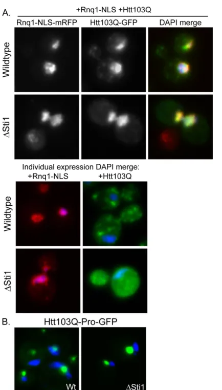
Figure 2.6 Sti1 is not required for Rnq1-NLS relocalization of Htt103Q to the nucleus ... 55
Figure 2.7 The StiF is a juxtanuclear compartment for amyloid-like proteins ... 56
Figure 2.8 Sti1 reorganizes complexes that contain Htt103Q and Hsp70 ... 57
Figure 2.9 Sti1 and Sis1 cooperate to regulate Rnq1 and Htt103Q toxicity ... 58
Figure 2.10 Model for StiF formation ... 59
Figure 2.11 Other cellular sorting factors are not necessary for Sti1’s mechanism of action ... 60
Figure 2.12 Sti1 attenuates Hsp104 shearing activity in the presence of GndHCl ... 61
Figure 3.1. Genomic fragments from high copy screen suppress Htt103Q-NLS toxicity ... 94
Figure 3.3 PolyQ-rich protein domain details ... 97
Figure 3.4 Nab3 is depleted by doxycycline and the polyQ-rich region of Pop2 promotes Htt103Q aggregation ... 98
Figure 3.5 Overexpression of Nab3 must be functional for Htt103Q-NLS toxicity suppression . 99 Figure 3.6 The polyQ-rich region of Cbk1 alters Htt103Q toxicity and aggregation ... 100
Figure 3.7 A short proline-rich stretch in the Pop2 polyQ domain is required for impact upon Htt103Q toxicity and aggregation ... 102
Figure 3.8 Cbk1 and Pop2 act in a Sti1-dependent manner ... 103
LIST OF ABBREVIATIONS
CD- conformational disease CHX- cycloheximide coIP-co-immuneprecpitation GF- genomic fragment
GFP- green fluorescent protein GndHCl- guanidinium hydrochloride
Htt103Q- Huntingtin with 103Q glutamine stretch IP-immunoprecipitation
IPOD- insoluble protein deposit
JUNQ- juxtanuclear quality control compartment mRFP- monomeric red fluorescent protein NLS- nuclear localization signal
NEF- nucleotide exchange factor ORF- open reading frame
CHAPTER 1
Amyloid in neurodegenerative diseases: Friend or foe?1
1.1 Overview
Accumulation of amyloid-like aggregates is a hallmark of numerous neurodegenerative disorders such as Alzheimer’s and polyglutamine disease. Yet, whether the amyloid inclusions found in these diseases are toxic or cytoprotective remains unclear. Various studies suggest that the toxic culprit in the amyloid folding pathway is actually a soluble oligomeric species which might interfere with normal cellular function by a multifactorial mechanism including aberrant protein-protein interactions. Molecular chaperones suppress toxicity of amyloidogenic proteins by inhibiting aggregation of non-native disease substrates and targeting them for refolding or degradation. Paradoxically, recent studies also suggest a protective action of chaperones in their promotion of the assembly of large, tightly packed, benign aggregates that sequester toxic protein species.
1.2 Introduction
Protein misfolding and the accumulation of amyloid aggregates are prominent features in a vast array of human diseases including numerous neurodegenerative disorders [1]. An amyloid fibril is an insoluble, highly ordered aggregate and the major component of extracellular plaques found in Alzheimer patient’s brains. Amyloid fibrils
are defined by a cross- structure where the sheets run perpendicular to the fibril axis
[2]. Amyloid can be distinguished from other disordered aggregates by several properties including insolubility in ionic detergent, protease resistance, and recognition by
diagnostic indicator dyes such as Congo Red [3]. Intracellular inclusions which exhibit similar characteristics are usually termed amyloid-like [2], but for the purposes of this review we will refer to amyloid plaques, fibrils, and amyloid-like aggregates all as amyloid unless otherwise noted. It remains highly controversial whether the amyloid deposits found in patients with amyloid disorders is the root problem as researchers first thought or if, as increasing evidence suggests, the large inclusions serve a protective cellular function [4, 5].
Amyloid diseases are associated with a broader family termed protein
conformational diseases, coined by Carrell and Lomas in 1997. They proposed that each disease occurs via a similar mechanism that involves the abnormal folding and
aggregation of specific disease associated proteins causing a toxic gain of function [6]. However, the precise reason behind aggregation of a disease protein causing toxicity remains unclear. An equally challenging enigma in neurodegenerative amyloidoses is the cause of selective vulnerability of certain neuronal populations. Each neurodegenerative disorder affects a specific subset of neurons even though the disease associated protein is often present in many cells throughout the brain and the rest of the body [7]. This
influence the fate of disease proteins and may account for selective vulnerability [10, 11]. Yet, the components that make up such collectives of protective or harmful interactions remain ambiguous. Herein, we discuss mechanisms for amyloid toxicity and explore how molecular chaperones act to modulate the proteotoxicity associated with formation of intracellular amyloid aggregates.
1.3 Formation of toxic species in amyloid diseases
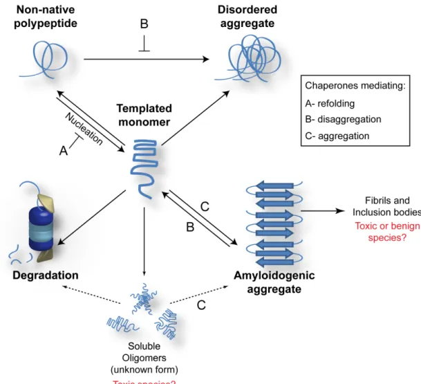
Although numerous neurodegenerative diseases are associated with the presence of amyloid-like aggregates, the toxic culprit in the aggregation pathway leading to amyloid formation remains elusive. Some researchers suggest that the inclusion bodies found in amyloid diseases (bottom, right in Fig 1) are the toxic species [12], yet there is often a negative correlation between neurotoxicity and existence of large amyloid aggregates[13, 14]. Accumulating literature suggests that a soluble, pre-fibrillar species of the disease protein (Soluble Oligomers in Fig 1) causes cytotoxicity and the fibrillar aggregates may be part of a cytoprotective mechanism whereby toxic species are sequestered [4, 5].
[16]. Although it is clear that directing a natively folded protein towards amyloid formation is linked to disease, the exact conformation of the species causing the primary toxic insult is still unknown. There are many neurodegenerative disorders characterized by amyloid inclusions, but some of the most highly studied are Alzheimer’s Disease (AD) and polyglutamine diseases such as Huntington’s Disease (HD). Below, we will discuss studies directed towards elucidating what the toxic amyloidogenic species might be in these two diseases.
1.3.1 Oligomeric amyloid assembly
The structural transition from a non-native fold to a pre-fibrillar conformation occurs via nucleated polymerization [15, 17]. The nucleation step of this process begins very slowly, perhaps due to an unfavorable energy barrier. After nucleation, however, polymerization occurs much more rapidly as the folding intermediate is capable of serving as a template or seed for further conformational switching [17]. Interestingly, it appears that all proteins which form amyloid may proceed through the same intermediate steps [18]. In this section, we will review recent advances in amyloid biology which suggest that there are common features in the mechanism for toxicity of structurally distinct proteins; these studies also highlight the toxic nature of soluble intermediates in the amyloid assembly pathway.
1.3.1.1 Polyglutamine diseases: Huntington’s Disease
associated with cleavage of an N-terminal exon 1 fragment containing the expanded polyQ tract from Htt. This is followed by nuclear accumulation of the cleaved fragment even though the wildtype form of Htt is localized mainly to the cytosol [20, 21].
Although aggregation intermediates have been hard to identify, polyQ length dependent formation of Htt oligomers has indeed been demonstrated both in vivo and in
vitro [22, 23]. Indeed, much evidence supports the hypothesis that there may be an
inverse relationship between Htt toxicity and aggregation. For instance, expression of Htt with an expanded glutamine stretch in cultured striatal neurons causes aggregation in a manner that does not correlate with cell death. In fact, suppression of intranuclear Htt inclusion formation is accompanied by increased cell death [14]. While this study
analyzed a total population of cells, Arrasate et al designed a series of experiments where they followed individual neurons. During the lifetime of the cells, the group tracked survival, Htt load, and Htt inclusion formation. This inventive experimental setup revealed that neuronal death correlates with increased polyQ expansion length and
amount of diffuse Htt within the cell. Additionally, Htt inclusion body formation reduced the amount of soluble Htt thereby increasing neuronal survival [24]. These studies
provide fundamental insight which supports the idea that the toxic Htt species is soluble rather than large intranuclear inclusions.
1.3.1.2 Alzheimer’s Disease
Amyloid aggregation in AD occurs with cleavage of APP fragment into Aβ42 which accumulates in extracellular amyloid plaques [27]. Aβ amyloid plaques found in human brains of AD patients have a low correlation with severity of AD [28]. A recent study using a mouse model of AD strongly supports the toxic oligomeric hypothesis [25]. AD symptoms were decreased upon reduction of insulin growth factor signaling which
correlated with formation of tightly packed A aggregates. As seems to be the case with
Htt, Aapparently may undergo oligomerization into a soluble, toxic conformer which
can be sequestered by protective cellular pathways into larger benign aggregates. In AD, the toxic soluble oligomer has been identified in various ways. Injection
of medium containing Amonomers and oligomers, but lacking amyloid fibrils, was able
to inhibit hippocampal long-term potentiation (LTP) in rats [4]. Pretreatment of the sample in order to destroy A monomer followed by injection still resulted in decreased
LTP. In another in vivo study, rats were injected with an oligomer specific antibody
which was able to block inhibition of LTP [26]. Altogether, these in vivo results suggest
that Aoligomers mediate toxicity or nucleate the formation of a toxic protein species.
Soluble intermediates in the amyloid assembly pathway for Aβ have a structure in
1.3.2 Interaction surfaces of toxic amyloid species
A broad array of protein interaction partners can affect aggregation both
positively and negatively, composing a vast protein homeostasis network which needs to be balanced to maintain cell viability [10]. For example, aberrant protein-protein
interactions can interfere with the normal function of various transcription factors, such as CREB Binding Protein and TATA Binding Protein, leading to transcriptional
dysregulation [29, 30]. Htt and various amyloid disease proteins might also deplete and/or inactivate other necessary cellular factors. These include but are not limited to ubiquitin proteosome machinery [31], ER associated degradation (ERAD) machinery [16], numerous glutamine or glutamine/asparagine (Q/N) rich proteins [32, 33], and large proteins predicted to be highly unstructured which may act as hubs or scaffolds [34]. In addition, coiled-coil (CC) domains were recently suggested to regulate aggregation and toxicity of Q/N rich amyloid proteins [35]. Increased CC propensity in regions of proteins that contain polyQ was found to increase occurrence of aggregation and toxicity. It’s possible that the CC structure may be an interaction domain which mediates the
nucleation event for pre-fibrillar -rich amyloid to occur in polyglutamine diseases.
Furthermore, there may be distinct chaperone machineries regulating the formation of the CC domain, imparting another level of control over the system. Although we are just beginning to understand mechanism for ‘nucleation’ of amyloid assembly, it seems that the nature of protein interactions are able to dictate how a non-natively folded protein is handled within the cell.
self-propagating proteinaceous particles [36] which, in yeast, are amyloid-like heritable genetic elements passed from mother to daughter cell [37]. Prion proteins can exist in their natively folded conformation or in their amyloid-like prion conformation. For instance, Rnq1 and Sup35 are the proteins which form [RNQ+] (also called [PIN+]) and [PSI+] prions, respectively. The [RNQ+] prion serves as a prime example of a protein which affects conformation of another protein. When [RNQ+] comes into contact with another amyloidogenic protein such as Sup35, the prion allows the interaction partner to assume its amyloid conformation, in this case [PSI+] [38, 39]. The Rnq1/Sup35
interaction is normally very weak, but nonetheless Rnq1 can serve as a nucleator of amyloid conversion and assembly [40]. In the context of HD, when Htt is expressed in yeast, toxicity only occurs in the presence of [RNQ+] [41]. Thus, Rnq1 serves as a nucleation factor not only for other yeast prions, but also for other amyloidogenic disease associated proteins. Htt assembly could actually be relocalized to the nucleus by tagging Rnq1 with a nuclear localization signal [42]. Relocalizing Htt assembly to the nucleus hindered amyloid assembly resulting in the accumulation of a soluble Htt species. This correlated with enhanced Htt-induced cell death. Consequently, the specific location of amyloid conversion impacts cytotoxicity perhaps by influencing aberrant protein-protein interactions that might disrupt the essential cellular functions mentioned above. While [RNQ+] as well as already templated amyloid proteins can serve as nucleators for
amyloidogenic proteins, further studies are needed to discover other cellular factors with this capability.
misfold and aggregate in specific, disease dependent, neuronal subpopulations. The primary amino acid sequence, which is different for each individual disease protein, may play a role in selective vulnerability. Second, the common amyloid toxicity mechanism seems to stem from soluble oligomeric species which may be the toxic culprit in various neurodegenerative amyloidosis. Finally, though it remains unclear whether the soluble amyloid is a folding intermediate or an off-pathway conformer, the toxic soluble species in various amyloid associated neurodegenerative disorders must adopt a -rich structure
[43, 44]. The toxic -rich structure may expose surfaces amenable to aberrant
intracellular interactions, but when packaged into large benign aggregates, these surfaces may be hidden.
1.4 Suppression of amyloid proteotoxicity by molecular chaperones
Molecular chaperones are a crucial part of PQC and work by maintaining the proper folding status of proteins within a cell. Chaperones recognize misfolded conformers which are then targeted for refolding, aggregation, or triaged towards degradation. They play a vital role in protecting the cell from conformational diseases such as amyloidogenic neurodegenerative disorders. Chaperones do this by either suppressing the initial oligomerization of disease proteins and disassembling disease protein aggregates or stimulating the conversion of toxic amyloid assembly intermediates into benign aggregates [5, 45-47]. In other words, chaperones can either inhibit or promote aggregation but the end goal is to eliminate the toxic soluble species.
1.4.1 Chaperone dependent suppression of aggregation
functions as a co-chaperone by binding non-natively folded proteins and delivering them to Hsp70 for refolding [48]. The diversity of Hsp40s polypeptide binding domain provides amazing substrate specificity to the Hsp70 [49, 50]. Various Hsp40/Hsp70 machineries act in different ways towards distinct disease protein substrates. This chaperone machinery has been highly studied as a mechanism which inhibits amyloid-like aggregate assembly of disease proteins [51, 52]. In yeast, overexpression of Hsp70, Ssa1, or Hsp40, Ydj1, alters aggregation of mutant Htt by preventing fibrillization [45]. Additionally, elevated expression of Ydj1 reduced aggregation and toxicity of Q/N rich amyloid aggregates [53]. Likewise in cell culture, increasing the amount of Hsp40 and Hsp70 has been shown to decrease formation of polyQ disease protein aggregates as well as alleviate toxicity [54, 55]. Thus it seems that the Hsp40/Hsp70 machinery may act to hold the disease protein in a soluble conformation in order to prevent accumulation of a toxic intermediate or byproduct of the amyloid aggregation pathway.
Molecular chaperones can also antagonize accumulation of amyloid assemblies by acting later in the folding pathway via solubilization of already formed aggregates. Hsp104, an AAA ATPase protein remodeling factor in yeast, is required for propagation of yeast prions via shearing of large amyloid-like aggregates into smaller seeds [38, 56]. Utilizing its solubilization activity, overexpression of Hsp104 in a yeast HD model was able to effectively fragment amyloid-like aggregates of expanded-polyQ Htt [57]. There is no Hsp104 in higher eukaryotes, however, exogenous expression of the chaperone in worm and rat models demonstrates that it retains its functional activity. Expression of Hsp104 in C. elegans reduced expanded polyQ protein aggregation and assuaged
and alter distribution of expanded polyQ proteins [59]. Though these are just a few examples, the Hsp40/Hsp70 and Hsp104 machines clearly can protect cells from amyloid disease proteins by suppressing aggregation thereby inhibiting formation of a toxic soluble species.
1.4.2 Chaperone mediated aggregation
Chaperone machinery has also been shown to promote aggregation of human amyloidogenic substrates. A human Hsp40, Hdj2, has been shown to increase aggregation of expanded polyQ Htt in Cos-7 cells [61]. In conjunction with
Hsp40/Hsp70 machinery, the chaperonin complex, TRiC, also alters amyloid aggregation while suppressing polyQ-mediated toxicity as shown in C. elegans [62], yeast [63], and
cell culture [64]. These studies demonstrate that depletion of TRiC leads to accumulation of a toxic, soluble, oligomeric Htt species. In fact, Behrends et al. utilized size exclusion chromatography to show that toxic Htt oligomers exhibited a molecular weight of
approximately 200 kDa. TRiC, in conjunction with Ssa1 and Ydj1, was able to shift the toxic soluble oligomers into non-toxic 500 kDa aggregates [63]. Thus, promotion of protective aggregation seems to be a mechanism whereby cells eliminate cytotoxic pools of Htt oligomer. Altogether, these studies demonstrate that molecular chaperones protect cells from proteotoxic insult by targeting misfolded proteins away from toxic oligomeric states; chaperones either enhance assembly into fibrillar benign aggregates, or
disassemble the oligomers into soluble monomeric forms which can be properly folded [65]. Incredibly, chaperones are able to carry out these opposing activities, both of which lead to promotion of a healthy and functional folding state of non-native proteins in the cellular milieu.
1.4.3 Chaperone assisted protein degradation
transfer of polyubiquitin chains to misfolded substrates, but also has inherent chaperone activity and acts as an Hsp70/Hsc70 co-chaperone [66, 67]. Indeed, CHIP has been shown to play a role in chaperone mediated degradation of expanded polyQ disease proteins. When CHIP was overexpressed, ubiquitination and turnover of polyQ disease substrates such as Htt increased, resulting in suppression of polyQ aggregation and cell death [68, 69]. CHIP’s chaperone activity is necessary in order to observe this
suppression. Elevated Hsc70 levels increased CHIP’s ability to suppress aggregation and cell death, thus CHIP works cooperatively with Hsc70.
Autophagy is another cellular clearance mechanism for neurodegenerative amyloid proteins which is independent of the ubiquitin-proteosome system. Autophagy, literally meaning “self-eating”, is a cellular process that involves compartmentalizing bulk cytosol, including cell components and proteins, which is sent to lysosomes to be degraded [70]. When autophagy was suppressed in mice via inhibition of ATG5, even without the presence of disease causing proteins, the mice displayed characteristics of neurodegeneration [71]. As a result, this study suggests that neuronal cells are constantly challenged by the formation of misfolded proteins and autophagy is required for the removal of these proteins. In the absence of this pathway, misfolded proteins accumulate and disrupt normal cellular function. In the context of neurodegenerative diseases, expanded polyQ disease proteins have indeed been shown to be degraded via autophagy [72]. Furthermore, recent evidence suggests that mutant Htt can specifically be targeted to autophagosomes by acetylation [73]. Altogether, these studies establish that
1.5 Concluding Remarks
Normal PQC is crucial to cell viability because it maintains the balance of non-native proteins targeted towards refolding or degradation. A shift in this equilibrium can be highly detrimental to the cell, leading to death. Specific mechanisms of proteotoxicity caused by misfolded protein accumulation remain unclear, but many cellular processes have been implicated. Neurodegenerative disorders characterized by amyloid inclusions seem to be caused by a multifactorial mechanism which may be centered around titration and inactivation of essential cellular components in processes such as transcription [74], degradation via the ubiquitin proteosome [31], and ER associated degradation (ERAD) [16] among others. Aberrant interactions arising from exposed hydrophobic surfaces within the amyloidogenic disease protein [33, 75] and/or mislocalization [42] can lead to cell death.
Not all cell types are equally affected by a single amyloidogenic protein, however. The equilibrium between a native and non-native conformation is cell type specific, and not all cell types equally respond to the same protein misfolding event. Some cell types may be less equipped than others and therefore unable to clear toxic amyloid species. Cellular mechanisms for clearance of these toxic amyloid species include chaperone dependent suppression or promotion of aggregation, chaperone dependent turnover, and clearance via autophagy.
1.6 Figures
Figure 1.1 Protein folding and amyloid formation.
Non-native proteins can be partitioned into several conformational fates including
refolding, ordered or disordered aggregate formation, and degradation. Amyloid is a type of very structured (or ordered) aggregate and forms through a poorly understood
REFERENCES
1. Chiti, F. and C.M. Dobson, Protein misfolding, functional amyloid, and human disease. Annu Rev Biochem, 2006. 75: p. 333-66.
2. Sipe, J.D., et al., Amyloid fibril protein nomenclature: 2010 recommendations from the nomenclature committee of the International Society of Amyloidosis.
Amyloid, 2010. 17(3-4): p. 101-4.
3. Ross, C.A. and M.A. Poirier, Opinion: What is the role of protein aggregation in neurodegeneration? Nat Rev Mol Cell Biol, 2005. 6(11): p. 891-8.
4. Walsh, D.M., et al., Naturally secreted oligomers of amyloid beta protein potently inhibit hippocampal long-term potentiation in vivo. Nature, 2002. 416(6880): p.
535-9.
5. Douglas, P.M., et al., Chaperone-dependent amyloid assembly protects cells from prion toxicity. Proc Natl Acad Sci U S A, 2008. 105(20): p. 7206-11.
6. Carrell, R.W. and D.A. Lomas, Conformational disease. Lancet, 1997. 350(9071):
p. 134-8.
7. Mazarei, G., et al., Expression analysis of novel striatal-enriched genes in Huntington disease. Hum Mol Genet, 2010. 19(4): p. 609-22.
8. Giorgini, F., et al., A genomic screen in yeast implicates kynurenine
3-monooxygenase as a therapeutic target for Huntington disease. Nat Genet, 2005.
37(5): p. 526-31.
9. Willingham, S., et al., Yeast genes that enhance the toxicity of a mutant huntingtin fragment or alpha-synuclein. Science, 2003. 302(5651): p. 1769-72.
10. Balch, W.E., et al., Adapting proteostasis for disease intervention. Science, 2008.
319(5865): p. 916-9.
12. Yang, W., et al., Aggregated polyglutamine peptides delivered to nuclei are toxic to mammalian cells. Hum Mol Genet, 2002. 11(23): p. 2905-17.
13. Kuemmerle, S., et al., Huntington aggregates may not predict neuronal death in Huntington's disease. Ann Neurol, 1999. 46(6): p. 842-9.
14. Saudou, F., et al., Huntingtin acts in the nucleus to induce apoptosis but death does not correlate with the formation of intranuclear inclusions. Cell, 1998.
95(1): p. 55-66.
15. Jahn, T.R., et al., Amyloid formation under physiological conditions proceeds via a native-like folding intermediate. Nat Struct Mol Biol, 2006. 13(3): p. 195-201.
16. Duennwald, M.L. and S. Lindquist, Impaired ERAD and ER stress are early and specific events in polyglutamine toxicity. Genes Dev, 2008. 22(23): p. 3308-19.
17. Jarrett, J.T. and P.T. Lansbury, Jr., Seeding "one-dimensional crystallization" of amyloid: a pathogenic mechanism in Alzheimer's disease and scrapie? Cell, 1993.
73(6): p. 1055-8.
18. Kayed, R., et al., Common structure of soluble amyloid oligomers implies common mechanism of pathogenesis. Science, 2003. 300(5618): p. 486-9.
19. Zoghbi, H.Y. and H.T. Orr, Glutamine repeats and neurodegeneration. Annu Rev
Neurosci, 2000. 23: p. 217-47.
20. Davies, S.W., et al., Formation of neuronal intranuclear inclusions underlies the neurological dysfunction in mice transgenic for the HD mutation. Cell, 1997.
90(3): p. 537-48.
21. DiFiglia, M., et al., Aggregation of huntingtin in neuronal intranuclear inclusions and dystrophic neurites in brain. Science, 1997. 277(5334): p. 1990-3.
22. Legleiter, J., et al., Mutant huntingtin fragments form oligomers in a
polyglutamine length-dependent manner in vitro and in vivo. J Biol Chem, 2010.
23. Takahashi, T., et al., Soluble polyglutamine oligomers formed prior to inclusion body formation are cytotoxic. Hum Mol Genet, 2008. 17(3): p. 345-56.
24. Arrasate, M., et al., Inclusion body formation reduces levels of mutant huntingtin and the risk of neuronal death. Nature, 2004. 431(7010): p. 805-10.
25. Cohen, E., et al., Reduced IGF-1 signaling delays age-associated proteotoxicity in mice. Cell, 2009. 139(6): p. 1157-69.
26. Klyubin, I., et al., Amyloid beta protein immunotherapy neutralizes Abeta oligomers that disrupt synaptic plasticity in vivo. Nat Med, 2005. 11(5): p.
556-61.
27. Haass, C. and D.J. Selkoe, Soluble protein oligomers in neurodegeneration: lessons from the Alzheimer's amyloid beta-peptide. Nat Rev Mol Cell Biol, 2007.
8(2): p. 101-12.
28. Terry, R.D., et al., Physical basis of cognitive alterations in Alzheimer's disease: synapse loss is the major correlate of cognitive impairment. Ann Neurol, 1991.
30(4): p. 572-80.
29. Riley, B.E. and H.T. Orr, Polyglutamine neurodegenerative diseases and regulation of transcription: assembling the puzzle. Genes Dev, 2006. 20(16): p.
2183-92.
30. Nucifora, F.C., Jr., et al., Interference by huntingtin and atrophin-1 with cbp-mediated transcription leading to cellular toxicity. Science, 2001. 291(5512): p.
2423-8.
31. Bence, N.F., R.M. Sampat, and R.R. Kopito, Impairment of the
ubiquitin-proteasome system by protein aggregation. Science, 2001. 292(5521): p. 1552-5.
32. Furukawa, Y., et al., Cross-seeding fibrillation of Q/N-rich proteins offers new pathomechanism of polyglutamine diseases. J Neurosci, 2009. 29(16): p. 5153-62.
33. Duennwald, M.L., et al., A network of protein interactions determines
34. Olzscha, H., et al., Amyloid-like aggregates sequester numerous metastable proteins with essential cellular functions. Cell, 2011. 144(1): p. 67-78.
35. Fiumara, F., et al., Essential role of coiled coils for aggregation and activity of Q/N-rich prions and PolyQ proteins. Cell, 2010. 143(7): p. 1121-35.
36. Prusiner, S.B., Novel proteinaceous infectious particles cause scrapie. Science,
1982. 216(4542): p. 136-44.
37. Shorter, J. and S. Lindquist, Prions as adaptive conduits of memory and inheritance. Nat Rev Genet, 2005. 6(6): p. 435-50.
38. Sondheimer, N. and S. Lindquist, Rnq1: an epigenetic modifier of protein function in yeast. Mol Cell, 2000. 5(1): p. 163-72.
39. Derkatch, I.L., et al., Prions affect the appearance of other prions: the story of [PIN(+)]. Cell, 2001. 106(2): p. 171-82.
40. Vitrenko, Y.A., et al., Propagation of the [PIN+] prion by fragments of Rnq1 fused to GFP. Curr Genet, 2007. 51(5): p. 309-19.
41. Meriin, A.B., et al., Huntington toxicity in yeast model depends on polyglutamine aggregation mediated by a prion-like protein Rnq1. J Cell Biol, 2002. 157(6): p.
997-1004.
42. Douglas, P.M., et al., Reciprocal Efficiency of RNQ1 and Polyglutamine Detoxification in the Cytosol and Nucleus. Mol Biol Cell, 2009.
43. Nekooki-Machida, Y., et al., Distinct conformations of in vitro and in vivo amyloids of huntingtin-exon1 show different cytotoxicity. Proc Natl Acad Sci U S
A, 2009. 106(24): p. 9679-84.
44. Zhang, Q.C., et al., A compact {beta} model of huntingtin toxicity. J Biol Chem,
2011.
45. Muchowski, P.J., et al., Hsp70 and hsp40 chaperones can inhibit self-assembly of polyglutamine proteins into amyloid-like fibrils. Proc Natl Acad Sci U S A, 2000.
46. Gokhale, K.C., et al., Modulation of prion-dependent polyglutamine aggregation and toxicity by chaperone proteins in the yeast model. J Biol Chem, 2005.
280(24): p. 22809-18.
47. Douglas, P.M., D.W. Summers, and D.M. Cyr, Molecular chaperones antagonize proteotoxicity by differentially modulating protein aggregation pathways. Prion,
2009. 3(2): p. 51-8.
48. Cyr, D.M., T. Langer, and M.G. Douglas, DnaJ-like proteins: molecular
chaperones and specific regulators of Hsp70. Trends Biochem Sci, 1994. 19(4):
p. 176-81.
49. Fan, C.Y., et al., Exchangeable chaperone modules contribute to specification of type I and type II Hsp40 cellular function. Mol Biol Cell, 2004. 15(2): p. 761-73.
50. Kota, P., et al., Identification of a consensus motif in substrates bound by a Type I Hsp40. Proc Natl Acad Sci U S A, 2009. 106(27): p. 11073-8.
51. Lotz, G.P., et al., Hsp70 and Hsp40 functionally interact with soluble mutant huntingtin oligomers in a classic ATP-dependent reaction cycle. J Biol Chem,
2010. 285(49): p. 38183-93.
52. Warrick, J.M., et al., Suppression of polyglutamine-mediated neurodegeneration in Drosophila by the molecular chaperone HSP70. Nat Genet, 1999. 23(4): p.
425-8.
53. Summers, D.W., et al., The type I Hsp40 Ydj1 utilizes a farnesyl moiety and zinc finger-like region to suppress prion toxicity. J Biol Chem, 2009. 284(6): p.
3628-39.
54. Jana, N.R., et al., Polyglutamine length-dependent interaction of Hsp40 and Hsp70 family chaperones with truncated N-terminal huntingtin: their role in suppression of aggregation and cellular toxicity. Hum Mol Genet, 2000. 9(13): p.
2009-18.
56. Chernoff, Y.O., et al., Role of the chaperone protein Hsp104 in propagation of the yeast prion-like factor [psi+]. Science, 1995. 268(5212): p. 880-4.
57. Cashikar, A.G., M. Duennwald, and S.L. Lindquist, A chaperone pathway in protein disaggregation. Hsp26 alters the nature of protein aggregates to facilitate reactivation by Hsp104. J Biol Chem, 2005. 280(25): p. 23869-75.
58. Satyal, S.H., et al., Polyglutamine aggregates alter protein folding homeostasis in Caenorhabditis elegans. Proc Natl Acad Sci U S A, 2000. 97(11): p. 5750-5.
59. Perrin, V., et al., Neuroprotection by Hsp104 and Hsp27 in lentiviral-based rat models of Huntington's disease. Mol Ther, 2007. 15(5): p. 903-11.
60. Lopez, N., R. Aron, and E.A. Craig, Specificity of class II Hsp40 Sis1 in maintenance of yeast prion [RNQ+]. Mol Biol Cell, 2003. 14(3): p. 1172-81.
61. Wyttenbach, A., et al., Effects of heat shock, heat shock protein 40 (HDJ-2), and proteasome inhibition on protein aggregation in cellular models of Huntington's disease. Proc Natl Acad Sci U S A, 2000. 97(6): p. 2898-903.
62. Nollen, E.A., et al., Genome-wide RNA interference screen identifies previously undescribed regulators of polyglutamine aggregation. Proc Natl Acad Sci U S A,
2004. 101(17): p. 6403-8.
63. Behrends, C., et al., Chaperonin TRiC promotes the assembly of polyQ expansion proteins into nontoxic oligomers. Mol Cell, 2006. 23(6): p. 887-97.
64. Tam, S., et al., The chaperonin TRiC blocks a huntingtin sequence element that promotes the conformational switch to aggregation. Nat Struct Mol Biol, 2009.
16(12): p. 1279-85.
65. Sakahira, H., et al., Molecular chaperones as modulators of polyglutamine
protein aggregation and toxicity. Proc Natl Acad Sci U S A, 2002. 99 Suppl 4: p.
16412-8.
67. Rosser, M.F., et al., Chaperone functions of the E3 ubiquitin ligase CHIP. J Biol
Chem, 2007. 282(31): p. 22267-77.
68. Jana, N.R., et al., Co-chaperone CHIP associates with expanded polyglutamine protein and promotes their degradation by proteasomes. J Biol Chem, 2005.
280(12): p. 11635-40.
69. Miller, V.M., et al., CHIP suppresses polyglutamine aggregation and toxicity in vitro and in vivo. J Neurosci, 2005. 25(40): p. 9152-61.
70. Levine, B. and D.J. Klionsky, Development by self-digestion: molecular
mechanisms and biological functions of autophagy. Dev Cell, 2004. 6(4): p.
463-77.
71. Hara, T., et al., Suppression of basal autophagy in neural cells causes neurodegenerative disease in mice. Nature, 2006. 441(7095): p. 885-9.
72. Ravikumar, B., R. Duden, and D.C. Rubinsztein, Aggregate-prone proteins with polyglutamine and polyalanine expansions are degraded by autophagy. Hum Mol
Genet, 2002. 11(9): p. 1107-17.
73. Jeong, H., et al., Acetylation targets mutant huntingtin to autophagosomes for degradation. Cell, 2009. 137(1): p. 60-72.
74. McCampbell, A., et al., CREB-binding protein sequestration by expanded polyglutamine. Hum Mol Genet, 2000. 9(14): p. 2197-202.
CHAPTER 2
The Hsp70/90 co-chaperone, Sti1, suppresses proteotoxicity by regulating spatial quality control of amyloid-like proteins2
2.1 Overview
Conformational diseases are associated with the conversion of normal proteins
into aggregation prone toxic conformers with structures similar to -amyloid. Spatial distribution of amyloid-like proteins into intracellular quality control centers can be beneficial, but cellular mechanisms for protective aggregation remain unclear. A high-copy suppressor screen in yeast was utilized to identify roles for the Hsp70 system in organization of toxic polyglutamine expanded Huntingtin (Htt103Q) into benign assemblies. The TPR-repeat co-chaperone Sti1 is reported to mediate the spatial organization of Htt103Q-GFP into benign foci. Under toxic conditions, Htt103Q accumulates in unassembled states and speckled cytosolic foci. Subtle modulation of Sti1 activity reciprocally impacts Htt toxicity and the packaging of Htt103Q into foci. Loss of Sti1 exacerbates Htt toxicity and hinders foci formation, whereas elevation of Sti1 suppresses Htt toxicity while organizing small Htt103Q foci into larger assemblies. Sti1 also suppresses cytotoxicity of the glutamine-rich yeast prion [RNQ+] while
reorganizing speckled Rnq1-mRFP into distinct foci. Htt103Q and Rnq1 are normally detected in peripheral IPOD compartments, and Sti1 inducible foci (StiF) are unique because they are perinuclear and do not colocalize with the aggresome. Sti1 aids in
suppression of proteotoxicity by redirecting amyloid-like proteins to accumulate in the StiF.
2.2 Introduction
Protein conformational diseases represent a collection of disorders in which structurally diverse proteins are converted to a common cytotoxic conformational state
that has features of -amyloid [1]. A large number of normal proteins are converted to an
amyloid-like state that is rich in -structure, detergent insoluble, and binds indicator dyes
such as thioflavin-T [2]. -amyloid is deposited in extracellular plaques in the brains of
Alzheimer’s patients [2-4], and there are also numerous instances where protein conformational disease is associated with intracellular accumulation of amyloid-like assemblies [1]. Flux of proteins through the amyloid assembly pathway is associated with disease, but whether the amyloid-like aggregates themselves or small oligomers of converted proteins are toxic is debatable [5-7]. There is mounting evidence that
molecular chaperones facilitate the packaging of toxic protein species into protein handling depots to suppress proteotoxicity [8, 9]. Yet, the cellular mechanisms for molecular chaperone function in protective protein aggregation remain unclear.
Hsp70 functions with specialized co-chaperones to suppress protein aggregation, refold clients, and promote protein degradation [10-13]. Hsp70 binds extended regions of polypeptide chains in an ATP-dependent reaction cycle that is regulated by Hsp40 [14-16]. Hsp40 serves as a substrate selector and helps stabilize Hsp70:polypeptide
complexes by converting Hsp70-ATP to Hsp70-ADP. Substrate release from Hsp70 is regulated by nucleotide exchange factors and tetratricopeptide repeat (TPR)
Hsp70/Hsp90 interaction domain that recognizes a conserved C-terminal EEVD motif [17], and specialized functional domains that dictate the fate of chaperone bound clients. Several examples of this are as follows: Sti1, whose mammalian homologue is HOP, contains 3 separate TPR domains which organize Hsp70 and Hsp90 into complexes and permit sequential action of these chaperones upon clients such as kinases and hormone receptors [20]; Sti1 also functions with Hsp70 and Hsp40 to modulate the biogenesis and function of prions in yeast and humans [21-23] ; CyP40 contains a cis-trans-prolyl-isomerase domain that facilitates conformational maturation of late stage protein folding intermediates [24]; CHIP is an ubiquitin ligase that regulates the heat stress response [25, 26] and targets misfolded proteins for proteasomal degradation [13, 27]. Thus, TPR-repeat co-chaperones of Hsp70/Hsp90 act downstream of Hsp40s and participate in multiple aspects of protein homeostasis.
In protein aggregates, hydrophobic surfaces that would be recognized by
components of the Hsp70 system are buried, so they escape recognition by protein quality control machines. Thus, aggregates are inefficiently cleared from cells and cause
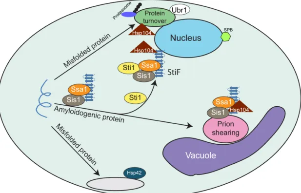
aggresomes [31, 38], and insoluble protein deposits (IPOD), which are located in the cell periphery [30, 37]. Protein oligomers and aggregates that are soluble in detergents accumulate in the spatially distinct juxtanuclear quality control (JUNQ) [30, 33] and peripheral compartments [32]. Ubiquitinated proteins that evade the proteasome and terminally misfolded proteins that escape recognition by protein quality control (PQC) E3 ligases are swept into the JUNQ [30]. Proteins in the JUNQ can be solubilized and degraded by the proteasome in a pathway that involves the PQC E3 ligase Ubr1 [34]. The peripheral compartment contains Hsp42, and holds proteins in a folding competent state [32]. The IPOD may function in protein disaggregation of detergent insoluble aggregates [37] whereas the peripherial compartment and JUNQ aid in refolding [32] and degradation [30] of detergent soluble protein assemblies, respectively. The aggresome resembles an inclusion body and may represent a long-term storage site for aberrant proteins [31, 38]. Thus, cells have the capacity to sequester potentially toxic protein species, but the mechanism for spatial segregation of protein handling centers is a mystery.
Although flux through protein aggregation pathways is associated with cell death, chaperone dependent sequestration of amyloid-like proteins into protein handling centers correlates with suppression of proteotoxicity [3, 6, 8, 40]. In yeast models,
overexpression of the yeast prion protein Rnq1 is toxic, but only if preexisting [RNQ+] prions are present to seed its conversion to an amyloid-like conformation [8, 41, 42]. The Hsp70, Ssa1, cooperates with the Hsp40, Sis1, to increase the efficiency by which seeded forms of Rnq1 assemble into amyloid-like [RNQ+] prions and thereby suppress
inclusions from Huntington’s Disease brains (Htt103Q), is toxic when expressed
ectopically in yeast, but only if [RNQ+] prions are present [43]. In yeast, the aggregation state of Htt103Q and its toxicity are inversely correlated with very tight, detergent
insoluble aggregates being benign and loose, detergent soluble aggregates being toxic [44]. Aggregation and toxicity of Htt103Q is influenced by its subcellular environment and also by the sequences of amino acids that flank its polyQ stretch [39, 44, 45]. Forms of Htt103Q that contain the polyproline region which follows the polyQ domain in full length Htt are benign due to their sequestration into the aggresome in a process that requires the ringed molecular chaperone p97 [38]. Forms of Htt103Q which lack the polyproline region are toxic as they accumulate in loose aggregates that are distributed throughout the cytosol in speckled foci, and tight aggregates that accumulate in the IPOD [30, 39]. Thus, studies of proteotoxicity of Htt103Q and Rnq1 in yeast provide an
excellent model system to uncover mechanisms for Hsp70 function in protein detoxification through protective protein aggregation.
coordinator of spatial PQC to organize molecular chaperone assemblies with their substrates as to drive protective aggregation of amyloidogenic proteins.
2.3 Results
2.3.1 The Hsp70/Hsp90 co-chaperone Sti1 suppresses Htt103Q toxicity
In order to identify cellular factors that defend against proteotoxicity, we carried out a high copy screen in Saccharomyces cerevisiae to discover proteins that suppressed
Htt103Q toxicity. A construct encoding the first 17 amino acids of the Htt gene followed
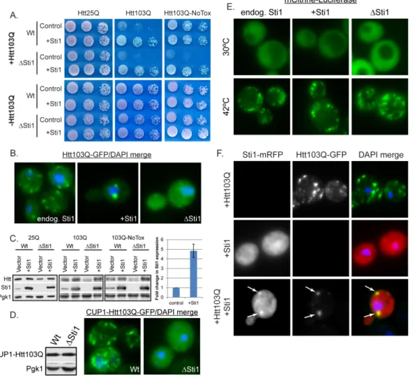
by 103 glutamines and a GFP moiety (Htt103Q) was integrated into a W303 strain under control of the galactose promoter. Htt103Q expression caused a growth defect in a glutamine length dependent manner [43] [44], and differential sensitivity to Htt103Q was exhibited by individual strains (Figure 2.1A). The yeast Hsp70/Hsp90 co-chaperone Sti1 [46] was identified as a high copy suppressor of the severe Htt103Q dependent growth defect. Expression of another TPR family co-chaperone, Sgt2, did not suppress Htt103Q toxicity (Figure 2.2A) indicating this activity is not a characteristic of TPR proteins in general. Additionally, deletion of Sti1 caused Htt103Q to be toxic in a strain where it was normally benign (Figure 2.1A right panels). Parallel growth assays using wildtype
and Sti1 Htt25Q strains demonstrate that this phenotype was not due to reduced growth
on galactose (Figure 2.1A left panels).
Elevation of Sti1 caused the subcellular localization of Htt103Q to become
Sti1 overexpression had no effect upon Htt103Q levels, yet deletion reduced expression (Figure 2.1C), likely due to delayed induction from the galactose promoter [47]. Deletion of Sti1 clearly sensitizes the cell to Htt103Q because even though there was reduced Htt103Q expression, the growth defect was exacerbated.
Sti1 may suppress Htt103Q toxicity by organizing it into foci, however, fewer
foci may be observed in the Sti1 strain since there is less Htt103Q being expressed. To
rule out this possibility, Htt103Q-GFP localization was examined when expressed to equivalent levels via copper inducible promoter in wildtype and Sti1 strains (Figure
2.1D). Again Sti1 was required for efficient organization of Htt103Q into foci. We also examined the influence of Sti1 expression upon temporal aspects of Htt103Q foci
accumulation. Over a 4 hour time course, Sti1 accelerated Htt103Q foci formation while reducing number of foci per cell (Figure 2.2B). Cycloheximide chase experiments demonstrate that once formed, the Sti1 dependent foci containing Htt103Q were stable for at least 5 hours (Figure 2.2C). These data provide a correlation between Sti1 action in suppression of Htt toxicity and the packaging of Htt103Q into perinuclear foci.
organize aggregated luciferase into foci, it appears to act specifically on aggregated forms of Htt103Q.
The TPR co-chaperone CHIP inhibits Hsp70 function in protein folding [27], so it is possible that Sti1 impacts Htt103Q assembly into foci by similarly interfering with Hsp70. This is unlikely, because Sti1 overexpression does not cause mCitrine-luciferase to aggregate (Figure 2.1E). Nevertheless, we examined in the impact of purified Sti1 on the ability of Ssa1 and Sis1 to refold denatured luciferase and found that Sti1 has no effect (Figure 2.2, D and E). Elevation of Sti1 does not appear to cause a change in Htt103Q aggregation by inhibition of normal Hsp70 folding activity.
If Sti1 has a direct effect upon Htt103Q, it should be present with Htt103Q foci. When expressed alone, Sti1 tagged with a monomeric RFP (mRFP) exhibited a diffuse localization pattern in the cytosol (Figure 2.1F). When co-expressed with Htt103Q, it redistributed the polyQ protein into distinct punctae, and Sti1-mRFP was indeed concentrated in foci with Htt103Q (Figure 2.1F, arrows). These data demonstrate that Sti1 promotes Htt103Q foci formation and this is beneficial to the cell, so there is an inverse relationship between Htt103Q aggregation and toxicity that is modulated by Sti1.
2.3.2 Sti1 interacts with high molecular weight forms of Htt103Q
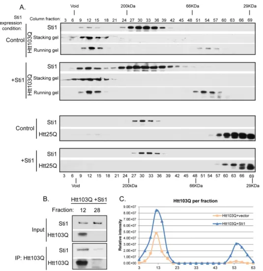
Western blot for levels of Htt103Q and Sti1 (Figure 2.3A-C). A portion of high
molecular weight SDS-resistant Htt103Q is retained at the stacking front of the gel [39], so both the stacking and running gel sections are shown. Htt expression was similar in the presence of endogenous and overexpressed Sti1 (Figure 2.1C) and under all
conditions, a pool of Sti1 (molecular weight of 67.6 KDa) migrated as a dimer [48] (Figure 2.3A). In addition to the dimeric form, endogenous Sti1 co-migrated with high molecular weight forms of Htt103Q. Yet, Htt103Q did not co-migrate in the fractions where the dimeric form of Sti1 was present (Figure 2.3A top panel/control). Importantly, endogenous Sti1 did not migrate in a high molecular weight fraction when
non-aggregation prone Htt25Q was expressed (Figure 2.3A bottom panel). Upon overexpression of Sti1, the high molecular weight pools of Sti1 and Htt103Q both increased proportionally (Figure 2.3A top panel/+Sti1, and 2.3C), yet this was not observed when Sti1 levels were elevated in the presence of Htt25Q (Figure 2.3A bottom panel/+Sti1). Sti1 was also present in immune complexes when high molecular weight Htt103Q was precipitated from column fraction 12, (Figure 2.3B). This interaction was specific because there was no Sti1 signal detected in the absence of Htt103Q (Figure 2.3B). While reorganizing foci and suppressing toxicity, Sti1 appears to interact with high molecular weight forms of Htt103Q.
2.3.3 Sti1 modulates assembly of amyloidogenic substrates at a perinuclear location Htt103Q is toxic to yeast upon undergoing a conformational change driven by interaction with the [RNQ+] prion [42, 43]. [RNQ+] prion biogenesis and toxicity is modulated by Hsp70 and Hsp40 [8, 49-51] and weak variants of the prion [PSI+] are
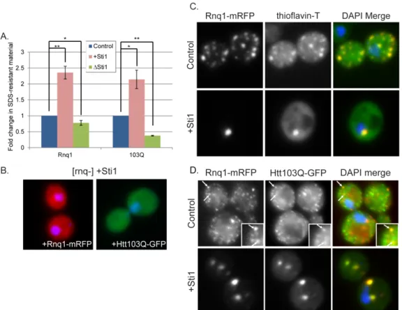
[RNQ+] prion toxicity and subcellular organization was evaluated. Just as with Htt103Q, an increase in Sti1 suppressed the toxicity associated with Rnq1 overexpression (Figure 2.4A). Under conditions utilized, Rnq1 expression is highly toxic, so deletion of Sti1 only led to a modest increase in growth defects caused by Rnq1 overexpression (Figure 2.4B). At endogenous levels of Sti1, Rnq1-mRFP foci were speckled throughout the cytosol, but upon overexpression of Sti1, Rnq1-mRFP became localized to one or two distinct juxtanuclear foci (Figure 2.4C). In contrast to what is observed with
Htt103Q-GFP, speckled Rnq1-mRFP foci were still detected in a Sti1 strain indicating that this
strain remains in the [RNQ+] prion state (Figure 2.4C, Sti1). Sti1 is not required for
maintenance of [RNQ+] prions and Sti1 overexpression does not eliminate [RNQ+] prions. Instead, suppression of Rnq1 and Htt103Q toxicity by Sti1 is associated with reorganization of multiple punctae containing these proteins into one or two distinct perinuclear foci.
We next sought to determine what conformational state of Rnq1 and Htt103Q is favored under conditions where Sti1 suppresses their proteotoxicty. Sti1 increased accumulation of oliogmeric SDS-resistant forms of Rnq1 and Htt103Q by more than two-fold (Figure 2.5A). Deletion of Sti1 reduced Htt103Q SDS-resistant material by over 50% while there was little effect upon Rnq1 (Figure 2.5A). Sti1 appears to act on the oligomeric assemblies of Htt and Rnq1 that only form in [RNQ+] prion strains because its overexpression has no effect upon localization of Htt103Q or Rnq1 in [rnq-] strains (Figure 2.5B).
support for this conclusion, we asked if Sti1 inducible foci stain positive for the amyloid specific dye thioflavin-T [2]. Rnq1-mRFP was used as a tool instead of Htt103Q-GFP because, upon binding amyloid, thioflavin-T emission is enhanced to a wavelength in the excitation range of GFP [53]. [RNQ+] prions have features of amyloid-like protein species as they bind thioflavin-T, whereas the dye does not bind to amorphous aggregates of Rnq1 that accumulate in a [rnq-] strain [8]. Speckled Rnq1-mRFP foci in both the cell periphery and in a perinuclear location were positive for thioflavin-T staining, as were the perinuclear Sti1 induced Rnq1-mRFP foci (Figure 2.5C).
To ascertain whether Htt103Q is also present in foci with amyloid-like [RNQ+] prions, we asked if they co-localize with each other in the presence and absence of Sti1 overexpression (Figure 2.5D). When Htt103Q-GFP and Rnq1-mRFP were co-expressed, a mixed population of speckled foci was detected with approximately 20% of foci
containing both proteins (Figure 2.5D, inset and arrows). Sti1 elevation resulted in redistribution of most of the Htt103Q and Rnq1 to one or two larger perinuclear foci that contain both proteins (Figure 2.5D).
not appear to be necessary for Rnq1 and Htt to interact. Instead, Sti1 may control the spatial organization of amyloid-like proteins into different protein handling centers.
2.3.4 Sti1 inducible foci are distinct protein quality control depots
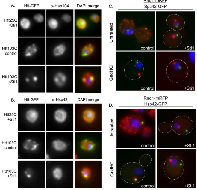
Sti1 inducible foci (StiF) are perinuclear and contain amyloid-like material, thus are distinct from the IPOD and JUNQ. To characterize the StiF in more detail, we determined whether these foci contain PQC machinery which would help protect cells from proteotoxic stress. At endogenous levels of Sti1, Hsp104 and Hsp42 were not detected in foci which contained Htt103Q-GFP (Figure 2.7, A and B). Yet, while Htt103Q-GFP and Hsp104 co-localized in the StiF (Figure 2.7A), Hsp42 was not typically present (Figure 2.7B).
Since the StiF is an Hsp104 positive inclusion, we next determined if StiF formation is impacted by modulation of Hsp104 activity. Here, we treated cells
containing endogenous or overexpressed Sti1 with guanidinium hydrochloride (GndHCl) for 4 hrs to inhibit Hsp104 activity [54, 55], and the effect this had on organization of Rnq1-mRFP into foci was monitored. To determine the nature of the foci observed, we employed strains containing a GFP tagged version of Hsp42 or Spc42, a spindle pole body component which marks the aggresome [31], which were under control of their endogenous promoter. Hsp104 inhibition condensed most of the Rnq1-mRFP material to one peripherally localized puncta, which co-localized with Hsp42, but not with Spc42 (Figure 2.7, C and D). Foci formed during Sti1 overexpression in the untreated samples did not co-localize with Spc42 revealing that the StiF is not the aggresome (Figure 2.7C). Furthermore, Htt103Q-GFP containing the proline-rich region is targeted in a p97
of Htt103Q-Pro (Figure 2.6B). Therefore, Sti1 is not promoting the accumulation of Htt103Q in the yeast aggresome.
Strikingly, Sti1 appears to divert proteins from the IPOD and promote their accumulation in the StiF, a previously unidentified protein handling depot. Rnq1 is considered to be an IPOD marker [30, 37] and the single Hsp42-GFP containing peripheral foci detected when Hsp104 is inhibited for 2 hrs appears to be the IPOD (Figure 2.7, C and D). Elevation of Sti1 in the presence of GndHCl prevents Rnq1-mRFP from accumulating in a peripheral foci and instead promotes its accumulation in the StiF.
These data demonstrate that organization of Rnq1 in speckled foci is highly sensitive to activity of Hsp104 and the Hsp70 system. Impairment of Hsp104 leads Rnq1-mRFP to accumulate in cytosolic foci. The effects of Sti1 on Rnq1-mRFP foci resemble those of Hsp104, except Sti1 promotes accumulation of amyloid-like proteins in StiF rather than a peripheral puncta. The StiF is a novel perinuclear quality control depot that contains Sti1, Hsp104, and amyloid-like proteins. It appears to be distinct from the aggresome as it does not form at the spindle pole body [38] and may serve as a
detoxification center for amyloid-like proteins.
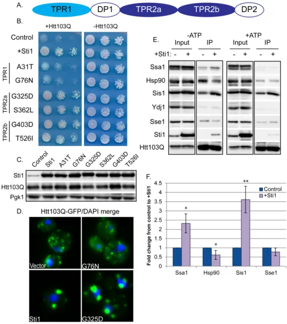
2.3.5 Sti1 reorganizes complexes that contain Htt103Q and Hsp70
Hence, point mutations in respective TPR domains were constructed and the ability of different TPR mutants to suppress Htt103Q toxicity and promote StiF formation was evaluated (Figure 2.8B). While mutations in TPR2a and TPR2b had no effect (Figure 2.8, B-D), mutations in TPR1 hindered the ability of Sti1 to redistribute Htt103Q to the StiF (Figure 2.8D). TPR1 mutants also lost their ability to suppress the Htt103Q growth defect (Figure 2.8B). Thus, StiF formation and suppression of Htt103Q toxicity requires functional domains of Sti1 that enable it to interact with the Hsp70/Hsp90 system.
To determine the impact Sti1 has upon interactions between Hsp70 and Hsp90 with Htt103Q, complex formation between these proteins was evaluated by
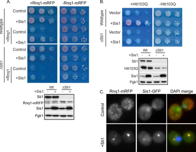
2.3.6 Sti1 and Sis1 cooperate to modulate amyloid toxicity
Sis1 interacts with oligomeric forms of Rnq1 to facilitate the propagation and impact the spatial organization of [RNQ+] prions [39, 49, 50]. Thus, Sti1 and Sis1 may cooperate to suppress growth defects caused by the presence of amyloid-like protein in the cytosol. We therefore investigated whether or not Sti1 is required for Sis1 to suppress Rnq1 and Htt103Q toxicity (Figure 2.9, A and B). Elevation of Sis1 diminished Rnq1 toxicity in a wild type strain, but its protective effect was severely diminished in a Sti1
strain, even though deletion of Sti1 had no effect on Sis1 expression. Similarly, elevated Sis1 also suppressed the growth defect caused by Htt103Q, and this protective effect was
reduced in a Sti1 strain (Figure 2.9B).
Further support for functional interaction between Sti1 and Sis1 in buffering the toxicity of amyloid-like proteins comes from the demonstration that Sti1 drives the redistribution of Sis1 to the StiF (Figure 2.9C). Under normal conditions, Rnq1-mRFP was found in speckled foci and Sis1-GFP was distributed between the cytosol and nucleus (Figure 2.9C and [39]). Upon elevation of Sti1, the localization pattern of Sis1 changed dramatically and a large pool of it was concentrated in the StiF with Rnq1-mRFP (Figure 2.9B). Data presented in Figure 2.7-2.9 suggests that Sti1 helps suppress proteotoxicity via stabilization of complexes between Hsp70, Sis1, and amyloid-like protein assemblies to promote their accumulation in the perinuclear StiF.
2.4 Discussion
templated Htt103Q. Amyloid-like protein accumulation in the StiF correlates with suppression of proteotoxicity caused by Rnq1 and Htt103Q. Partitioning of aberrant proteins between the StiF and other protein handling centers is controlled by interactions between Sti1 and the Hsp70 chaperone system. Loss of Sti1 exacerbates Htt
proteotoxicity while increases in Sti1 levels suppress it by increasing protein accumulation in the StiF. Sti1 is identified as an important component of the
Hsp70/Hsp90 system that acts in spatial PQC to promote protective protein aggregation. Sti1 serves to reorganize speckled foci containing amyloid-like forms of Rnq1 and Htt103Q into the StiF (Figure 2.10). The StiF appears to be distinct from other misfolded protein deposition sites because it contains amyloid-like protein species, is perinuclear, and does not co-localize with an aggresome marker. The StiF is one of five cytosolic protein handling centers, most of which have been characterized in both yeast and humans to function in protein homeostasis [30, 31, 33, 38]. The aggresome and the IPOD are responsible for storage of detergent insoluble and amyloid-like protein species [30, 38], whereas the peripheral compartment and JUNQ handle detergent soluble protein aggregates [30, 32]. The detection of substrate accumulation in these sites often requires gross protein overexpression and/or inhibition of the proteasome. Importantly, amyloid-like proteins are present in the StiF under conditions of modest protein expression and normal flux through the proteasome. Thus, amyloid-like proteins appear to transit through the StiF under normal growth conditions.
punctae and diffuse assemblies. Elevation of Sti1 accelerates the sequestration of amyloid-like proteins into the StiF and increases detergent insoluble oligomers of [RNQ+] and Htt103Q over 2 fold. This may occur by diversion of amyloidogenic proteins away from the IPOD where they could be sheared by Hsp104, possibly generating toxic oligomers [37, 58]. Shunting amyloid-like proteins away from the aggresome to the StiF is protective because this prevents sequestration of essential proteins into aggregates. For example, [RNQ+] prions sequester Spc42 away from its normal function in the spindle pole body leading to cell death [59], so increasing flux of [RNQ+] prions to the StiF would be protective. Accumulation of amyloid-like protein species in the JUNQ is cytotoxic as it inhibits normal PQC function [30, 33-35], so diverting these proteins to the StiF would assure normal function of the ubiquitin
proteasome system. The fate of proteins that enter the StiF is not entirely clear, yet these foci are highly stable, so they may serve as a long-term storage vault for otherwise toxic proteins.
How does modulation of Sti1 alter spatial partitioning of amyloid-like aggregates? Sti1 promotes association of Ssa1 and Sis1 with Htt103Q while reducing the presence of Hsp90, thus stabilizing the Hsp70:substrate bound complex. Sti1 interaction with the Hsp70 system seems to allow the co-chaperone to directly affect the substrate’s fate. In support of this concept, Sti1 was found in complex with a high molecular weight
Sti1’s cooperation with Sis1 in spatial distribution of amyloid-like proteins is consistent with observations that Sis1 impacts the subcellular localization of prions and misfolded proteins [36, 39]. During heat stress, Sis1 is proposed to interact with the aggregate sorting factor Btn2, targeting misfolded proteins to a juxtanuclear location [36]. Sis1 function is also implicated in targeting of [RNQ+] prions to the nucleus [8, 39] in a process that may involve Cur1 [36]. Levels of Btn2 and Cur1 during heat stress are thought to regulate the relative cytosolic concentrations of Sis1, thereby dictating whether misfolded protein accumulates with Sis1 at a juxtanuclear site or with Hsp42 at a
peripheral cytosolic site [36]. Thus, there are protein sorting factors that interact with Sis1 and impact targeting of protein aggregates to different protein handling centers. Cur1, Btn2, and Hsp42 were deleted from strains expressing Htt103Q, but were
dispensable for Sti1 dependent accumulation of Htt103Q in the StiF and suppression Htt toxicity (Figure 2.11). Nevertheless, the existence of such spatial PQC factors suggests the possibility that Sti1 plays a similar role in an alternate pathway that may be specific for amyloid-like proteins.
but Sti1 overexpression does not cure yeast of [RNQ+] prions (Figure 2.12A, left panel). However, when Hsp104 is inhibited, Sti1 elevation accelerates the rate of [RNQ+] prion curing caused by inhibition of Hsp104 (Figure 2.12). Thus, Sti1 is not an inhibitor or Hsp104. Instead, it appears to interact with Sis1 and possibly other spatial PQC factors to direct amyloid-like proteins to the StiF, and this sequesters pools Rnq1 and Htt103Q away from the machinery that would normally break them into smaller foci.
The co-chaperones CHIP and BAG3 target Hsp70 clients for degradation via the ubiquitin proteasome or autophagy, respectively [10, 13, 61]. Sti1 is now reported to enable Hsp70 to function in spatial PQC by targeting amyloid-like proteins to the StiF. Sti1 is stress inducible, and, in humans, CHIP and Sti1 differentially bind to Hsp70 and Hsp90 based on changes in phosphorylation [62]. Thus, under normal and stress
2.5 Methods
Strains and plasmids
Yeast strains and plasmids are listed in Tables S1 and S2. All strains harbored Rnq1 in its [RNQ+] prion form unless otherwise indicated. The generation of isogenic
[rnq-] strains was accomplished via sequential passage of cells on plates containing 3mM
guanidinium-HCl. Strains were transformed with plasmids and cultured in synthetic media as described previously [8]. Constructs containing a copper inducible promoter (CUP1) were induced at a low constitutive level from basal levels of copper in media unless specified that media was supplemented with additional CuSO4. This is the case for all Rnq1-mRFP microscopy unless otherwise indicated. For Sti1 overexpression experiments, Sti1 was always expressed from its endogenous promoter on a high copy plasmid.
Expression of Htt103Q
Proline rich region following the polyQ stretch. Expression of Htt103Q-Pro was induced with 2% galactose for 24 h from a low copy plasmid and galactose inducible promoter.
Toxicity assays
Yeast samples were normalizedto the same OD600, then 5 fold serial diluted in a sterile 96-well plate. Each dilution was plated in 10uL spots on appropriate synthetic dropout media. Most images of yeast grown on glucose were scanned at 48h and galactose at 72h. Lysates for Western blots from toxicity assays were prepared using cultures picked from control growth plates after they were scanned. Rnq1 toxicity was assessed on plates containing 2% glucose with 500uM CuSO4 and Htt toxicity plates contained 2% galactose.
SDS-PAGE and Western Blotting
Lysates were prepared from pellets of yeast by either mechanical disruption via glass bead breaking (for SDD-AGE and co-IP) as described previously [8, 39] or an alkali lysis method as described previously [34] (for samples which were directly run on SDS-PAGE gels). When using mechanical disruption, the lysis buffer used was 0.1% Triton X-100, 75mM Tris pH7.4, 150mM NaCl, 1mM EDTA, 1mM PMSF, and Sigma protease inhibitor cocktail. Standard separation techniques were used to analyze lysates via SDS-PAGE. Protein was transferred to nitrocellulose for 75-90 min at 110V, and probed using the indicated antibodies. Antibodies used in this study are listed in Table S3.
Fluorescence microscopy