ASSOCIATIONS BETWEEN BIOCHEMICAL MARKERS OF CARTILAGE METABOLISM AND GAIT BIOMECHANICS IN HEALTHY PARTICIPANTS
Hope C. Davis
A thesis submitted to the faculty at the University of North Carolina at Chapel Hill in partial fulfillment of the requirements for the degree of Master of Art in the Department of
Exercise and Sport Science (Exercise Physiology).
Chapel Hill 2016
Approved by:
ABSTRACT
Hope C. Davis: Associations Between Biochemical Markers of Cartilage Metabolism and Gait Biomechanics in Healthy Participants
(Under the direction of Anthony C. Hackney)
ACKNOWLEDGEMENTS
TABLE OF CONTENTS
LIST OF TABLES………...……xiii
LIST OF ABBREVIATIONS……….………...ix
CHAPTER 1………...1
Introduction………1
Purpose………..4
Research Question.………..……...5
Research Hypotheses………..……5
Definition of Terms……….………..5
Knee Osteoarthritis………...…………5
Cartilage Metabolism………...5
Gait Biomechanics………....6
Assumptions………..……6
Delimitations………...7
Limitations………..7
Significance of the Study……….………8
CHAPTER 2: LITERATURE REVIEW………..….9
Part I………...…..10
Cartilage Metabolism……….……11
Local Metabolic Factors……….12
Systemic Metabolic Factors………..14
Biomarkers of Cartilage Metabolism………15
Collagen Turnover………..15
Collagen Synthesis……….16
Collagen Degradation………...…….16
Part II………...……….17
Mechanical Loading at the Knee………..17
Biomarkers of Mechanical Loading………..18
Mechanosignaling………...…18
Summary……….……19
CHAPTER 3: METHODOLOGY………...…20
Participants………..…20
Instrumentation………20
Procedures………..…21
Session I………..……21
Session II………...22
Blood Draw and Handling Procedures………22
Gait Biomechanics Assessment………...……22
Data Analysis………..…23
Subject Characteristics……….………….25
Peak Vertical Ground Reaction Force………..………..26
Type II Collagen Turnover……….26
Associations Between Cartilage Metabolism and Peak Vertical Ground Reaction Force……….……27
Post Hoc Analysis……….………..27
Outlier Removal………..28
Transformed Data………...……28
Associations with Physical Activity and CPII:C2C……….……29
CHAPTER 5: DISCUSSION………..31
Association Between Joint Loading and Cartilage Turnover………...……33
Physical Activity and Biomarkers of Cartilage Metabolism………..35
Limitations………37
Conclusion………...……39
APPENDIX 1: IRB APPROVED CONSENT FORM………..40
APPENDIX 2: IRB APPROVED STORED SPECIMENS CONSENT FORM...……….…47
APPENDIX 3: TEGNER ACTIVITY LEVEL SCALE………..…..…….52
LIST OF TABLES
Table 1 – Descriptive Data for Subjects………..26
Table 2 – Kinetic Data (Normalized to BW) for Subjects……….26
Table 3 – Biomarker Data for Subjects………...27
Table 4 – Biomarker Data for Corrected Subjects…….………28
Table 5 – Transformed (Base-10 log) Biomarker Data………....29
LIST OF ABBREVIATIONS
ACL Anterior cruciate ligament
ACLr Anterior cruciate ligament reconstruction C2C Collagen type-II cleavage product
CPII Collagen type-II C-propeptide
ELISA Enzyme-linked immunosorbent assay IL Interleukin
KOA Knee Osteoarthritis
MMP Matrix metalloproteinases MPa Megapascal
MRI Magnetic resonance imaging OA Osteoarthritis
PA Physical activity TLR Toll-like receptors
CHAPTER 1
Introduction
Over the past several decades, musculoskeletal injuries have become an increasing concern in the general population. (Bjorklund 1998). Specifically,
osteoarthritis (OA) is one of the most prevalent musculoskeletal diseases, and it will continue to rise with the increasing age of the population (Bjorklund 1998). In 2008, an estimated 27 million adults suffered from OA (Lawrence et al., 2008), and by 2020 it is predicted to be the fourth leading cause of disability (Bjorklund 1998). About 80% of OA patients suffer from limited mobility (Guccione et al., 1994), and an estimated 25% cannot perform activities of daily living without assistance (Guccione et al., 1994). Furthermore, OA is a major healthcare cost in the United States, with total knee replacements (i.e. a common treatment for end stage OA) estimating over $28 billion dollars (Murphy et al., 2012). Job-related OA costs are $3.4 to $13.2 billion per year; with adults suffering from KOA (KOA) reporting more than 13 lost workdays per year due to health (Buckwalkter et al., 2004). KOA is so debilitating because the knee joint is a significant shock absorber for any type of walking, climbing, or load-bearing activity.
KOA affects both young and older populations, although the risk factors
KOA (Heidari, 2011). Also, individuals who exhibit aberrant biomechanics during gait may have a higher risk of developing KOA (Maly, 2008; Baliunas et al., 2002). Knee adduction moment is one common variable assessed in KOA patients (Maly, 2008). High adduction moments have been associated with the disease (Maly, 2008).
Currently, KOA is diagnosed after confirmation of radiographic damage within the joint. This damage is characterized by joint space narrowing due to osteophyte formation (Kijowski et al., 2006). Further bone sclerosis and subchondral cysts are used assess the severity of OA (Kijowski et al., 2006). By the time radiographic evidence is present, the patient already has irreversible cartilage breakdown and bone remodeling.
Therefore, the development of early intervention techniques to will be essential in decreasing the physical and financial burden associated with OA.
Current research assesses biochemical and biomechanical markers in OA patients in hopes of finding new methods of detection for high-risk populations. Clinically, OA is diagnosed once these irreversible changes in the bone and cartilage are radiographically present (Kijowski, 2006). However, there is evidence that
biochemical changes alter cellular metabolism at the knee prior to radiographic evident changes (Garnero et al., 2001).
event can lead to cartilage damage, the cardinal characteristic of KOA. Weight gain (metabolic syndrome), aging, and acute joint trauma such as anterior cruciate ligament (ACL) injury (Lohmander et al., 1994) can all trigger this local metabolic change.
Proinflammatory cytokines can affect joint metabolism for years after the initial injury (Lohmander et al., 1994). This metabolic alteration can lead to cartilage, bone, and synovium breakdown, all increasing the risk of KOA.
Although biochemical changes potentially begin the cascade of events leading to OA, biomechanical changes can progress OA and increase the severity of it. Several decades ago studies examined cartilage damage as a result of aberrant loading in animal models. Results showed that repetitive and aberrant loading in rabbits can lead to subchondral bone thickening and cartilage fibrillations (Farkas et al., 1987; Radin et al., 1984). In human studies evidence suggests that patients with KOA have altered peak vertical ground reaction forces (vGRF) compared to healthy subjects (Gyory et al., 1976; Stauffer et al., 1977; Radin et al., 1991). Many studies currently focus on aberrant gait as not only a result but also a cause of KOA (Childs, 2003). Examining associations between biomechanics and biochemical markers could identify at risk patients before KOA symptoms begin.
Serum markers of collagen turnover and proteoglycan breakdown can be used to assess joint metabolism (Attur et al., 2013). Certain markers may indicate if an
individual is more likely to develop KOA or if OA will further progress (Attur et al., 2013). Collagen type-II C-propeptide (CPII) detects collagen synthesis, while cartilage
degradation is detected by serum concentrations of collagen type-II cleavage product (C2C; Cahue et al., 2007). The ratio of CPII: C2C describes collagen turnover (Cahue et al., 2007). Additionally, proteoglycan, another structural macromolecule in cartilage, can be indicative of knee joint cellular metabolism (Larsson et al., 2012). Proteoglycan release into the synovial fluid may be associated with cartilage catabolism (Lohmander et al., 1999) and another sign of KOA progression. Although there is not one
biochemical marker that can diagnose or predict KOA, evaluating several different markers can help determine metabolic processes of the knee joint prior to radiographic changes.
Although there is evidence that shows biochemical and biomechanical changes following joint trauma, there is little research done on serum biomarkers in a healthy population. Assessment of cartilage markers in asymptomatic subjects will add to the body of knowledge of OA biochemical markers found in serum and biomechanical markers.
Purpose
Research Question
1. Will there be an association between collagen turnover and peak vGRF? Research Hypotheses
1. There will be a negative association between collagen turnover and peak vGRF. Definition of Terms
Knee Osteoarthritis
KOA is one of the most prevalent causes of disability in the United States
(Lawrence et al., 2008). It is characterized first by articular cartilage breakdown, causing the femur and tibia to rub painfully against each other during weight bearing activities (Neogi & Zang, 2013). Subchondral bone thickening and general pain of the joint are also symptoms of the disease (Radin, 1984). While the current study focuses on articular cartilage, recent literature suggests that changes to other surrounding tissues such as synovial fluid (Benito et al., 2005) and bone (Ishijima et al., 2011) may
accompany or occur prior to cartilage breakdown. Although there are many risk factors, two discussed in this thesis are altered cartilage metabolism (Berenbaum, 2013) and aberrant gait (Stauffer et al., 1977).
Cartilage Metabolism
that contributes to the strength of cartilage (Poole et al., 2002). Both type II collagen and proteoglycan are regulated by synthesis and degradation; however, in a diseased joint the resulting ratio can decrease causing loss of articular cartilage, the hallmark
characteristic of KOA (Lohmander et al., 1992). Gait Biomechanics
Gait biomechanics include alignment of the lower extremity as well as joint torque moments that occur during the different phases of human gait. Peak vertical ground reaction force (vGRF) is a commonly used parameter to assess gait mechanics (Childs et al., 2004). Although it is still unclear if aberrant mechanics is a cause of OA, several studies demonstrate that alterations in mechanical loading can cause cartilage
fibrillations (Radin et al., 1973). Assumptions
1. Participants will be honest in medical history questionnaire.
2. All participants will have no history of ACL, meniscus or other knee injuries. 3. Participants will not engage in unnecessary physical activity during the 24-hours
prior to the two sessions. Participant will still be allowed to walk to and from class/work and complete activities of daily living.
Delimitations
1. Participants must be healthy males or females between the ages of 18 and 35. 2. They must be physically active according to current physical activity guidelines
(US Department of Health and Human Services, 2008).
3. Participants will be excluded if they have history of orthopedic injury or surgery on either lower limb or any current fractures or activity restrictions in the past 6 months.
4. Participants will be excluded if they have osteoporosis, are post-menopausal, are undergoing hormone replacement therapy, have growth hormone or androgen deficiency, hypothyroidism, chronic renal failure, rheumatoid arthritis, or have a BMI over 30.
5. Participants will be excluded if they have a current musculoskeletal condition limiting gait, a central nervous system disorder or balance issue, a need of an external device for walking, history of concussion, or a heart condition limiting exercise capacity.
6. Participants will be excluded if they are on blood thinner medication, are pregnant, have a history of blood infection, or a self-reported fear of needles. Limitations
1. Assumption must be made that serum markers are indicative of synovial fluid markers.
Significance of Study
With present technology, KOA is diagnosed after irreversible changes in the joint have been made. A patient already suffers from pain and potentially limited mobility, which greatly decreases his or her quality of life. Markers of cartilage breakdown correlate with KOA severity and progression (Cahue et al., 2007). Additionally, it is known that aberrant mechanical loading can cause cartilage fibrillations (Radin et al., 1973). The findings from this research project could suggest that cartilage breakdown is associated with a higher loading in the joint in a healthy population. If this finding exists, future studies can focus on earlier detection and even prevention of KOA. Perhaps OA prevention programs could focus on lowering reaction forces placed on the knee joint during physical activities. Additionally, monitoring patients’ and athletes’ cartilage metabolism biomarkers could prevent KOA or at least OA progression. Currently, KOA costs the United States millions of dollars each year in health care and missed
workdays (Murphy et al., 2012; Buckwalter et al., 2004). By far the most efficient way to lower these costs is to focus on early detection and prevention. By assessing
CHAPTER 2: LITERATURE REVIEW
This review will examine properties of tibiofemoral joint tissue physiology and mechanical loading of articular cartilage, as well as alterations that can increase the risk of knee osteoarthritis (OA). Articular cartilage surrounds the femur and tibia that make up the tibiofemoral joint, providing a strong and protective layer between the bones. Articular cartilage is composed primarily of collagen, proteoglycans, and chondrocytes (Buckwalter et al., 2005). Chondrocytes are the only type of cell that resides in the cartilage, and they are responsible for all aspects of cartilage metabolism.
Chondrocytes maintain the ratio of synthesis to degradation for all macromolecules in the cartilage (Buckwalter et al., 2005). Proinflammatory cytokines can increase both local and systemic inflammation, both of which alter the cartilage metabolism
(Berenbaum, 2013). Biochemical markers in serum can indicate levels of
Part I
Articular Cartilage Composition
The tibiofemoral joint is used in all weight-bearing activities, and therefore its structure is a critical shock absorber during walking, jumping, running, and other dynamic movements. A type of connective and force-absorbing tissue called articular hyaline cartilage surrounds the femur, tibia, and patella. This strong, hydrophilic structure allows for fluid joint motion.
Articular cartilage is made up of structural macromolecules, chondrocytes, and water (Buckwalter et al., 2005). Compared to other tissues, cartilage has a low
metabolic activity; however, interactions between different components contribute to maintaining cartilage health in the correct ratios (Buckwalter et al., 2005). The structural macromolecules collagen and proteoglycan weave together to form a strong and tensile material that is smooth and resistant to force (Buckwalter et al., 2005). Collagens make up the majority of the dry weight of cartilage, and although there are several different types, type II collagen is most prevalent (Buckwalter et al., 2005). Proteoglycans are made of a protein core and at least one glycosaminoglycan chain, which contains at least one carboxylate or sulfate group (Buckwalter et al., 2005). These groups are negatively charged and repel similarly charged molecules while attracting cations. This property pulls water into the extracellular matrix, which gives articular cartilage the ability to compress and de-compress when loads are applied to the joint (Buckwalter et al., 2005). Articular cartilage contains both large aggregating and smaller
as aggrecans, make up the majority of proteoglycan matter in cartilage (Buckwalter et al., 2005). These aggrecans link together to stay anchored within the matrix. This helps prevent deformation of the meshwork during mechanical loading.
Proteoglycans and collagen, as well as several other types of smaller proteins, are coordinated in a highly ordered structure to form functioning articular cartilage. Chondrocytes, the only type of cell inside the articular cartilage, are responsible for regulating and maintaining structure. Chondrocytes synthesize new collagen and proteoglycans, as well as breakdown dysfunctional cartilage (Buckwalter et al., 2005). Cartilage Metabolism
Articular cartilage, especially in the knee joint, undergoes a wide range of
stressors throughout a human’s lifetime. Mechanical loads can induce different amounts of stress and compressive strains based on the duration and type of mechanical loading occurring at the joint (Marasovic et al., 2009). Ability to resist these forces depends on maintaining a proper ratio of synthesis to degradation of macromolecules. This turnover is a critical component of healthy cartilage metabolism.
Chondrocytes are the sole cell found in articular cartilage, and they make up approximately 1% of cartilage’s total volume (Buckwalter et al., 2005), making
MMPs and aggrecanases contribute to proteoglycan cleavage (Pollard et al., 2008). The cardinal characteristic of OA is loss of articular cartilage that surrounds the patella, femur and tibia (Poole et al., 2002). One prevalent theory is that OA is initiated in part by a change in the cellular metabolism of articular cartilage, where synthesis of new molecules cannot keep up with an increase in degradation (Pollard et al., 2008).
Increased cleavage of proteoglycans leads to increased levels of aggrecan fragments in surrounding synovial fluid and even serum (Pollard et al., 2008). This enhanced
cleavage can be the result of many mechanisms, including inflammation and joint trauma (Pollard et al., 2008). The remaining aggrecan has room to expand and swell, causing stretching of the collagen framework (Pollard et al., 2008). After this stretching, type II collagen is significantly more vulnerable to mechanical damage, and these
combined effects can lead to increased cartilage breakdown due to decreased strength of the tissue (Pollard et al., 2008). Additionally, chondrocytes become vulnerable to necrosis in this stage (Pollard et al., 2008). Chondrocyte death will lead to further disruption of the cellular matrix, and will eventually lead to an irreversible situation if not corrected early on (Pollard et al., 2008).
Local Metabolic Factors
Joint swelling is a common symptom of KOA, and it can be the result of local upregulation of proinflammatory mediators. One potential mechanism for local
breakdown (Pollard et al., 2008). After breakdown occurs, fragments are released into the joint where they make contact with the synovium. This invasion of foreign particles into the synovium can cause synovial cells to produce additional cytokines, starting an inflammatory feed forward loop that can be difficult to inhibit (Berenbaum, 2013). This evidence suggests that local inflammation in the synovial tissue may be a driver in the OA process rather than a result of previous cartilage breakdown (Berenbaum, 2013). Innate immunity may also trigger local inflammation in the knee joint. Synovial fluid from patients with early cartilage damage show increased fibroblast-like synoviocyte
responses to several receptor ligands of toll-like receptors (TLR; Nair et al., 2012). Binding of these ligands initiates intracellular signaling for inflammatory gene
transcription (Nair et al., 2012). Additionally, increased levels of specific interleukin (IL-15) proteins exist in early OA patients compared to late stage OA patients (Scanzello et al., 2009). Although mechanisms are not well-established, the literature suggests that several different proinflammatory mediators facilitate the upregulation of degradative enzymes.
Eventually, the shift of cartilage metabolism towards breakdown becomes
irreversible and OA develops and progresses. Although not well established, increased MMP expression may play an important role in the eventual cartilage fibrillation
(Tchetina, 2011). Additionally, many cytokines not only increase degradation, but also inhibit synthesis processes important to maintaining cartilage turnover ratios (Sokolove et al., 2013). Although the exact mechanism is undetermined, local metabolic
Systemic Metabolic Factors
While local metabolic alterations play an important part in early OA development, evidence also suggests that systemic cytokine activity can progress OA via cartilage breakdown (Berenbaum, 2013). Inflammatory events that occur peripherally can influence cytokine upregulation inside of the knee joint tissue (Berenbaum, 2013). Several studies found increased levels of cytokines in OA patients’ serum compared to controls (Berenbaum, 2013). One study showed that peripheral blood leukocytes (PBL) of OA patients with increased levels of IL-1B expression experienced a higher rating of pain and increased dysfunction (Attur et al., 2011), putting these patients at a greater risk of OA progression (Attur et al., 2011). Likewise, leptin and adipokines, hormones with proinflammatory properties, have been studied in KOA. Leptin becomes biologically functional by binding to specific receptors that enhances the synthesis for MMP
production (Gabay & Berenbaum, 2009). Adiponectin acts similarly to leptin when it makes contact with articular cartilage. Adiponectin receptors are present on the surface of chondrocytes, and when bound facilitate an increase in additional cytokines as well as MMPs (Gabay & Berenbaum, 2008). Other evidence shows an increased risk of hand OA in obese patients (Berenbaum, 2013). While mechanical loading would not be an issue, systemic factors such as increased adipokines like leptin and adiponectin could play a role in increased risk of hand OA (Berenbaum, 2013).
alterations lead to the upregulation of MMPs via increased production by chondrocytes (Berenbaum, 2013; Gabay & Berenbaum, 2009; Tchetina, 2011).
Biomarkers of Cartilage Metabolism Collagen Turnover
Collagen turnover is defined as the ratio of collagen synthesis to degradation (Buckwalter et al., 2005). Collagen must be degraded as it ages in order to make room for new collagen. In a healthy, functional joint, there is both collagen degradation, as well as new collagen synthesis at a fairly constant ratio (Buckwalter et al., 2005). MMPs are the main collagenases responsible for cleaving mature collagen. Once collagen is cleaved, it is considered waste and moved out of the articular cartilage (Poole et al., 2002). For several decades, studies have looked at varieties of cleaved collagen product in hopes of finding a biochemical marker that is an indicator of cartilage metabolism. Specifically, studies have focused on markers of collagen turnover
(Garnero et al., 2002; Cahue et al., 2007). Garnero et al. measured levels of cartilage synthesis and degradation markers individually and in ratio. Results showed that the ratio of synthesis and degradation markers, or turnover, could predict OA progression in subjects better than individual markers (Garnero et al., 2002). Cahue et al. employed similar methods and found an elevated ratio of degradation to synthesis (as reported by the author) that was associated with the likelihood of KOA progression between
that turnover measurements may be better at predicting differences between subgroups of OA patients. Individual markers of collagen synthesis and degradation were
significantly different between radiographic OA patients and patients with no OA.
However, when looking at collagen turnover, a difference was also discernable between pre-radiographic OA patients and patients without OA (Cibere et al., 2009). Examining turnover ratios could potentially be a method to find OA before it is radiographically evident (Cibere et al., 2009).
Collagen Synthesis
C-propeptide of type II procollagen (CPII) is a widely used serum biomarker to detect levels of cartilage synthesis (Nelson et al., 1998). The precursor to newly formed collagen is known as procollagen. The two ends of procollagen are cleaved by
collagenases as a final step in the production of type II collagen. These fragments are detected in the CPII biomarker (Cahue et al., 2007). Generally with increased collagen degradation, there is an accompanying increase in synthesis (Nelson et al., 1998). However, in pathological cartilage this new collagen is quickly cleaved, and the ratio of synthesis to degradation still decreases (Cibere et al., 2009; Cahue et al., 2007). The decreased ratio is a reflection of arthritic conditions that favor degradation over
synthesis (Cahue et al., 2007). Collagen Degradation
Serum cartilage type II collagen cleavage product (C2C) is a common biomarker used to detect collagenase-mediated cleavage generated neoepitopes of type II
2011; Poole et al., 2002). MMPs 1, 8, and 13 are known to cleave mature type II collagen in both healthy and pathological collagen (Billinghurst et al., 1997). Positive correlations were observed between C2C and transverse proton relaxation time of cartilage (King et al., 2004). Alone, differences in C2C concentrations can be detected between OA and healthy patients (Cahue et al., 2007; Ishijima et al., 2011), but the ratio of synthesis to degradation provides a more sensitive measure and can distinguish between healthy and pre-radiographic OA participants (Cibere et al., 2009).
Part II
Mechanical Loading at the Knee
moderate loading is critical for joint health, overloading can lead to cartilage degradation (Sun, 2010). Likewise, results from several studies indicate that mechanical
overloading may stimulate signaling pathways of several proinflammatory cytokines and induce overexpression of MMPs (Sun, 2010). Increased or aberrant mechanical
loading could potentially alter joint tissue physiology, possibly leading to cartilage breakdown (Sun, 2010). However, most studies assessing associations between
mechanical and physiological parameters focus on pathological populations. Examining a healthy population is a necessary step to gain insight on how these parameters
influence one another.
Biomarkers of Mechanical Loading
Although many biomarkers of mechanical loading exist in the literature, one of the most robust and basic markers is peak vertical ground reaction force (vGRF). vGRF is the vertical component of force exerted by the subject while in contact with the ground (Marasovic et al., 2009). In healthy gait, vGRF exhibits an M shaped curve as a result of the initial heel strike and final toe off (Marasovic et al., 2009). One risk factor of KOA is overloading of the joints (Radin et al., 1973), and many injury prevention programs focus on decreasing vGRF during walking, running, and jumping (Padua & DiStefano, 2009). Although it is known that vGRF is altered after onset of KOA, there is little in the literature of associations in vGRF and cartilage metabolism in healthy populations. Mechanosignaling
Controversy over the initiation of OA from mechanics versus cartilage
the debate in the future. Any aberrant mechanical stress applied to a joint can initiate intracellular signaling at the site of loading. Eventually this signaling could lead to overexpression of inflammatory mediators in the synovium (Berenbaum, 2013). By examining associations between mechanical loading and biochemical measurements, great progress could be made in early detection of OA. Examining associations between biomechanical and biochemical markers in a healthy population will contribute to the growing body of knowledge currently available on this topic.
Summary
CHAPTER 3: METHODOLOGY
Participants
Thirty healthy, moderately active people from the University community between the ages of 18-35 will be recruited for this study. To achieve a power of 0.80 with α< 0.05, an estimated 26 subjects will be required to detect significance (r=0.50; Lachin, 1981). Participants will sign a consent form (Appendix 1) after being thoroughly informed of the experimental protocol and any risks or rewards related to the study. Participants will be excluded from the study if they do not meet all of the criteria listed in the delimitations found in Chapter 1. Participants will agree over email to abstain from any unnecessary physical activity (i.e. anything other than walking to class or doing activities of daily living) 24 hours prior to the session.
Instrumentation
Gait analysis will be conducted using two Bertec force plates (40x60cm,
Asheville, NC USA). Serum CPII and C2C will be assessed using enzyme-linked immunosorbent assay (ELISA) kits (IBEX technologies, Montreal, Quebec, Canada). Procedures
Session I
Participants will report to the Sports Medicine Research Laboratory (SMRL) in
Fetzer Hall on UNC Chapel Hill’s campus for testing. Prior to participation, each
participant will read and sign an informed consent form (Appendix 1). If the participant
has any questions about the procedures or the study in general, he/she will have the
option to ask the researcher. After consent is obtained, the investigator will screen the
participants to see if they meet inclusion/exclusion criteria. If subjects meet the criteria,
practice gait trials will be performed to obtain walking speed and familiarize the subject with the force plates. Subjects will walk forward along a 6m (~20ft) walkway containing two force plates at a comfortable, self-selected “normal” speed. The plates are
staggered so that an entire stance phase for both limbs can be obtained in a single walking trial. At least 5 practice trials will be performed to determine the average preferred speed and ensure subjects can consistently strike the force plate mounted in the walkway without noticeably altering their gait (i.e. “aiming” for the force plate). Gait speed will be monitored via an infrared timing system. The next session will then be
scheduled.
Session II
gain information about the participant’s physical activity. Each participant will then
undergo a blood draw and gait trial described below.
Blood Draw and Handling Procedures
Participant will lie down quietly for 30 minutes prior to the blood draw. The blood sample will be collected via vacutainer from the antecubital vein using a 21-gauge needle and immediately stored on ice. The sample will be centrifuged at 4° C and 4000 rpm for 10 minutes. Plasma will be separated from blood samples and pipetted equally into 4, 1.5 mL cryovials and stored at -80° C in a freezer until later analysis at the end of the study. These procedures follow the guidelines of Hackney and Viru for working with biological specimens (Hackney & Viru, 2008). Sera will be assessed for markers of collagen synthesis (CPII) and collagen degradation (C2C).
Gait Biomechanics Assessment
Subjects will perform the analysis with bare feet and tight spandex shorts
provided by the laboratory. Men will be shirtless while women will wear a sports bra or a tank top based on preference. Subjects will walk forward along a 6m (~20ft) walkway containing two force plates at a comfortable, self-selected “fast” speed while
biomechanical data are collected.The plates are staggered so that an entire stance phase for both limbs can be obtained in a single walking trial. Gait speed will be monitored via an infrared timing system to ensure each trial is within ±5% of the
previously determined speed. Kinetic measurements will be taken with each trial.
averaged for statistical analysis. Ground reaction forces will be sampled at 1,200 Hz.
The stance phase will be defined as the interval from ground contact (vGRF> 20 N) to toe off (vGRF<20 N). For data analysis, peak vGRFs will be extracted from the first 50% of the stance phase of gait. The bodyweight of each participant will be converted into Newtons to normalize peak vGRF was normalized to bodyweight/ seconds.
Statistical Analysis
This study will employ a descriptive laboratory-based design. It will examine associations between biomarkers of type II collagen turnover and mechanical loading at the knee joint in healthy subjects. The null hypothesis is that there will be no
association between any of the biomarkers and mechanical loading. The research hypothesis is that there will be a moderate positive association between the different biological markers and kinetic markers.
SPSS statistical software will be used to analyze data (version 19.0, Chicago, IL, USA). Significance for all data will be set at α < 0.05. Shapiro-Wilk test will be used to assess normality (Ghasemi & Zahediasl, 2012).
Correlation of variables within normal distribution will be associated using
Chapter 4: Results
Due to the number of experimental sessions, stringency of the inclusion criteria, and length of the protocol, biomarker and kinetic outcome measures were collected on 24 out of the 30 initially enrolled subjects. Of the six who did not complete the study, two subjects had preexisting lower extremity conditions unknown at the time of enrollment and were asked to withdraw from the study. One subject withdrew after enrollment but prior to data collection due to injury. One subject withdrew after enrollment but prior to data collection due to scheduling conflicts. Two subjects were unable to complete the biomechanics session due to time constraints.
Subject Characteristics
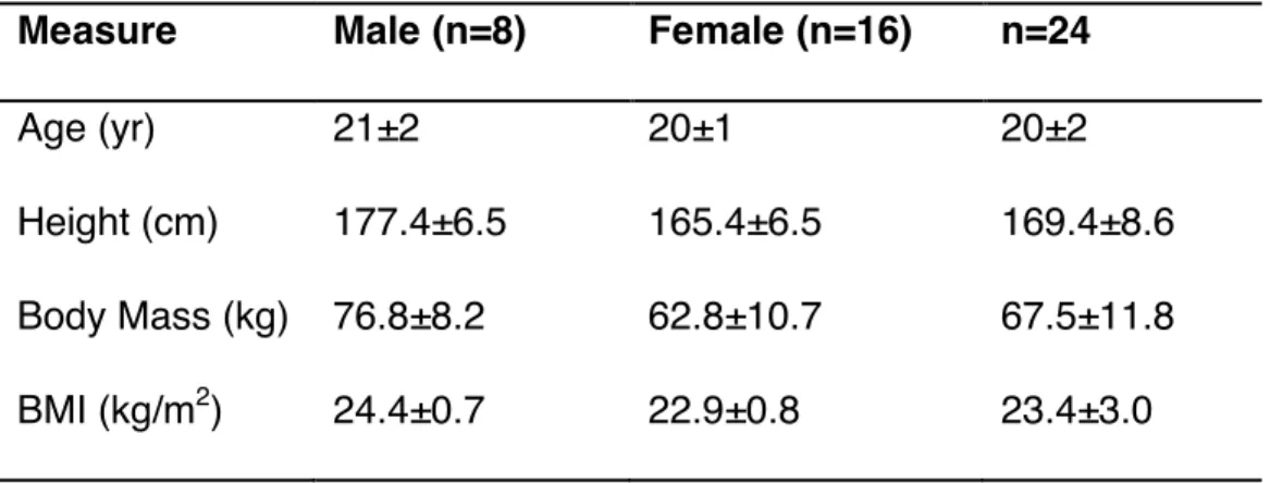
Table 1: Descriptive Data for Subjects
Peak Vertical Ground Reaction Force
Shapiro-Wilk test confirmed normal distribution of peak vGRF on the dominant limb (W=0.080; in this test W>0.05 is considered normal) and non-dominant limb (W=0.187). There was a significant difference between peak vGRF normalized to body weight (BW) between the dominant limb (1.08±0.08 BW) and non-dominant limb
(1.09±0.07 BW; p = 0.048) using a Paired samples t-test. The average limb symmetry index (non-dominant/dominant) was 1.01±0.03 as shown in Table 2.
Table 2. Kinetic Data (Normalized to BW) for Subjects
Measure Mean±SD
Peak vGRF dominant limb (BW) 1.08±0.08 Peak vGRF non-dominant limb (BW) 1.09±0.07
Limb Symmetry Index 1.01±0.03
Type II Collagen Turnover
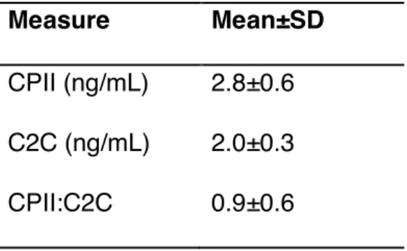
CPII, a marker of type II collagen synthesis, was not normally distributed (W<0.001) with a mean value of 12538.9±58857.7 ng/mL. C2C, the type II collagen
Measure Male (n=8) Female (n=16) n=24
Age (yr) 21±2 20±1 20±2
Height (cm) 177.4±6.5 165.4±6.5 169.4±8.6 Body Mass (kg)
BMI (kg/m2)
76.8±8.2 24.4±0.7
62.8±10.7 22.9±0.8
degradation marker, was normally distributed (W=0.565) with an average value of 106.3±54.1 ng/mL. Due to the synthesis marker (CPII), the turnover ratio was also skewed (W<0.001) with an average value of 115.2±529.1. All data is shown in Table 3. The mean intra-assay coefficient of variation of the CPII ELISA was 2.7%, and the mean coefficient of variation of the C2C ELISA was 18.1%.
Table 3. Biomarker Data for Subjects
Measure Mean±SD
CPII (ng/mL) 12538.9±58857.7
C2C (ng/mL) 106.3±54.1
CPII:C2C 115.2±529.1
Associations Between Cartilage Metabolism and Peak Vertical Ground Reaction
Force
No significant associations existed between the CPII:C2C ratio and peak vGRF (rho=0.185; p=0.386) using Spearman’s correlation analysis. Additionally, no significant associations were found between individual CPII or C2C biomarkers and peak vGRF (rho=0.110, p=0.609; r= -0.115, p=0.592) using Spearman’s and Pearson correlation analysis, respectively.
Post Hoc Analysis
vGRF and the biomarker ratio (CPII:C2C) were examined with correlation analysis. Outlier Removal
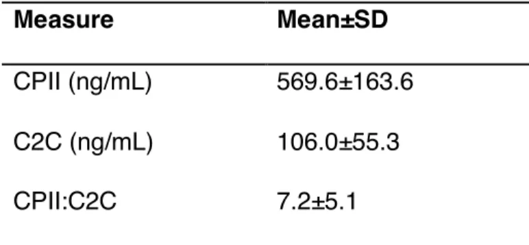
Subject 3 was considered an outlier due to a CPII concentration of 288911.38 ng/mL (i.e., greater than three standard deviations above the mean response; Howell, 1998). After removal of this subject, CPII:C2C mean and standard deviation was lowered substantially, as shown below in Table 4. CPII was normally distributed
(W=0.365) with an average value of 569.6±163.6 ng/mL. C2C was normally distributed (W=0.508) with an average of 106.0±55.3 ng/mL. CPII:C2C ratio, however, was still not normally distributed (W=0.000) with an average value of 7.2±5.1.
Table 4. Biomarker Data for Corrected Subjects
Measure Mean±SD
CPII (ng/mL) 569.6±163.6 C2C (ng/mL) 106.0±55.3
CPII:C2C 7.2±5.1
With the outlier removed, there was no significant correlation between CPII:C2C and peak vGRF (rho=0.079, p=0.720) using Spearman’s analysis.
Transformed Data
All biomarkers were subsequently transformed to a base-10 log scale value, but the outlier value was not removed. These data are shown below in Table 5. Both CPII and C2C were not normally distributed (W=0.000, W=0.000), but CPII:C2C was
Table 5. Transformed (Base-10 log) Biomarker Data
There was a correlation between CPII:C2C and peak vGRF which trended towards significance (r=0.363, p=0.081) using Pearson product moment correlations. Associations with Physical Activity and CPII:C2C
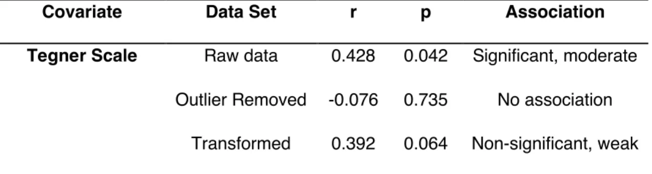
Further explorative analysis showed an association between physical activity (PA) level and the biomarker ratio. PA was assessed via Tegner scale (on a 1-10 scale). The average Tegner scale for the raw data and the raw data with the outlier removed was 4.2±1.1, and neither were normally distributed (W=0.001, W=0.002). Partial correlations analysescontrolling for the Tegner scale was conducted within the three data sets (raw, outlier removed, transformed). There was a significant, moderate partial correlation between peak vGRF and CPII:C2C using raw data
(r=0.428, p=0.042), no significant partial correlation once the outlier was removed (r=-0.076, p=0.735), and a partial correlation trending toward significance using the transformed data (r=0.392, p=0.064). As noted, not all data was normally distributed, but a nonparametric partial correlation analysis does not exist, therefore the parametric partial correlationapproachwas utilized. These results are shown below in Table 6.
Measure Mean±SD
Table 6. Partial Correlations of Peak vGRF and CPII:C2C Controlled for PA
Covariate Data Set r p Association
Tegner Scale Raw data 0.428 0.042 Significant, moderate Outlier Removed -0.076 0.735 No association
Chapter 6: Discussion
The purpose of this study was to examine associations between biochemical markers of cartilage metabolism and peak vGRF in young, healthy individuals. The research hypothesis stated that increases in peak vGRF in the first 50% of stance phase of gait would associate with a decrease in synthesis-to-degradation biomarker ratio. To test this hypothesis, gait kinetics and resting serum samples were measured in 24 young, healthy subjects.
The primary finding of the study showed a small non-significant association between peak vGRF and CPII:C2C. It was noted previously that the data was not normally distributed due to an extremely high CPII value (i.e., single individual). With the removal of this outlier, there was no correlation between peak vGRF and CPII:C2C. However, this data was still not normally distributed due to several moderately high CPII:C2C values that substantially skewed data but could not qualify as outliers (i.e. these values were ~2 standard deviations above the normal). Additionally, due to the small sample size, removal of further subjects would lower statistical power
The association between peak vGRF and CPII:C2C does change based on the data set analyzed (raw data vs. removed outlier vs. transformed), and this wide range of CPII:C2C values may be due in part to the variability of the study population. This study enrolled participants of widely varying PA levels, from individuals who strength trained very regularly to half-iron man athletes to individuals that merely met the required guidelines of 20 minutes of activity three times a week. In an effort to control for this variance in PA level and its potential impact on outcomes, partial correlations were analyzed with Tegner activity level scale as a covariate. Although the Tegner scale does not take into account the type of activity performed and focuses more on competition level rather than amount of training, it is a common survey used to assess functionality after knee surgery and is transferable to a healthy population (Briggs et al. 2009). After controlling for PA with the Tegner activity level scale, there was a moderate and
significant correlation between peak vGRF and CPII:C2C. However, this data also appears influenced by several very high CPII:C2C values of a few individuals within the data set. Once again, in an effort to maintain statistical power and normalize data, a partial correlation was conducted controlling for PA using the transformed base-10 log data on all data values. Using the transformed data, there was a moderate correlation approaching significance between peak vGRF and CPII:C2C.
early OA and can predict OA progression better than synthesis or degradation markers alone (Garnero et al., 2002; Cahue et al., 2007). This study provides evidence that increased peak vGRF in the first 50% stance phase of gait may be associated with higher levels of type II collagen synthesis to degradation ratio. Increased PA activity levels may be associated with higher synthesis-to-degradation turnover of type II collagen as well.
Association Between Joint Loading and Cartilage Turnover
Although it is outside the scope of this study to determine the causation of peak vGRF and CPII:C2C within this cohort of healthy individuals, it is well researched that cyclic loading occurring during normal and healthy gait is a critical component of extracellular matrix metabolic activity. Throughout the past several decades, studies have examined cyclic loading both in vitro and in vivo to investigate metabolic response in articular cartilage (Ikenoue et al., 2003; Saadat et al., 2006; Steinmeyer and Knue, 1997).
synthesis within the matrix may be more dependent on frequency and duration of the load rather than the total magnitude of the load. Reducing frequency from 0.5 Hz to 0.001 Hz resulted in decreased inhibition of proteoglycan biosynthesis, and reduced proteoglycan release accompanied an increase in loading duration (Steinmeyer and Knue, 1997). Type II collagen was not measured during this study, which was most likely due to the much slower turnover rate of collagen compared to proteoglycan (Bank et al., 1998). This increase in proteoglycan biosynthesis as well as type II collagen synthesis was found in a similar study examining human articular cartilage explants (Ikenoue et al., 2003). By applying intermittent hydrostatic pressure four hours a day for four days, proteoglycan signal levels increased as the amount of load increased from one to ten MPa. At higher loads of five and ten MPa, type II collagen signal levels were upregulated as well (Ikenoue et al., 2003). This study suggests that higher loading, as long as the loading is not abnormal or injurious in nature, may contribute to increased synthesis of cartilage macromolecules.
synthesis, thereby increasing the synthesis-to-degradation ratio.
Although studies examining associations of loading and cartilage metabolism in young, healthy human subjects are limited, studies have examined similar parameters in both ACL reconstruction (ACLr) subjects and subjects with KOA. In one cohort of ACLr subjects, higher peak vGRF in the injured limb was moderately associated with
CPII:C2C (Pietrosimone et al., 2016). It was hypothesized that higher amounts of cartilage synthesis were necessary to mitigate the peak vGRF during gait, and it was noted there were no significant differences in peak vGRF between limbs. Because biomarkers were collected from serum rather than synovial fluid, CPII:C2C ratios are representative of all physiological type II collagen rather than CPII:C2C specific to knee articular cartilage. It is possible that in this ACLr sample higher peak vGRF resulted in higher CPII:C2C ratios in both the injured and uninjured limb. It has been shown that subjects with KOA generally have a lower GRF than healthy controls, although this may be a result of decreased walking speed (Zeni and Higginson, 2009). Additionally,
several studies have shown that a decrease in collagen synthesis compared to
degradation is a good predictor of OA progression (Cahue et al., 2007; Garnero et al., 2002). Overall, these findings tentatively suggest that an increase in joint loading may associate with an increased stimulus for collagen synthesis rather than degradation in healthy human cartilage.
Physical Activity and Biomarkers of Cartilage Metabolism
running or sports that involved running, and it was observed that these subjects tended to have higher ratios of CPII:C2C. Previous research has shown benefits of load bearing activities (i.e., walking and running) including improved blood circulation to tissue fluid and increased lubrication of joints from synovial fluid (Renstrom, 1991; Simkin et al., 1990). While most researchers agree that immobilization or under-loading of a joint tends to weaken cartilage (Foley et al., 2007) and excessive loading can lead to cartilage loss (Roos and Dahlberg, 2005), there seems to be mixed opinions on what constitutes excessive loading.
Limitations
While this current study provided new insight of loading and cartilage metabolism in a healthy population, there were limitations to acknowledge. The current study might be underpowered with only 24 subjects completing all aspects of the experimental sessions (hence the low correlation coefficients and significance level found in some analysis); however, this low level of association may be what exists physiologically. The subjects, while healthy and similar in age, had wide variation in the amount and type of PA each subject participated in. In the future, it may be more useful to limit the study to subjects that participate in a certain amount of weight-bearing activities. This study did not control for amount of PA completed the day of the session. Although participants were asked to refrain from exercising the day of the study and sat for 30 minutes prior to data collection, participants walked through campus to get to the testing laboratory. Additionally, some subjects reported to the laboratory first thing in the morning, some in the afternoon, and others at night. This most likely influenced CPII:C2C ratio
considering many subjects walked to and from numerous classes the day of their testing session.
Another limitation may have been that this study focused on degradation only in articular cartilage rather than other parts of the knee. The literature suggests that changes to other surrounding tissues such as synovial fluid (Benito et al., 2005) and bone (Ishijima et al., 2011) may accompany or occur prior to cartilage breakdown. PA was quantified through the Tegner activity level scale. This scale is a 1-10 rating based on the type of PA in which the subject participates. The Tenger scale emphasizes the level of competition rather than the amount of PA completed, which is not always relevant in a university setting. Many subjects in various studies may maintain a high training load despite not participating at a varsity or competitive level. Additionally, no distinction is made for the type of PA. It is important to recognize that loading is very different for a rower, swimmer, or runner despite the competition level (O’Kane et al., 2006). Future studies assessing the association between PA and cartilage biomarkers may benefit from a different survey to assess PA level.
Conclusion
The data presented in the results of this study can be interpreted several different ways. Analyzing the raw biomarker data, there was no correlation between peak vGRF and CPII:C2C. However, by using a base-10 log transformation, the data was
normalized and a correlation existed between peak vGRF and the transformed
CPII:C2C. The discussion of this study focused on the transformed biomarker data in order to analyze a normally distributed data set. Using the transformed biomarker data, it was found that the ratio of serum markers of type II collagen synthesis to degradation is associated with peak vGRF in the first 50% stance phase of gait in healthy, young adults. The positive association between these markers in the present study agrees with data found in previous research (Ikenoue et al., 2003; Saadat et al., 2006). This data contrasts with the research hypothesis, which stated that peak vGRF and
APPENDIX 1: IRB APPROVED CONSENT FORM
University of North Carolina at Chapel Hill Consent to Participate in a Research Study Adult Participants
Consent Form Version Date: ______________ IRB Study # 15-1003
Title of Study: Disability, Biomechanical, Biochemical and Diagnostic Ultrasound Outcomes Following Anterior Cruciate Ligament Reconstruction: A Case-Control Study
Principal Investigator: Brian Pietrosimone
Principal Investigator Department: Exercise and Sport Science Principal Investigator Phone number: 962-3617
Principal Investigator Email Address: [email protected]
Co-Investigators: Troy Blackburn, Anthony Hackney, Yvonne Golightly, Darin Padua
Study Contact Telephone Number: 843-2014 Study Contact Email: [email protected]
_________________________________________________________________
What are some general things you should know about research studies?
You are being asked to take part in a research study. To join the study is voluntary.
You may refuse to join, or you may withdraw your consent to be in the study, for any reason, without penalty.
Research studies are designed to obtain new knowledge. This new information may help people in the future. You may not receive any direct benefit from being in the research study. There also may be risks to being in research studies. Deciding not to be in the study or leaving the study before it is done will not affect your relationship with the researcher, your health care provider, or the University of North Carolina-Chapel Hill. If you are a patient with an illness, you do not have to be in the research study in order to receive health care.
Details about this study are discussed below. It is important that you understand this information so that you can make an informed choice about being in this research study.
You will be given a copy of this consent form. You should ask the researchers named above, or staff members who may assist them, any questions you have about this study at any time.
What is the purpose of this study?
The purpose of this research study is to determine how different types of physical activities (i.e. walking/running/jumping) affect indicators of knee cartilage health in individuals following ACL injury and reconstruction compared to healthy control participants.
Are there any reasons you should not be in this study?
If you have undergone ACL reconstruction surgery, you should not be in this study if you: 1) Are not between the ages of 18-35
2) Have injured the ACL in BOTH of your legs
3) Have re-injured your ACL or required a second/revision ACL surgery following your initial ACL injury
4) Received your ACL reconstruction surgery less than 6 months ago 5) Have injured either of your legs within the last 6 months
6) Have not been cleared by your physician to return to physical activity 7) Do not currently participate in at least 20 minutes of physical activity 3x per week 8) Are currently pregnant
If you are a healthy individual who has not undergone ACL reconstruction surgery, you should not be in this study if you
1) Have a history of lower extremity surgery or ligamentous knee injury 2) Have chronic ankle instability or balance disorders
3) Are not physically active
4) Have concussion or head injury in the past 6 months 5) Have a history of cardiac condition or stroke
6) Are currently pregnant
How many people will take part in this study?
There will be approximately 60 people in this research study. Specifically, there will be 30 participants following ACLR and 30 Healthy participants.
How long will your part in this study last?
If you agree to participate, you will be asked to report to the Sports Medicine Research Laboratory in Fetzer Hall on the UNC-Chapel Hill campus for 3 testing sessions separated by 1 week. Each testing session will last approximately 1.5 hours.
What will happen if you take part in the study?
Session 1
Disability Outcomes
When you arrive to the laboratory, you will complete several electronic forms that will evaluate your knee function and physical activity level.
Blood Draw
You will perform a 5 minute “warm up” on a stationary bicycle. We will then place sensors on your legs, pelvis, and trunk that will allow us to measure movements of your joints, as well as sensors on the front and back of your legs to measure muscle activity. The areas where we will place some of these sensors will be shaved, lightly abraded, and cleaned with alcohol to improve adhesion to the skin and signal quality.
1) Gait Biomechanics Assessment – you will be asked to walk forward along a 20 foot
walkway at a comfortable, self-selected “fast” speed while movements of your legs are collected. You will be asked to walk over a device that will measure how hard you are stepping on the ground. You will perform at least 5 practice trials to ensure that you are comfortable walking with the sensors. You will then perform 5 trials that will be recorded
2) Landing Biomechanics Assessment – You will be asked to stand on top of a 12 inch
box that is placed half of your height away from the devices (force plates) in the ground that measure the force you apply to the ground during landing. During each trial, you will be asked to jump off the box using both legs, land with one foot on each force plate, and immediately jump vertically as high as you can. You will be allowed to practice this landing as many as 3 times to ensure that you are
comfortable with the task. You will then perform 5 trials that will be recorded.
3) Squatting Assessment – You will be asked to stand with your feet shoulder-width apart and your arms raised over your head. You will then descend into a double-leg squat as far as you can comfortably go, and then return to the starting position while motion patterns of your legs are measured. You will perform 5 trials of this task.
4) Voluntary Activation - You will be asked to perform maximal contractions of the quadriceps muscle on the front of your thigh. You will be seated and asked to kick out your leg against a device that measures how much force you can produce. During the contraction, a very brief (less than 1/1,000th of a second) pulse of electricity will be sent to your quadriceps muscle. This pulse of electricity will allow us to measure muscle function and is similar to a strong “carpet shock”. You will complete a warm up that will help you become comfortable and familiar with the electrical stimulus, after which 2 trials will be recorded. The stimulator used in this study is not FDA approved.
5) Meter Walk Test – You will be asked to complete 5 trials where you will walk 40-meters. You will be timed to see how fast that you can complete the 40-meter walk.
Ultrasound Outcomes
You will then lay on a table with your knee bent. We will use an ultrasound unit to take a picture of the inside of your knee to measure the size of your joint cartilage.
Loading Condition
Session 2
Disability Outcomes - Same as above
Blood Draw – Same as above
Biomechanics Outcomes – Same as above
Ultrasound Outcomes – Same as above
Loading Condition – You will then complete one of the other loading conditions for 30 minutes. We will then re-measure your knee cartilage size following this loading condition.
Session 3
Ultrasound Outcomes - Same as above
Loading Condition – You will then complete the last of the three loading condition for 30 minutes. We will then re-measure your knee cartilage size following this loading condition.
What are the possible benefits from being in this study?
Research is designed to benefit society by gaining new knowledge. You will not benefit personally from being in this research study. However, identifying early function,
biomechanical, and cartilage healthy changes following ACL reconstruction will be important in preventing the future development of osteoarthritis within this patient population.
What are the possible risks or discomforts involved from being in this study?
The quadriceps contractions during the voluntary activation testing may infrequently result in muscle soreness or, in extremely rare cases, muscle injury. The electrical stimulation used during the Muscle Function Assessment, though extremely brief (less than 1/1000th of a second), may cause discomfort – but not pain – during testing.
Similarly, the physical tasks you will perform (Landing, Squatting, and Walking) carry the extremely rare potential for musculoskeletal injury. However, because you are physically active, these risks are minimal and are not different from the risks you experience with normal physical activity/exercise. Additionally, there is a very minimal risk that preparing the skin (i.e. shaving, lightly abrading, and cleaning) for sensor placement will result in discomfort.
be conducted by research assistants trained in phlebotomy under the supervision of a nationally certified phlebotomist.
There may be uncommon or previously unknown risks. You should report any problems to the researchers.
What if we learn about new findings or information during the study?
You will be given any new information gained during the course of the study that might affect your willingness to continue your participation.
How will information about you be protected?
Any information obtained in connection with this research study that can be linked to you will remain confidential. You will be identified only by a subject identification number. A code list that associates your name and information with a specific subject identification number will be kept under key-card access on a password-protected computer in the Neuromuscular Research Laboratory. Only the research team will have access to this information.
Participants will not be identified in any report or publication about this study. Although every effort will be made to keep research records private, there may be times when federal or state law requires the disclosure of such records, including personal information. This is very unlikely, but if disclosure is ever required, UNC-Chapel Hill will take steps allowable by law to protect the privacy of personal information. In some cases, your information in this research study could be reviewed by representatives of the University, research sponsors, or government agencies (for example, the FDA) for purposes such as quality control or safety.
What will happen if you are injured by this research?
All research involves a chance that something bad might happen to you. This may include the risk of personal injury. In spite of all safety measures, you might develop a reaction or injury from being in this study. If such problems occur, the researchers will help you get medical care, but any costs for the medical care will be billed to you and/or your insurance company. The UNC-Chapel Hill has not set aside funds to pay you for any such reactions or injuries, or for the related medical care. You do not give up any of your legal rights by signing this form.
What if you want to stop before your part in the study is complete?
You can withdraw from this study at any time, without penalty. The investigators also have the right to stop your participation at any time. This could be because you have had an unexpected reaction, or have failed to follow instructions, or because the entire study has been stopped.
Will you receive anything for being in this study? You will not receive anything for being in this study
Will it cost you anything to be in this study? It will not cost you anything to be in this study.
What if you have questions about this study?
you have questions about the study (including payments), complaints, concerns, or if a research-related injury occurs, you should contact the researchers listed on the first page of this form.
What if you have questions about your rights as a research participant?
All research on human volunteers is reviewed by a committee that works to protect your rights and welfare. If you have questions or concerns about your rights as a research subject, or if you would like to obtain information or offer input, you may contact the Institutional Review Board at 919-966-3113 or by email to [email protected].
Participant’s Agreement:
I have read the information provided above. I have asked all the questions I have at this time. I voluntarily agree to participate in this research study.
______________________________________________________ Signature of Research Participant
____________________ Date
______________________________________________________ Printed Name of Research Participant
______________________________________________________ Signature of Research Team Member Obtaining Consent
____________________ Date
______________________________________________________ Printed Name of Research Team Member Obtaining Consent
APPENDIX 2: IRB APPROVED STORED SPECIMENS CONSENT FORM
University of North Carolina at Chapel Hill
Consent for Storing Biological Specimens With Identifying Information
______________________________________________________________________ Consent Form Version Date: ______________
IRB Study # 15-1003
Title of Study: Disability, Biomechanical, Biochemical and Ultrasound Outcomes Following Anterior Cruciate Ligament Reconstruction: A Case-Control Study Principal Investigator: Brian Pietrosimone
Principal Investigator Department: Exercise and Sport Science Principal Investigator Phone number: 962-3617
Principal Investigator Email Address: [email protected]
Co-Investigators: Darin Padua, Troy Blackburn, Anthony Hackney, Yvonne Golightly, Jeffrey Spang
Study Contact Telephone Number: 843-2014 Study Contact Email: [email protected]
______________________________________________________________________ What are some general things you should know about research?
Research is designed to gain scientific information that may help other people in the future. You may not receive any direct benefit from participating. There also may be risks.
You may refuse to take part in research. If you are a patient with an illness, you do not have to be in research in order to receive treatment.
Details are discussed below. It is important that you understand this information so that you can make an informed choice. You will be given a copy of this consent form. You should ask the researchers named above, or staff members who may assist them, any questions you have about this study at any time.
What is the purpose of this specimen repository or “biobank?”
Research with blood, tissue or body fluids (specimens) can help researchers
store some of the serum found in your blood in a freezer. If a new marker of interest may provide further information regarding the development of knee arthritis we may test the serum that we have stored. We will not perform any genetic research with your serum in the future.
How will the specimens be collected?
Blood will be collected from a vein the front of your elbow using a needle by a trained investigator. The blood will be stored in small tubes after collection. By agreeing to have your specimens stored, you will allow your medical facts, specimens and results from your participation in the UNC ACL study, but not your name, to be used in future studies.
What will happen to the specimens?
If you agree, after collecting your blood, we will store your samples for future testing of biomarkers that might partly cause osteoarthritis related to injuries of the joint. The specimens will be stored with a number and not your name for an indefinite period of time. We will be able to match a specimen to an individual through use of a code that will be separate from the storage place of the samples. The specimen codes will be stored on a password protected computer in the Neuromuscular Research Laboratory that is only accessible by the study personnel. We will keep your identity unknown to any other researchers and we will keep your records private to the extent allowed by law.
We may use the blood samples for scientific research or teaching purposes. We may also share the biomarkers with other researchers who work with us. Dr. Pietrosimone will carefully review study proposals from other researchers interested in working with us and all studies will be submitted to the Institutional Review Board for approval. Some of the blood or other specimens you give in this study may be sent to pharmaceutical or other commercial companies for future studies.
Your samples may be used in our research to help other researchers invent or find something new. The aim is to use the finding and inventions to develop new products that may improve public health. At times, such findings or inventions may have a value if they are made or sold. The government, the university, our research team or partners may get a patent on these. They may also license these. You would not share in any money or other things that the government, the university, our research team or
partners might get for what someone may invent or find using your study samples. We have no reason to believe your samples will be used to invent or find something new, but we want you to know what will happen in case it does.
What are the possible benefits to you?
Benefits to you are unlikely. Studies that use specimens from this repository may provide additional information that will be helpful in understanding the biochemical changes following ACL injury
What are the possible risks or discomforts involved with the use of your specimens?
There is a risk of breach of confidentiality.
Will there be any cost to you for storage of the specimens?
There will be no cost to you for the storage and use of the specimens for research purposes.
Will you receive anything for the use of your specimens?
You will not receive anything for the storage of your specimens in this study.
Who owns the specimens?
Any blood, body fluids, or tissue specimens obtained for this purpose become the exclusive property of the University of North Carolina at Chapel Hill. This organization may retain, preserve or dispose of these specimens and may use these specimens for research that may result in commercial applications. There are no plans to compensate you for any future commercial use of these specimens. Dr. Brian Pietrosimone, as the Principal Investigator of the “Disability, Biomechanical, Biochemical and Diagnostic Ultrasound Outcomes Following Anterior Cruciate Ligament Reconstruction: A Case-Control Study”, study, is the custodian of any blood specimens obtained for the purpose of this study or any future studies in collaboration with other researchers,
pharmaceutical or other commercial companies. How will information about you be protected?
No one will be identified in any report or publication about these future studies.
Although every effort will be made to keep research records private, there may be times when federal or state law requires the disclosure of such records, including personal information. This is very unlikely, but if disclosure is ever required, UNC-Chapel Hill will take steps allowable by law to protect the privacy of personal information. In some cases, your information in this research study could be reviewed by representatives of the University, research sponsors, or government agencies for purposes such as quality control or safety.
The specimens may be shared with researchers at this or other institutions. Research studies may be done at many places at the same time. Your personal identifying information will not be sent to other researchers.
You will not be identified in any report or publication about research using your