Address for correspondence Bartoš Vladimír, MD, PhD, MSc. Department of Pathology,
Faculty Hospital, Žilina, V. Spanyola 43, Žilina, 012 07, Slovakia
E-mail: [email protected]
Original Article
Basal cell carcinoma of the skin: Topographic
distribution and clinicopathological differences with
regards to the extent of sunlight exposure
Introduction
Basal cell carcinoma (BCC) of the skin is recently the most commonly diagnosed cancer in
white population.1,2 It exhibits a very
heterogeneous histomorphology, and an extremely broad spectrum of subtypes and variants have been described to date.3 Among
them, the superficial, nodular and infiltrative ones are the most frequent and the most important from clinical perspective.
It is well-known that the main cause of BCC development is an excessive exposure to
Bartoš Vladimír, Milada Kullova*
Department of Pathology, Faculty Hospital in Žilina, V. Spanyola 43, Žilina, 012 07, Slovakia * Department of Dermatovenereology, Faculty Hospital in Žilina, V. Spanyola 43, Žilina, 012 07, Slovakia
Abstract
Objective To analyze a topographic distribution of cutaneous basal cell carcinoma (BCC) and clinicopathological differences of disease with regards to the extent of sunlight exposure.Methods A total of 1,065 BCC cases from 815 patients were investigated. The topographic regions of the body affected were merged into the sun-protected, intermittently sun-exposed, and permanently sun-exposed sites.
Results BCCs occurred most commonly in the permanently sun-exposed sites (63.8%), followed by intermittently sun-exposed sites (30.8%), and sun-protected sites (5.4%). There was higher proportion of the men in the parts of the body intermittently exposed to sunlight and vice versa, higher percentage of the women in the sun-protected, as well as, in the permanently sun-exposed parts. There was a statistically significant trend towards an increased age with rising extent of sunlight exposure. Superficial BCC correlated positively with the intermittently sun-exposed sites and negatively with the permanently sun-exposed sites. Nodular BCC was related to the permanently sun-exposed sites and negatively with the intermittently sun-exposed sites. Infiltrative BCC was linked to permanently sun-exposed sites, while it was completely absent in the body regions, the skin of which was usually protected from UVR. A proportion of BCCs with aggressive growth phenotype positively correlated with rising extent of sunlight exposure.
Conclusion Considerable clinicopathological variations in BCCs depending on locations and corresponding solar exposure levels were confirmed. With respect to the body sites, from which the lesions arise, this neoplasia may have distinct etiopathogenesis and biology. Probably, different patterns of sun exposure are independent risk factors for certain histological BCC subtypes and hence prognosis of this malignancy.
Key words
sunlight, hence the vast majority of lesions arise on habitually sun-exposed parts of the body, especially on the head. According to pattern of ultraviolet radiation (UVR) exposure, three distinct forms are defined: a) intermittent (recreational); b) chronic (occupational); and c) cumulative (total) exposure.4 This categorization
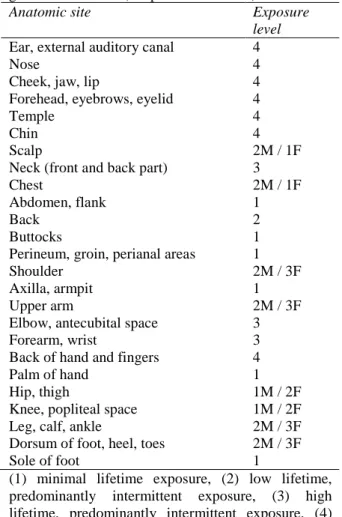
has been proven to be of great importance in terms of their impact in the development of various skin malignancies, such as BCC, squamous cell carcinoma (SCC) and malignant melanoma. With regards to detailed anatomic locations and with respect to gender differences, following four categories (levels) of solar UVR exposure exist (Table 1): a) minimal lifetime exposure; b) low lifetime, predominantly intermittent exposure; c) high lifetime, predominantly intermittent exposure; and c) maximal (chronic + intermittent) lifetime exposure.
Although UVR is the primary extrinsic etiologic factor in BCC carcinogenesis, this association is not straightforward.5 Compared with cutaneous
SCC, the relationship between chronic UVR exposure and BCC is relatively modest5 and
even association between morphologic markers of cutaneous photo-damage and an increased risk of BCC has been proven only moderate.6
Approximately one quarter of all BCCs occur on anatomic sites that are not habitually exposed to the sun, such as the trunk.2 Hence, there are
suggestions, that the pathophysiology of BCC arising on distinct (exposed vs sun-protected) parts of the body and developing into different subtypes, as well as, the patterns of UVR exposure that give rise to lesions arising on distinct body sites differ.2,6-9 Many studies
done till present time have analyzed a relationship between anatomic distribution of BCCs and other clinicopathological parameters.7-14 However, to the best of our
Table 1. A summary of the sun exposure levels corresponding to detailed body sites with respect to gender differences (adopted from ref. 15).
Anatomic site Exposure
level Ear, external auditory canal
Nose
Cheek, jaw, lip
Forehead, eyebrows, eyelid Temple
Chin Scalp
Neck (front and back part) Chest
Abdomen, flank Back
Buttocks
Perineum, groin, perianal areas Shoulder
Axilla, armpit Upper arm
Elbow, antecubital space Forearm, wrist
Back of hand and fingers Palm of hand
Hip, thigh
Knee, popliteal space Leg, calf, ankle
Dorsum of foot, heel, toes Sole of foot
4 4 4 4 4 4 2M / 1F 3 2M / 1F 1 2 1 1 2M / 3F 1 2M / 3F 3 3 4 1 1M / 2F 1M / 2F 2M / 3F 2M / 3F 1
(1) minimal lifetime exposure, (2) low lifetime, predominantly intermittent exposure, (3) high lifetime, predominantly intermittent exposure, (4) maximal (both chronic + intermittent) exposure. M – male, F - female
knowledge, none of them investigated their association with the levels of solar exposure mentioned above, as it was carried out in cutaneous malignant melanoma.15 This
categorization scheme seems to be more precise, since it takes into account differences in UVR exposure within a specific parts of the body, as well as, certain gender-related variations.
BCC phenotype, especially its histomorphology and biological behaviour.
Methods
For the purpose of the study, we retrospectively reviewed all consecutive primary BCCs of the skin, that were histologically diagnosed at the Department of Pathology in Faculty Hospital in Zilina (Slovakia) during 9-years period (January 2007 – December 2015). Recurrent lesions, as well as, subsequent re-excisions after incomplete tumor removal were excluded. The study group totally consisted of 1,065 BCC cases from 815 subjects. The development of a new tumor in a patient already included in the file was considered as a new case. The specimens were derived from a variety of clinical sources,
especially surgery, dermatology,
otorhinolaryngology and ophthalmology departments. Biopsy material was fixed in buffered formalin, embedded in paraffin blocks and stained with hematoxylin and eosin. If necessary, immunohistochemical staining
methods were also applied. The
histopathological classification of BCC subtypes was done using the latest World Health Organization classification system of skin cancers.16 Further, all the cases were simply categorized into non-aggressive and aggressive growth phenotypes. The latter included the infiltrative, morpheic, micronodular and metatypical (basosquamous) subtypes. Mixed BCCs exhibiting any proportion of these histomorphologic patterns were definitively categorized as an aggressive growth phenotype. The anatomical sites affected by tumors were basically classified as follows: head and neck, trunk, upper extremities, and lower extremities. In both genders, solar exposure levels were determined based on the criteria describing in
Table 1. For the sake of simplicity, we decided to merge the levels 2 and 3 (both with predominantly intermittent exposure) into a
single category and subsequently, we defined three separate topographic regions of the body for statistical evaluation: a) sun-protected sites (corresponding with the level 1); b) intermittently sun-exposed sites (corresponding with the levels 2 and 3); and c) permanently sun-exposed sites (corresponding with the level 4). Clinical data of the patients needed for the study were obtained from their medical records. Data were collected in a databank, using a software SPSS Statistics. For the statistical analysis, chi-square test was employed and P value < 0.05 was considered significant.
Results
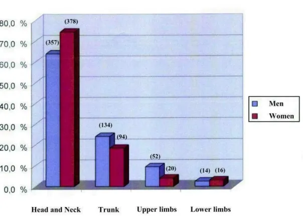
Basic Clinical and Histopathological Data A total of 1,065 lesions from 815 subjects in the age rage between 25-97 years were evaluated in the study. Out of them, 395 (48.4%) were men, and 420 (51.6%) were women. The mean age of participants was of 69.8±12.7 years, with a mean of 70.2 years for females and 69.5 years for males. The number of lesions per patient ranged from 1 to 17, with a mean of 1.3. Topographic distribution of BCCs was as follows: the head and neck (735 cases, 69.0%); the trunk (228 cases, 21.4%); the upper extremities (72 cases, 6.8%); and the lower extremities (30 cases, 2.8%). There were striking gender differences depending on sites affected. Women had tumors located more frequently on the head and neck (74.4% vs. 64.0%), as well as, on the lower limbs (3.1% vs. 2.5%) compared with men, respectively. Conversely, men had lesions situated more commonly on the trunk (24.1% vs. 18.5%, p = 0.02) and on the upper limbs (9.4% vs 4.0%) compared with women, respectively (Figure 1).
Figure 1 Topographic distribution of BCCs in women and men. (number in the brackets indicates number of the cases).
Table 2. An overview of the clinicopathologiocal parameters of BCC investigated in relation to anatomic sites of the body differently exposed to sunlight.
Clinicopathologic parameter Sun-protected sites
Intermittently sun-exposed sites
Permanently sun-exposed sites
Number of tumors 57 (5.4%) 328 (30.8%) 680 (63.8%)
Gender
• corresponding to men • corresponding to women
17 (29.8%) 40 (70.2%)
209 (63.7%) 119 (36.3%)
331 (48.6%) 349 (51.4%) Mean age
≤ 60 years ≥ 61 years
62.8 y 26 (45.6%) 31 (54.4%)
68.2 y 88 (26.8%) 240 (73.2%)
71.3 y 126 (18.5%) 554 (81.5%) Main histologic subtypes
• superficial • nodular • infiltrative
14 (24.5%) 18 (31.5%) 0
95 (29.0%) 92 (28.0%) 12 (3.6%)
26 (3.8%) 315 (46.3%) 64 (9.4%) Histologic phenotype
• non-aggressive • aggressive
49 (86.0%) 8 (14.0%)
244 (74.4%) 84 (25.6%)
411 (60.4%) 269 (39.6%)
Infiltrative BCC consisted of 77 cases. Approximately one third of the cases (369/1065,
Distribution of affected body sites with different sunlight exposure levels and their correlation with clinicopathological parameters
In our series, the tumors occurred most commonly in the permanently sun-exposed sites of the body (680 cases, 63.8%), followed by intermittently sun-exposed site
30.8%), and
sun-5.4%). It generally suggests that BCC development has the strongest association with a cumulative exposure to sunlight, although as will be discussed below, given complex etiopathogenesis of disease and probably distinct pathogenetic mechanisms in certain BCC subtypes, this finding should be considered only simplified. Several differences in the clinicopathological parameters depending on given solar exposure levels were found (Table 2).
By correlating gender with the locations of tumor, there was significantly higher proportion of the men in the sites of the body intermittently exposed to sunlight (63.7%, p < 0.0001) and vice versa, higher percentage of the women in the sun-protected sites (70.2%, p < 0.001), as well as, in the permanently sun-exposed sites (51.4%, p < 0.01). In summary, in men and women, the sun-protected, the intermittently sun-exposed, and the permanently sun-exposed sites included 17 (3.0%) and 40 (7.9%) cases, 209 (37.6%) and 119 (23.4%) cases, and 331 (59.4%) and 349 (68.7%) cases, respectively.
As for age, there was a statistically significant trend (p < 0.001) towards an increased age with the extent of sunlight exposure, starting from the sun-protected (mean age of 62.8 years) and ending in the chronically sun-exposed sites (mean age of 71.3 years). No association between age and gender was found (p = 0.7).
Considering the basic histological subtypes, superficial BCC correlated positively with the intermittently sun-exposed sites (p < 0.0001) and negatively with the permanently sun-exposed sites (p < 0.0001). Conversely, nodular BCC was strongly related to the permanently sun-exposed sites (p < 0.0001) and negatively associated with the intermittently sun-exposed sites (p < 0.0001). Infiltrative BCC was linked to the permanently sun-exposed sites of the body (p
.001), while it was completely absent in the body regions, the skin of which was usually protected from UVR (p 03). Among all three main histologic tumor subtypes, infiltrative BCC was the only one that exhibited a continuous increase in proportion with rising extent of solar exposure. After merging cancers into non-aggressive and aggressive (at least focally infiltrative) growth phenotype, a significant relationship (p < 0.001) with solar exposure levels was found. Likewise infiltrative BCC subtype, percentages of BCCs with any aggressive growth features increased progressively with rising extent of solar exposure, being only 14.0% in the sun-protected sites and reaching nearly 40% in the permanently sun-exposed sites of the body.
Discussion
Although UVR has been accepted as the principal causal factor for cutaneous BCC, a lot of questions still remain regarding the relationship between sun exposure and tumor development. It is well-known that BCCs arise most frequently in the head and neck, which comprises 68.4%-89.9% of all cases, depending on individual published papers.8,10-14 In the
locations, which corresponded to the highest solar exposure level 4 found in our study (63.8%), this reinforces cumulative sun exposure as a main risk factor for BCC. However, relative high percentages of lesions arising in the intermittently sun-exposed sites of the body (nearly one third of all cases) clearly suggest that discontinuous episodes of intense sunlight exposure play an important role in tumor pathogenesis. Indeed, many epidemiological and clinical studies17-21 have confirmed such
hypothesis and found that an intermittent solar exposure rather than chronic one was more prejudical etiologic determinant in BCC carcinogenesis. For example, large European study Helios19 revealed that, in contrast with
SCC, the risk of BCC developing reached a maximum at 8,000-10,000 hours of cumulative sun exposure received during life, and subsequently declined slightly with higher levels. Outdoor recreational activities (main source of intermittent sunlight exposure), even for relatively short periods, were associated with an increased risk of cutaneous BCC. Similar findings were published by Kricker et al.20,21
They also showed that beyond a certain level of sun exposure, risk of BCC development did not increase further,20 as well as, that a particular
amount of sun exposure delivered in infrequent intense increments increased risk of BCC more than a similar dose delivered more continuously over the same total period of time.21 In the study
published by Pelluchi et al.18, direct associations
were observed with recreational sunlight exposure and severe sunburn episodes, along with some differences in risk of tumor development between distinct BCC subtypes and anatomic locations. Even Kennedy et al.17 found
that the painful sunburns at a young age were linked to an increased risk of BCC. However, in contrast with these observations, the results of recent epidemiological study from Iannacone et al.22 have suggested that the sunlight exposure
was associated with an equal risk of BCC
development regardless of the pattern, which was predominantly received. Therefore, not completely clear data about the real role of sunlight and the importance of different pattern (intermittent vs. continuous) of solar exposure in the evolution of different subtypes of BCC justify the relevance of another research in the future.
A large proportion of BCCs arising on the skin, which is partially or totally covered from the sunlight all through the year and conversely, the rare involvement of typical habitually sun-exposed areas as the dorsum of the hands indicate the UVR is surely not the only causative factor for BCC development. Among other etiologic factors involved in the genesis of this disease, genetic susceptibility seems to have the greatest impact. Previous studies have demonstrated23,24 that the development of truncal
BCCs, which typically manifest some specific clinicopathologic features such as predominant superficial histologic subtype, presentation with multiple primary lesions, and younger age of patients at the time of diagnosis, was proven particularly in genetically susceptible individuals. This finding indicates that a genetic background may significantly determine or modulate skin response to UVR. A reduced DNA repair capacity for UVR-induced DNA damage has been established as an independent risk factor for the development of BCC.25 It is
possible that people who develop BCCs in sun-protected areas of the body may have a decreased DNA repair capacity.
An important observation in our present study has been that almost all available clinicopathological parameters changed depending on solar exposure levels. In accord with other reports10-13 we have confirmed
with data in the literature.11-13 This is probably
due to greater sun exposure of this body part during their work outside or outdoor leisure activities. Conversely, the women had more often affected the head and neck regions, as well as, the lower extremities, although the strength of this association was not as great as that of the trunk for men. The propensity of women to develop BCCs on the legs has been reported in other studies,11,12 perhaps due to the fact that they
more frequently wear clothes (short skirts), that expose this region of their body. Hence, a considerable predominance of the males in intermittently sun-exposed parts is the result of their frequent trunk involvement.
As expected, we have shown a tendency towards a rising age with the extent of sunlight exposure. This association was foreseeable, because total solar UVR dose increases, as patient ages.
Regarding histological subtypes, superficial BCC occurred only sporadically in the permanently sun-exposed sites of the body (3.8% of cases), while it was the most frequently found in the sites intermittently exposed to sunlight (29.0% of cases), especially in the trunk. On the other hand, infiltrative BCC subtype was strongly related to chronically sun-exposed sites of the body (9.4% of cases), while it was completely absent in anatomic regions, the skin of which was usually protected from UVR. Nodular BCC was linked to permanently sun-exposed sites (46.3% of cases) and negatively associated with intermittently sun-exposed sites (28.0% of cases). Hence, there is clear relationship of superficial BCC with intermittent and of infiltrative and nodular BCC with cumulative sunlight exposure. Furthermore, we have found a close association between the patterns of solar exposure and BCC growth phenotype, as proportion of BCCs with aggressive growth features increased progressively with rising extent of solar
exposure. Based on these observations it might be assumed that a prolonged cumulation of UVR influence a pathogenesis of BCC by driving tumor progression and aggressive biological behaviour. These findings are consistent with the experiences of another authors,1,8,9,12 who have
also reported that aggressive growth variants of BCCs were more frequently present in the chronically sun-exposed parts of the body. Since there is a relative lack of detailed data in this field, an additional research will be valuable to further characterize sunlight exposure-response relationships in development of different BCC subtypes.
In conclusion, we have shown considerable clinicopathological variations in cutaneous BCCs depending on their anatomic locations and corresponding solar exposure levels. With respect to the body sites, from which the lesions arise, this neoplasm may have distinct pathogenesis and biology. Probably, different patterns of sun exposure are independent risk factors for certain histological BCC subtypes and hence prognosis of this malignancy. Understanding how UVR response may differ in various skin cancers would be important for educating the public on safe sunlight behaviors.
References
1. Crowson AN. Basal cell carcinoma: biology, morphology and clinical implications. Mod Pathol. 2006;19 (Suppl 2):S127-S147. 2. Khalesi M, Whiteman DC, Rosendahl C, Johns
R, Hackett T, Cameron A et al. Basal cell carcinomas on sun-protected vs. sun-exposed body sites: a comparison of phenotypic and environmental risk factors. Photodermatol Photoimmunol Photomed. 2015;31:202-11. 3. Kazakov DV, McKee PH, Michal M,
Kacerovska D. Cutaneous Adnexal Tumors, 1st edition. Philadelphia: Lippincott Williams & Wilkins; 2012.
7216 controls. Int J Epidemiol. 2009;38 :814-30.
5. Armstrong BK, Kricker A. The epidemiology of UV induced skin cancers. J Photochem Photobiol B. 2001;63:8-18.
6. Khalesi M, Whiteman DC, Doi SA, Clark J, Kimlin MG, Neale RE. Cutaneous markers of photo-damage and risk of basal cell carcinoma of the skin: a meta-analysis. Cancer Epidemiol Biomarkers Prev. 2013; 22:1483-9.
7. Scrivener Y, Grosshans E, Cribier B. Variations of basal cell carcinomas according to gender, age, location and histopathological subtype. Br J Dermatol. 2002;147:41-7. 8. Bastiaens MT, Hoefnagel JJ, Bruijn JA,
Westendorp RG, Vermeer BJ, Bouwes
Bavinck JN. Differences in age, site
distribution, and sex between nodular and superficial basal cell carcinoma indicate different types of tumors. J Invest Dermatol. 1998;110:880-4.
9. McCormack CJ, Kelly JW, Dorevitch AP. Differences in age and body site distribution of the histological subtypes of basal cell carcinoma. A possible indicator of differing causes. Arch Dermatol. 1997;133:593-6. 10. Vantuchová Y, Čuřík R. Hodnocení
bazocelulárního karcinómu vzhledem k histologickému typu, věku, pohlaví a lokalizaci. Čes-Slov Derm. 2007;82:140-5. 11. Souza CF, Thomé EP, Menegotto PF, Schmitt
JV, Shibue JR, Tarlé RG. Topography of basal cell carcinoma and their correlations with gender, age and histologic pattern: a retrospective study of 1042 lesions. An Bras Dermatol. 2011;86:272-7.
12. Ferreira FR, Pevide Bda, C, Rodriques RF, Nascimento LF, Lira ML. Differences in age and topographic distribution of the different histological subtypes of basal cell carcinoma, Taubaté (SP), Brazil. An Bras Dermatol. 2013;88:726-30.
13. Mantese OAS, Gomides ADM, Berbert VCLA, Rocha A. Basal cell carcinoma – analysis of 300 cases observed in Uberlândia - MG, Brazil. An Bras Dermatol. 2006;81 :136-42.
14. Mateoiu C, Georgescu CV, Fota G, Simionescu C. Histopathological study of basal cell carcinomas. Curr Health Sci J. 2009;35:119-23.
15. Bulliar JL, De Weck D, Fisch T, Bordoni A, Levi F. Detailed site distribution of melanoma and sunlight exposure: aetiological patterns from a Swiss series. Ann Oncol. 2007;18 :789-94.
16. LeBoit P, Burg G, Weedon D, Sarasin A. World Health Organization Classification of
Tumours, Pathology and Genetics of Skin tumours. Lyon: IARC Press; 2006.
17. Kennedy C, Bajdik CD, Willemze R, De Gruijl FR, Bouwes Bavinck JN; Leiden Skin Cancer Study. The influence of painful sunburns and lifetime sun exposure on the risk of actinic keratoses, seborrheic warts, melanocytic nevi, atypical nevi, and skin cancer. J Invest Dermatol. 2003;120:1087-93.
18. Pelucchi C, Di Landro A, Naldi L, La Vecchia C; Oncology Study Group of the Italian Group for Epidemiologic Research in Dermatology (GISED). Risk factors for histological types and anatomic sites of cutaneous basal cell carcinomas: an Italian case-control study. J Invest Dermatol. 2007;127: 935-44.
19. Rosso S, Zanetti R, Martinez C, Tormo MJ, Schraub S, Sancho-Garnier H et al. The multicentre south European study 'Helios'. II: Different sun exposure patterns in the aetiology of basal cell and squamous cell carcinoma of the skin. Br J Cancer. 1996;73:1447-54.
20. Kricker A, Armstrong BK, English DR, Heenan PJ. A dose-response curve for sun exposure and basal cell carcinoma. Int J Cancer. 1995;60:482-8.
21. Kricker A, Armstrong BK, English DR, Heenan PJ. Does intermittent sun exposure cause basal cell carcinoma? A case-control study in Western Australia. Int J Cancer. 1995;60:489-94.
22. Iannacone MR, Wang W, Stockwell HG, O'Rourke K, Giuliano AR, Sondak VK et al. Patterns and timing of sunlight exposure and risk of basal cell and squamous cell carcinomas of the skin – a case-control study. BMC Cancer. 2012;12:417.
23. Ramachandran S, Lear JT, Ramsay H, Smith
AG, Bowers B, Hutchinson PE et al.
Presentation with multiple cutaneous basal cell carcinomas: association of glutathione S-transferase and cytochrome P450 genotypes with clinical phenotype. Cancer Epidemiol Biomarkers Prev. 1999;8:61-7.
24. Lear JT, Tan BB, Smith AG, Bowers W, Jones PW, Heagerty AH et al. Risk factors for basal cell carcinoma in the UK: case-control study in 806 patients. J R Soc Med. 1997;90:371-4. 25. Wang LE, Li C, Strom SS, Goldberg LH,