O R I G I N A L I N V E S T I G A T I O N
Open Access
Insulin resistance and exercise tolerance in heart
failure patients: linkage to coronary flow reserve
and peripheral vascular function
Martin Snoer
1*, Tea Monk-Hansen
1, Rasmus Huan Olsen
1, Lene Rørholm Pedersen
1, Lene Simonsen
2,
Hanne Rasmusen
1, Flemming Dela
3and Eva Prescott
1Abstract
Background:Insulin resistance has been linked to exercise intolerance in heart failure patients. The aim of this study was to assess the potential role of coronary flow reserve (CFR), endothelial function and arterial stiffness in explaining this linkage.
Methods:39 patients with LVEF < 35% (median LV ejection fraction (LVEF) 31 (interquartile range (IQ) 26–34), 23/39 of ischemic origin) underwent echocardiography with measurement of CFR. Peak coronary flow velocity (CFV) was measured in the LAD and coronary flow reserve was calculated as the ratio between CFV at rest and during a 2 minutes adenosine infusion. All patients performed a maximal symptom limited exercise test with measurement of peak oxygen uptake (VO2peak), digital measurement of endothelial function and arterial stiffness (augmentation index), dual X-ray absorptiometry scan (DEXA) for body composition and insulin sensitivity by a 2 hr
hyperinsulinemic (40 mU/min/m2) isoglycemic clamp.
Results:Fat free mass adjusted insulin sensitivity was significantly correlated to VO2peak (r = 0.43, p = 0.007). Median CFR was 1.77 (IQ 1.26-2.42) and was correlated to insulin sensitivity (r 0.43, p = 0.008). CFR (r = 0.48, p = 0.002), and arterial stiffness (r =−0.35, p = 0.04) were correlated to VO2peak whereas endothelial function and LVEF were not (all p > 0.15). In multivariable linear regression adjusting for age, CFR remained independently associated with VO2peak (standardized coefficient (SC) 1.98, p = 0.05) whereas insulin sensitivity (SC 1.75, p = 0.09) and arterial stiffness (SC −1.17, p = 0.29) were no longer associated with VO2peak.
Conclusions:The study confirms that insulin resistance is associated with exercise intolerance in heart failure patients and suggests that this is partly through reduced CFR. This is the first study to our knowledge that shows an association between CFR and exercise capacity in heart failure patients and links the relationship between insulin resistance and exercise capacity to CFR.
Keywords:Coronary flow reserve, Heart failure, Exercise capacity, Insulin sensitivity, Arterial stiffness
Background
The link between insulin resistance and heart failure is complex. Several studies have shown that insulin resist-ance is common in heart failure of both ischemic and non-ischemic origin. The exact mechanisms are not known but potentially include both causal and secondary associations. Insulin resistance has been linked to
reduced exercise tolerance in heart failure and mechan-isms may include both central and peripheral vascular dysfunction [1].
In the absence of significant coronary artery stenosis, coronary flow reserve (CFR) is a measure of microvascu-lar function. CFR has been shown to be reduced in patients with classic risk factors of cardiovascular disease such as hypertension [2], obesity [3] and diabetes [4,5] and is a strong independent marker of poor prognosis in patients with ischemic heart disease [6]. The role of cor-onary flow reserve in heart failure is less well described. * Correspondence:snoer@dadlnet.dk
1Dept Cardiology, Bispebjerg University Hospital, Copenhagen, Denmark Full list of author information is available at the end of the article
In idiopathic dilated cardiomyopathy CFR has been shown to be reduced [7,8] and in some studies it was also a prognostic marker of future outcome [7,9]. The relationship between insulin resistance and CFR has not been examined in patients with chronic heart failure.
In healthy young volunteers CFR has been related to VO2peak [10,11]. VO2peak is one of the most important
prognostic factors in CHF patients [12] and exercise training in heart failure has been shown to improve peak oxygen consumption, muscle strength, symptoms and quality of life and has become part of current guideline recommendations. Effects are thought to be primarily through peripheral vascular and muscular mechanisms [13]. Few studies have examined CFR in relation to VO2peak in heart failure patients. Recently, however, in
a small randomized trial, 4 months of exercise training of 13 patients with primary heart failure improved both
VO2peak and CFR [14]. This study added mechanistic
insight to explain the apparent benefit of exercise train-ing in heart failure and may also be a link to explain improved prognosis following exercise training in heart failure patients [15]. A possible causal web linking these factors is depicted in Figure 1.
Peak oxygen uptake, vascular function and metabolic function are all important prognostic markers in heart failure. The aim of this study was to assess the potential role of coronary flow reserve, endothelial function and arterial stiffness in explaining the link between insulin resistance and exercise intolerance.
Methods Patients
The study was a local substudy based on patients screened for inclusion to the Smartex-HF trial [16] in which patients with chronic systolic heart failure were rando-mized to different modalities of exercise training. The patients were recruited from the heart failure outpatient
clinic at Bispebjerg University Hospital, Copenhagen, Denmark. Inclusion criteria were a LVEF < 35%, clinically stable (no signs of worsening for at least 6 weeks), mini-mum 3 months of optimal medical treatment, and if CRT or revascularization was performed, this should be more then 6 months prior to inclusion. All patients had been examined with either coronary angiography or cardiac CT as part of their heart failure examination program and were revascularized according to guidelines. All patients had to have a LAD without significant sten-osis (<50%) in order to perform coronary flow mea-surements. Angiography was not repeated prior to the enrolment in to the study. However, the participants were excluded from the study if there were signs of ischemia or ventricular arrhythmias during the maximal symptom lim-ited bicycle exercise test. The study complied with the Declaration of Helsinki and was approved by the science ethics committee for the Capital Region of Denmark (HC-2008-108). All participants gave informed written consent.
Cardiopulmonary exercise test
All patients underwent an upright bicycle (Via Sprint 150P, Ergoline, Bitz, Germany) exercise test with breath-by-breath gas exchange measurement (Jaeger, Mastersc-reen CPX, Cardinal Health, Würzburg, Germany). After 3 minutes of rest on the bicycle the test was initiated using either a protocol starting at 20 watt with a 10 watt increase pr. minute or starting at 40 watts with a 20 watt increase pr. minute, to ensure optimal exercise time based on an expectation of the patients exercise cap-acity. The patients were encouraged to continue until maximal exhaustion. A leveling off of oxygen uptake despite increasing workload and a respiratory exchange ratio (RER) > 1.05 were used as criteria for maximal oxy-gen uptake. The mean of the 3 highest consecutive 10-second measurements before exercise terminations were used for determining VO2peak.
Echocardiography
A complete transthoracic echocardiography was performed using Philips IE33 (Philips Medical Systems, Andover, MA, USA) with an S5 probe with the patients in the left supine position. Left ventricle end diastolic (EDV) and end sys-tolic (ESV) volumes and ejection fraction (LVEF) were cal-culated from apical 2- and 4-chamber views using the biplane Simpson model. Left ventricular mass (LVM) was calculated using the formula LVM = 0.8*(1.04*(LVEDD + PWTd + SWTd)3-(LVEDD)3) + 0.6 g, where LVEDD is left ventricle end diastolic diameter, PWTd is posterior wall thickness in diastoly and SWTd is septum wall thickness in diastoly, and indexed (LVMi) to body surface area
(BSA) calculated by Du Bois’ formula (BSA = 0.007184 x
weight kg0.425x height cm0.725). Figure 1A proposed linkage (non-exhaustive) between insulin
Coronary flow reserve
CFR can be measured non-invasively using transthoracic Doppler echocardiography with a high success rate [17] and this technique has been validated against invasive measurements [18] with good results. CFR was mea-sured using a high frequency broadband transducer (S8, Philips). All patients were instructed to abstain from caf-feine for 12 hours before the examination and oral use of dipyridamole was paused for 72 hours. With the pa-tient in the left supine position the LAD was located as distal as possible using color Doppler either in an apical modified two chamber view or middistal using a modi-fied short axis view. Coronary flow velocity (CFV) was measured using pulsed wave Doppler with a sample size of 3–4 mm, at rest and during a 2 minute infusion of ad-enosine at 0.14 mg kg-1min-1. The solution was diluted so that the infusion rate was kept at 10 ml min-1. Before and during the adenosine infusion care was taken to maintain the position and angle of the probe, so mea-surements were done on the same segment of the LAD at the same angle. During the infusion the scale of flow velocity was changed in order to obtain optimal curves for offline measurement. CFR was calculated as the ratio between peak diastolic CFV during adenosine infusion and during rest using a mean of 3 consecutive cardiac cycles (10 cycles if the patient had atrial fibrillation). Analyses were done offline by an investigator blinded to the other examinations. Blood pressure and ECG were monitored before and during the adenosine infusion. Interobserver variability of CFR was tested on all sub-jects and the mean difference in CFR was 0.07 with lim-its of agreement ±0.21. The coefficient of variation (CV), calculated as the within-subject standard deviation divided by the mean of the observations, was 5.5%. Intraobserver variability was tested on 10 randomly selected examinations with a mean difference of 0.03, limits of agreement ±0.29 and CV 7.5%. Figure 2 shows measurement of CFR
Insulin sensitivity
Insulin sensitivity was measured using an isoglycemic hyperinsulemic glucose clamp, with an insulin infusion
rate at 40 mU/min/m2. Subjects met in the morning
after a 12-hour overnight fast and without unusual strenuous activity for 3 days before the clamp. Blood glucose concentrations were measured in arterialized blood drawn from a catheter inserted in a dorsal hand vein. Arterialisation of the blood was achieved by placing the hand in a heating pad. Fasting blood glucose concen-tration was determined as the average of two blood
sam-ples, taken at t =−15 min and 0 min. Insulin was
initiated by a bolus injection, and then infused continu-ously for 120 minutes. Glucose was measured every 5 minutes using a handheld glucose-measuring device
(Accu-chek Inform, Roche Diagnostics, Indianapolis, USA). Insulin sensitivity was defined as the steady-state average glucose infusion rate during the final 30 minutes of the clamp (M-value). Metabolic clearance rate of glu-cose was calculated as the M-value divided by the pre-vailing blood-glucose concentration in order to facilitate comparisons between individuals at the steady-state condition.
Body composition
A whole body dual x-ray absorptiometry scan (DEXA) (Lunar DPX-IQ, GE Lunar Corp, Madison, WI) was per-formed for estimation of body composition (fat mass and fat free mass).
Vascular function
Flow mediated vasodilation measurement was performed using Endo-PAT 2000 (Itamar, Israel) which measures arterial pulsatile volume changes in the fingertip before and after upper arm occlusion. The examination was performed in the morning in a fasting state. After 5 min-utes of baseline measurements upper arm occlusion on one arm was sustained for 5 minutes using a blood pres-sure cuff inflated to a minimum of 200 mmHg, while the other arm served as a control. After the release of the cuff the hyperemic response was recorded and the reactive hyperemia index (RHI) was calculated as a measure of endothelial function using the automatic software taking the relative difference between basic and hyperemic blood flow on the occluded arm and dividing it with the response on the control arm, to correct for any autonomic changes in vascular diameter.
Augmentation index is derived from pulse wave ana-lyses and is a measure of arterial stiffness. A higher aug-mentation index indicates more arterial stiffness [19]. Data was collected using the Endo-PAT 2000, where pulse waveforms from the digital probes obtained before upper arm occlusion were used to calculate the heart rate so the results are normalized for heart rate at 75 bpm and were calculated using the Endo-PAT 2000 software.
Statistics
Unless stated otherwise values are expressed as median and inter quartile range for continuous variables and as number (percent) for categorical variables. Continuous
variables were compared using Students unpaired t-test
statistically significant. All analyses were performed in STATA 11.1 (StataCorp. 2009. Stata Statistical Software: Release 11. College Station, Texas, USA).
Results
Patient characteristics are displayed in Table 1. A total of 39 patients (33 men), without significant stenosis of LAD were included and had a successful measurement of CFR.
Median LVEF was 31 (IQ range 26–34) and 23/39 were
of ischemic origin.
Exercise capacity
Median VO2peak was 16.2 (14.6-19.4) using total body
mass and 23.5 (20.1-27.4), when using fat free mass in the calculation. No ventricular arrhythmias or signs of ische-mia were seen during the test. Median test duration was 7.2 minutes (6.0-8.7) and maximum watt achieved was 90
watts (70–120). Median RER was 1.14 (1.07-1.19).
VO2peak (FFM adjusted) was higher in the patients with
non-ischemic vs. ischemic heart failure (median 26.2 vs. 22.5, p = 0.05). There were no differences between patients with or without diabetes and atrial fibrillation.
Insulin sensitivity
Median M-value was 4.8 mg/min/kg (3.6-5.6) and glu-cose clearance was 4.2 ml/min/kg (3.3-5.1) when using total body mass and 6.8 mg/min/kg FFM (4.9-8.1) and 6.0 ml/min/kg FFM (4.7-7.2) respectively when using fat free mass. When comparing patients with insulin sensi-tivity above and below the median value 6.8 (Table 2) the patients with higher insulin sensitivity had
signifi-cantly higher CFR and tended to have higher VO2peak
(p = 0.09), but otherwise there were no differences.
Coronary flow reserve
Median CFR was 1.77 (1.26-2.42), and there were no sig-nificant differences in CFR between patients of ischemic and non-ischemic origin (1.57 vs. 1.99, p = 0.48), patients with or without type 2 diabetes (1.59 vs. 1.88, p = 0.31), with or without mitral regurgitation (1.67 vs. 1.88, p = 0.24) or between NYHA group II and III (1.78 vs. 1.42, p = 0.48). There were also no differences in patients with or without previous myocardial infarction, percu-taneous coronary intervention (PCI) and coronary artery bypass graft (CABG) operation regardless of whether the LAD was involved or not (all p > 0.05). When comparing high and low CFR divided by the median (Table 2),
patients with higher CFR had higher VO2peak and
insu-lin sensitivity, but did not differ with regards to periph-eral vascular factors. The patients with higher CFR tended to have lower EDV, ESV and LVMi, but this was not statistically significant.
Table 3 shows correlations between the different mea-surements. VO2peak was correlated to insulin sensitivity
(r = 0.43, p = 0.007), CFR (r = 0.48, p = 0.002), augmentation index (r =−0.35, p = 0.04) and age (r =−0.38, p = 0.02) (Fig-ure 3). There was a correlation between CFR and insulin sensitivity (r = 0.43, p = 0.008) and augmentation index (−0.45, p = 0.007). The relationship between insulin sensi-tivity, CFR and VO2peak was still present when removing
the 8 patients with type 2 diabetes (r = 0.48, p = 0.006 and r = 0.49, p = 0.006 respectively). There were no significant
correlations between resting (r =−0.29, p = 0.07) and
hyperemic (r = 0.29, p = 0.07) CFV and VO2peak. Insulin
Unlike augmentation index endothelial function mea-sured by Endo-Pat 2000 was not correlated with CFR (r =−0.10, p = 0.56), insulin sensitivity (r = 0.13, p = 0.45) or VO2peak (−0.24, p = 0.16). Left ventricular ejection
fraction was not correlated to CFR (r = 0.18, p = 0.26),
VO2peak (r = 0.23, p = 0.15) or insulin sensitivity
(r = 0.14, p = 0.41).
In multivariable linear regression adjusting for age (Table 4), CFR remained independently associated with VO2peak (SC 1.98, p = 0.05) whereas insulin sensitivity
(SC 1.75, p = 0.09) and arterial stiffness (SC −1.17,
p = 0.29) were no longer associated with VO2peak. The
same results were achieved when using glucose clear-ance instead of insulin sensitivity.
Discussion
The present study confirms that exercise intolerance is related to insulin resistance in heart failure patients. The study further shows that coronary flow reserve, a measure
of coronary microvessel function, is correlated to both exercise capacity and insulin resistance and supports a mediating role of CFR in explaining the link between insu-lin resistance and exercise intolerance. Peripheral endothe-lial function and arterial stiffness were not associated with exercise capacity and, notably, left ventricular function was not related to either insulin resistance, exercise cap-acity, coronary- or peripheral vascular function.
Previous studies has shown a reduced CFR in patients with type 2 diabetes compared to patients without dia-betes and an association between CFR and the severity of diabetes measured as HbA1c [3] and fasting glucose [5]. Only two studies have examined the relationship be-tween the degree of insulin resistance and CFR in patients without diabetes, with one study showing an in-verse relationship between CFR and HOMA index in 45 women with suspected coronary artery disease and angiographically normal coronary arteries [20] and an-other showing correlation between CFR and insulin sen-sitivity measured using a hyperinsulinaemic euglycemic clamp in obese patients [21]. Patients with chronic heart failure are known to have a high degree of insulin resist-ance and many develop diabetes. The mechanisms of this are unclear, but a common denominator in type 2 diabetes and heart failure is physical inactivity. In the present study patients with type 2 diabetes did not have significantly reduced CFR, although with a larger study population there might have been a difference, but insu-lin sensitivity was strongly correlated with CFR, indicat-ing pre-diabetes microvascular damage.
Exercise intolerance is the most prevailing symptom in chronic systolic heart failure and VO2peak is an
import-ant prognostic factor in this patient group. However, the link between the degree of cardiac dysfunction and exer-cise capacity is poorly understood, and previous studies
have shown a poor correlation between VO2peak and
LVEF [22]. In concordance, we found no correlation be-tween these two parameters. We found a positive
correl-ation between CFR and VO2peak, which has been
shown previously in healthy young men but not in patients with heart failure [10,11]. It is possible that impaired microvascular function contributes to explain the link between cardiac dysfunction and the impair-ment inVO2peak.
Insulin resistance is highly prevalent in heart failure [23]. In accordance with previous studies we found a positive correlation between insulin sensitivity and VO2peak in heart failure patients [1,24], which remained
unaffected when leaving out patients with diabetes. The underlying mechanism for the relationship between in-sulin sensitivity and VO2peak is not completely
under-stood. We showed that when adjusting for the effect of CFR on VO2peak, the relationship to insulin sensitivity
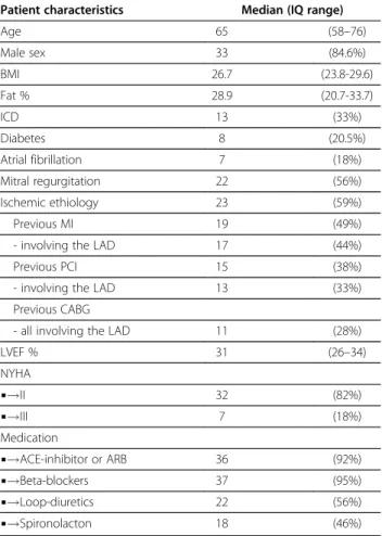
was weakened and no longer statistically significant. A Table 1 Baseline patient characteristics. Values are
median (IQ range) or number (%) as indicated
Patient characteristics Median (IQ range)
Age 65 (58–76)
Male sex 33 (84.6%)
BMI 26.7 (23.8-29.6)
Fat % 28.9 (20.7-33.7)
ICD 13 (33%)
Diabetes 8 (20.5%)
Atrial fibrillation 7 (18%)
Mitral regurgitation 22 (56%)
Ischemic ethiology 23 (59%)
Previous MI 19 (49%)
- involving the LAD 17 (44%)
Previous PCI 15 (38%)
- involving the LAD 13 (33%)
Previous CABG
- all involving the LAD 11 (28%)
LVEF % 31 (26–34)
NYHA
▪!II 32 (82%)
▪!III 7 (18%)
Medication
▪!ACE-inhibitor or ARB 36 (92%)
▪!Beta-blockers 37 (95%)
▪!Loop-diuretics 22 (56%)
▪!Spironolacton 18 (46%)
possible explanation is that increasing insulin resistance influences the cardiac microcirculation resulting in a reduced CFR, which leads to an impaired exercise cap-acity (Figure 1).
Our study did not show any relationship between CFR and peripheral endothelial function measured using flow mediated vasodilation. While a relationship has been shown between CFR and flow mediated vasodilation using brachial ultrasound in other patient populations [25], this is not well examined in heart failure patients. In one study of patients with dilated cardiomyopathy no correlation between CFR using echocardiography and flow mediated vasodilation was found [26]. Similarly, a study comparing patients with non-ischemic heart
failure to healthy controls found that flow mediated vasodilation and CFR measured using positron emission tomography were correlated in the healthy controls but not in the patients with heart failure [27]. Interestingly, in a small study of dogs with pacing induced heart fail-ure, CFR was reduced with progression of heart failfail-ure, but coronary endothelial function was preserved until a late stage of heart failure [28]. Thus our results are con-sistent with the literature in finding no association between CFR and endothelial function.
The present study shows a negative correlation between CFR and augmentation index, which is as measure of stiff-ness in the large arteries. Only one previous study has related CFR to augmentation index [29]. In this study of Table 2 Measurements for patients divided into groups with low and high CFR and insulin sensitivity by the median
Coronary flow reserve Insulin sensitivity
Low High Low High
CFR < 1.77 CFR > 1.77 Insulin sensitivity Insulin sensitivity
n = 19 n = 20 n = 19 n = 20
Exercise capacity
VO2peak (ml/min/kg FFM) 22.1 (19.1-25.5) 26.3 (21.1-29.7)* 22.1 (19.9-26.4) 25.5 (20.3-29.5)
RER 1.14 (1.07-1.19) 1.11 (1.03-1.19) 1.12 (1.07-1.19) 1.13 (1.06-1.16)
Vascular function
RHI 1.51 (1.34-1.91) 1.59 (1.37-1.92) 1.54 (1.40-1.67) 1.62 (1.33-2.36)
Augmentation Index 12.4 (−1.6-32.8) 11.0 (−5.7-16.6) 12.8 (−3.4-27.6) 8.1 (−6.3-21.2)
Glucose metabolism
Insulin sensitivity (mg/min/kg FFM) 5.8 (4.4-6.9) 7.2 (6.4-9.2)* 4.9 (3.5-6.3) 8.1 (7.1-9.2)}
Glucose clearence (ml/min/kg FFM) 5.1 (4.4-5.9) 6.6 (5.6-8.9) 4.7 (2.8-5.8) 7.2 (6.3-9.3)}
Microvascular function
CFV rest (cm/s) 29.7 (23.0-33.0) 22.7 (18.5-27.3)} 30.0 (22.0-33.0) 23.0 (19.0-25.3)*
CFV stress (cm/s) 35.0 (29.0-43.0) 63.3 (43.0-72.2)} 41.3 (32.3-63.6) 47.3 (37.0-65.7)
CFR 1.26 (1.04-1.55) 2.39 (2.09-3.09)} 1.41 (1.04-2.14) 2.10 (1.70-3.09)}
Echocardiography
LVEF (%) 29.5 (23–34) 31.5 (27–33) 31 (26–33) 31 (26–34)
EDV (ml) 183 (144–245) 153 (112–219) 183 (113–255) 172 (138–219)
ESV (ml) 132 (95–176) 104 (75–159) 127 (77–176) 122 (90–155)
LVMi (g/m2) 131 (110–148) 104 (82–159) 128 (92–148) 126 (89–152)
CFR: coronary flow reserve, VO2peak: peak oxygen uptake, RER: Respiratory exchange ratio, RHI: Reactive hyperaemia index, FFM: Fat free mass, LVEF: left ventricular ejection fraction, EDV: left ventricle end diastolic volume, ESV: left ventricle end systolic volume, LVMi: left ventricle mass index * p < 0.05,}p < 0.01 Values are expressed as median (IQ range).
Table 3 Correlations and p-values between different measurements
Age Augmentation index RHI Insulin sensitivity LVEF CFR
VO2peak −0.37 (0.02) −0.35 (0.04) −0.24 (0.16) 0.43 (0.007) 0.23 (0.15) 0.48 (0.002)
CFR −0.18 (0.27) −0.45 (0.007) −0.10 (0.56) 0.43 (0.008) 0.18 (0.26)
LVEF 0.05 (0.76) 0.12 (0.48) 0.08 (0.65) 0.14 (0.41)
Insulin sensitivity −0.14 (0.41) 0.12 (0.50) 0.13 (0.45)
RHI −0.03 (0.85) 0.44 (0.008)
Augmentation index 0.14 (0.44)
asymptomatic patients with risk factors for ischemic heart disease, the patients with higher augmentation index and pulse wave velocity had a lower CFR. In a normal elastic aorta the pulse wave reflects from the periphery and returns to the heart in diastole, which improves the dia-stolic filling of the coronary arteries. With aorta stiffening the pulse wave returns during systole resulting in increased afterload and myocardial oxygen demand which could mean, that a reduced CFR in heart failure patients without significant coronary artery stenosis might not only
be due to impaired coronary microcirculation, but that a reduced diastolic filling can be a participating factor. Insu-lin sensitivity and augmentation index were independently associated with CFR, suggesting that they influence CFR through different mechanisms. However, this finding remains to be confirmed in other studies.
Previous studies have shown that about 50% of asymp-tomatic patients with type 2 diabetes have signs of dia-stolic dysfunction [30] and that diadia-stolic dysfunction is associated with insulin resistance in patients with sus-pected coronary artery disease without diabetes [31] without systolic heart failure. In patients with systolic heart failure, patients with type 2 diabetes have signifi-cantly higher E/e’ than patients without diabetes [32]. Although we did not include measures of diastolic func-tion the present study, a possible mechanism between insulin sensitivity and diastolic dysfunction might be impaired coronary microvascular function.
A reduced CFR has been shown to be an independent predictor of poor outcome in patients with dilated cardio-myopathy [8] and in a mixed population of heart failure patients [33]. In the present study we find a relationship between CFR, VO2peak and insulin sensitivity, which also
Figure 3Scatter plots with linear regression lines between VO2peak and CFR (upper left), VO2peak and insulin sensitivity (upper right), insulin sensitivity and CFR (lower left) and augmentation index and FR (lower right).
Table 4 Univariate and multivariable linear regression with VO2peak as the dependent factor
Univariate beta p Multiple beta p
Age −0.23 0.02 −0.18 0.04
Insulin sensitivity (SC) 2.86 0.007 1.75 0.09
CFR (SC) 3.07 0.002 1.98 0.05
RHI (SC) −2.01 0.16 Ns
Augmentation index (SC) −2.21 0.04 Ns
LVEF 0.25 0.15 Ns
are well known prognostic risk factors in heart failure, suggesting that these risk factors are closely linked. Im-proving VO2peak and insulin sensitivity through exercise
training could also improve CFR as has been shown in a recent study of heart failure patients [14]. CFR has also been improved through other interventions such as weight loss in obese women [34] and after treatment with a beta-1 receptor blocker in dilated cardiomyopathy [35]. However, it has not yet been shown whether improve-ment in CFR through e.g. exercise training is followed by an improvement in prognosis for heart failure patients. An ongoing study will determine if CFR can improve after different modalities of exercise training in heart failure patients, and if any changes will correspond to changes in peak oxygen uptake, metabolic fitness and in augmenta-tion index.
Limitations: Although the study is limited in size this is somewhat counteracted by the precision of the mea-sures used: insulin sensitivity and glucose clearance were assessed by hyperinsulinemic isoglycemic clamp, the gold standard, and adjusted to fat free mass from DEXA
scan. VO2peak was a true maximum test as indicated by
the high RER values. The cross-sectional nature of the study impedes causal inference and any conclusions regarding potential effects of intervention must therefore remain speculative. The patients had all had a LAD without significant stenosis at a previous coronary angi-ography or coronary CT-scan, but the examination was not repeated at the time of the study, meaning that some of the patients potentially could have developed a signifi-cant LAD-stenosis. All of the patients did however per-form a maximal symptom limited cardiopulmonary exercise test at enrolment in to the study without chest pain or signs of ischemia on the ECG minimizing the chances of a significant LAD-stenosis.
Conclusion
Our study confirms that insulin resistance is associated with reduced exercise tolerance in heart failure patients and suggests that this is partly due to reduced coronary flow reserve. This is the first study to our knowledge that shows an association between CFR and exercise capacity in heart failure patients and links the relation-ship between insulin resistance and exercise capacity to CFR.
Abbreviations
AI: Augmentation index; BSA: Body surface area; CFR: Coronary flow reserve; CFV: Coronary flow velocity; DEXA: Dual X-ray absorptiometry; EDV: End diastolic volume; ESV: End systolic volume; FFM: Fat free mass;
ICD: Implantable cardioverter-defibrillator; IQ: Inter quartile; LAD: Left anterior descending artery; LVEF: Left ventricular ejection fraction; LVMi: Left Ventricular Mass index; NYHA: New York Heart Association; RER: Respiratory exchange ratio; RHI: Reactive hyperemia index; SC: Standardized coefficient; VO2peak: Peak Oxygen uptake.
Competing interests
The authors declare that they have no competing interests.
Authors’contribution
MS participated in the design of the study, collection of data, statistical analysis and drafting the paper. TM participated in the design of the study, collection of data and contributed to discussion. RO participated in analyzing the data and contributed to discussion. LP participated in analyzing the data and contributed to discussion. LS reviewed/edited the manuscript and contributed to discussion. HR reviewed/edited the manuscript and contributed to discussion. FD participated in the design of the study, reviewed/edited the manuscript and contributed to discussion. EP participated in the design of the study, statistical analysis reviewed/edited the manuscript and contributed to discussion. All authors read and approved the final manuscript.
Funding
This work was supported by The Danish Research Council.
Author details
1Dept Cardiology, Bispebjerg University Hospital, Copenhagen, Denmark. 2
Department of Clinical Physiology and Nuclear Medicine, Bispebjerg University Hospital, Copenhagen, Denmark.3Xlab Center for Healthy Aging, Department of Biomedical Science, Faculty of Health Sciences, University of Copenhagen, Copenhagen, Denmark.
Received: 7 August 2012 Accepted: 8 August 2012 Published: 13 August 2012
References
1. AlZadjali MA, Godfrey V, Khan F, Choy A, Doney AS, Wong AK, Petrie JR, Struthers AD, Lang CC:Insulin resistance is highly prevalent and is associated with reduced exercise tolerance in nondiabetic patients with heart failure.J Am Coll Cardiol2009,53:747–753.
2. Hamouda MS, Kassem HK, Salama M, El Masry M, Shaaban N, Sadek E, Khandheria BK, Seward JB, Elhendy A:Evaluation of coronary flow reserve in hypertensive patients by dipyridamole transesophageal Doppler echocardiography.Am J Cardiol2000,86:305–308.
3. Ahmari SAL, Bunch TJ, Modesto K, Stussy V, Dichak A, Seward JB, Pellikka PA, Chandrasekaran K:Impact of Individual and Cumulative Coronary Risk Factors on Coronary Flow Reserve Assessed by Dobutamine Stress Echocardiography.Am J Cardiol2008,101:1694–1699.
4. Kranidis A, Zamanis N, Mitrakou A, Patsilinakos S, Bouki T, Tountas N, Anthopoulos P, Raptis S, Anthopoulos L:Coronary microcirculation evaluation with transesophageal echocardiography Doppler in type II diabetics.Int J Cardiol1997,59:119–124.
5. Yokoyama I, Ohtake T, Momomura S, Yonekura K, Woo-Soo S, Nishikawa J, Sasaki Y, Omata M:Hyperglycemia rather than insulin resistance is related to reduced coronary flow reserve in NIDDM.Diabetes1998,47:119–124. 6. Sicari R, Rigo F, Cortigiani L, Gherardi S, Galderisi M, Picano E:Additive
prognostic value of coronary flow reserve in patients with chest pain syndrome and normal or near-normal coronary arteries.Am J Cardiol
2009,103:626–631.
7. Rigo F, Gherardi S, Galderisi M, Sicari R, Picano E:The independent prognostic value of contractile and coronary flow reserve determined by dipyridamole stress echocardiography in patients with idiopathic dilated cardiomyopathy.Am J Cardiol2007,99:1154–1158.
8. Rigo F, Gherardi S, Galderisi M, Pratali L, Cortigiani L, Sicari R, Picano E:The prognostic impact of coronary flow-reserve assessed by Doppler echocardiography in non-ischaemic dilated cardiomyopathy.Eur Heart J
2006,27:1319–1323.
9. Rigo F, Ciampi Q, Ossena G, Grolla E, Picano E, Sicari R:Prognostic value of left and right coronary flow reserve assessment in nonischemic dilated cardiomyopathy by transthoracic Doppler echocardiography.J Card Fail
2011,17:39–46.
10. Kiviniemi TO, Snapir A, Saraste M, Toikka JO, Raitakari OT, Ahotupa M, Hartiala JJ, Scheinin M, Koskenvuo JW:Determinants of coronary flow velocity reserve in healthy young men.Am J Physiol Heart Circ Physiol
11. Hägg U, Wandt B, Bergström G, Volkmann R:Gan Lm: Physical exercise capacity is associated with coronary and peripheral vascular function in healthy young adults.Am J Physiol Heart Circ Physiol2005,289:H1627–H1634. 12. Myers J, Gullestad L, Vagelos R, Do D, Bellin D, Ross H, Fowler MB:Clinical,
hemodynamic, and cardiopulmonary exercise test determinants of survival in patients referred for evaluation of heart failure.Ann Intern Med
1998,129:286–293.
13. Piña IL, Apstein CS, Balady GJ, Belardinelli R, Chaitman BR, Duscha BD, Fletcher BJ, Fleg JL, Myers JN, Sullivan MJ:Exercise and Heart Failure. Circulation2003,107:1210–1225.
14. Santos JM, Kowatsch I, Tsutsui JM, Negrao CE, Canavesi N, Carvalho FC, Mady C, Ramires JA, Mathias W Jr:Effects of exercise training on myocardial blood flow reserve in patients with heart failure and left ventricular systolic dysfunction.Am J Cardiol2010,105:243–248. 15. Davies EJ, Moxham T, Rees K, Singh S, Coats AJ, Ebrahim S, Lough F, Taylor
RS:Exercise training for systolic heart failure: Cochrane systematic review and meta-analysis.Eur J Heart Fail2010,12:706–715.
16. Stoylen A, Conraads V, Halle M, Linke A, Prescott E, Ellingsen O:Controlled study of myocardial recovery after interval training in heart failure: SMARTEX-HF - rationale and design.Eur J Prev Cardiol2012,19:813–821. 17. Dimitrow PP, Galderisi M, Rigo F:The non-invasive documentation of
coronary microcirculation impairment: role of transthoracic echocardiography.Cardiovasc Ultrasound2005,3:18.
18. Caiati C, Montaldo C, Zedda N, Montisci R, Ruscazio M, Lai G, Cadeddu M, Meloni L, Iliceto S:Validation of a new noninvasive method (contrast-enhanced transthoracic second harmonic echo Doppler) for the evaluation of coronary flow reserve: comparison with intracoronary Doppler flow wire.J Am Coll Cardiol1999,34:1193–1200.
19. Janner JH, Godtfredsen NS, Ladelund S, Vestbo J, Prescott E:Aortic augmentation index: reference values in a large unselected population by means of the SphygmoCor device.Am J Hypertens2010,23:180–185. 20. Eroglu S, Sade LE, Bozbas H, Haberal A, Ozbicer S, Demir O, Muderrisoglu H:
Association of serum adiponectin levels and coronary flow reserve in women with normal coronary angiography.J Cardiovasc Risk2009,
16:290–296.
21. Kondo I, Mizushige K, Hirao K, Nozaki S, Tsuji T, Masugata H, Kohno M, Matsuo H:Ultrasonographic assessment of coronary flow reserve and abdominal fat in obesity.Ultrasound Med Biol2001,27:1199–1205. 22. Gardin JM, Leifer ES, Fleg JL, Whellan D, Kokkinos P, Leblanc MH, Wolfel E,
Kitzman DW:Relationship of Doppler-Echocardiographic left ventricular diastolic function to exercise performance in systolic heart failure: the HF-ACTION study.Am Heart J2009,158:S45–S52.
23. Ingelsson E, Sundstrom J, Arnlov J, Zethelius B, Lind L:Insulin resistance and risk of congestive heart failure.JAMA2005,294:334–341.
24. Suskin N, McKelvie RS, Burns RJ, Latini R, Pericak D, Probstfield J, Rouleau JL, Sigouin C, Solymoss CB, Tsuyuki R,et al:Glucose and insulin abnormalities relate to functional capacity in patients with congestive heart failure.Eur Heart J2000,21:1368–1375.
25. Campuzano R, Moya JL, Garcia-Lledo A, Tomas JP, Ruiz S, Megias A, Balaguer J, Asin E:Endothelial dysfunction, intima-media thickness and coronary reserve in relation to risk factors and Framingham score in patients without clinical atherosclerosis.J Hypertens2006,24:1581–1588. 26. Dini FL, Ghiadoni L, Conti U, Stea F, Buralli S, Taddei S, De Tommasi SM:
Coronary flow reserve in idiopathic dilated cardiomyopathy: relation with left ventricular wall stress, natriuretic peptides, and endothelial dysfunction.J Am Soc Echocardiogr2009,22:354–360.
27. Stolen KQ, Kemppainen J, Kalliokoski KK, Karanko H, Toikka J, Janatuinen T, Raitakari OT, Airaksinen KEJ, Nuutila P, Knuuti J:Myocardial perfusion reserve and peripheral endothelial function in patients with idiopathic dilated cardiomyopathy.Am J Cardiol2004,93:64–68.
28. Saito T, Maehara K, Tamagawa K, Oikawa Y, Niitsuma T, Saitoh S, Maruyama Y:Alterations of endothelium-dependent and -independent regulation of coronary blood flow during heart failure.Am J Physiol Heart Circ Physiol
2002,282:H80–H86.
29. Saito M, Okayama H, Nishimura K, Ogimoto A, Ohtsuka T, Inoue K, Hiasa G, Sumimoto T, Higaki J:Possible link between large artery stiffness and coronary flow velocity reserve.Heart2008,94:e20.
30. Magnusson M, Jovinge S, Shahgaldi K, Israelsson B, Groop L, Melander O:
Brain natriuretic peptide is related to diastolic dysfunction whereas urinary albumin excretion rate is related to left ventricular mass in asymptomatic type 2 diabetes patients.Cardiovasc Diabetol2010,9:2.
31. Dinh W, Lankisch M, Nickl W, Scheyer D, Scheffold T, Kramer F, Krahn T, Klein RM, Barroso MC, Futh R:Insulin resistance and glycemic abnormalities are associated with deterioration of left ventricular diastolic function: a cross-sectional study.Cardiovasc Diabetol2010,9:63. 32. Willemsen S, Hartog JWL, Hummel YM, van Ruijven MHI, van der Horst ICC,
van Veldhuisen DJ, Voors AA:Tissue advanced glycation end products are associated with diastolic function and aerobic exercise capacity in diabetic heart failure patients.Eur J Heart Fail2011,13:76–82. 33. Anantharam B, Janardhanan R, Hayat S, Hickman M, Chahal N, Bassett P,
Senior R:Coronary flow reserve assessed by myocardial contrast echocardiography predicts mortality in patients with heart failure.Eur J Echocardiogr2011,12:69–75.
34. Coppola A, Marfella R, Coppola L, Tagliamonte E, Fontana D, Liguori E, Cirillo T, Cafiero M, Natale S, Astarita C:Effect of weight loss on coronary circulation and adiponectin levels in obese women.Int J Cardiol2009,
134:414–416.
35. Erdogan D, Gullu H, Caliskan M, Ciftci O, Baycan S, Yildirir A, Muderrisoglu H:
Nebivolol improves coronary flow reserve in patients with idiopathic dilated cardiomyopathy.Heart2007,93:319–324.
doi:10.1186/1475-2840-11-97
Cite this article as:Snoeret al.:Insulin resistance and exercise tolerance in heart failure patients: linkage to coronary flow reserve and peripheral vascular function.Cardiovascular Diabetology201211:97.
Submit your next manuscript to BioMed Central and take full advantage of:
• Convenient online submission • Thorough peer review
• No space constraints or color figure charges
• Immediate publication on acceptance
• Inclusion in PubMed, CAS, Scopus and Google Scholar
• Research which is freely available for redistribution