CELLULAR & MOLECULAR BIOLOGY LETTERS http://www.cmbl.org.pl
Received: 09 July 2008 Volume 14 (2009) pp 57-69 Final form accepted: 16 September 2008 DOI: 10.2478/s11658-008-0035-4 Published online: 06 October 2008 © 2008 by the University of Wrocław, Poland
# The content of this Mini review was first presented in a shortened form at the 12th Mejbaum-Katzenellenbogen Seminar “Membrane Skeleton. Recent Advances and Future Research Directions”, June 15-18, 2008, Zakopane, Poland
* Author for correspondence; e-mail: [email protected], tel.: (734) 647 2720, fax: (734) 763 1166
Abbreviations used: AIS – axon initial segments; CAM – cell adhesion molecule; EGFR – epidermal growth factor receptor; FGFR – fibroblast growth factor receptor; GF – giant fiber; GPI – glycosyl phosphoinositol; NMJ – neuromuscular junction; RTK – receptor tyrosine kinase; SAP – synapse-associated proteins; TTMn - tergotrochanteral motorneuron; VUM – ventral unpaired median
Mini review
THE INTERACTION BETWEEN L1-TYPE PROTEINS AND
ANKYRINS - A MASTER SWITCH FOR L1-TYPE CAM FUNCTION #
MICHAEL HORTSCH1*, KAKANAHALLI NAGARAJ1,2
and TANJA A. GODENSCHWEGE3
1Department of Cell and Developmental Biology, University of Michigan, Ann
Arbor, MI 48109, USA, 2present address: Department of Applied Zoology,
Kuvempu University, Shankaraghatta, Shimoga, India, 3Florida Atlantic
University, Department of Biological Sciences, Boca Raton, FL 33431, USA
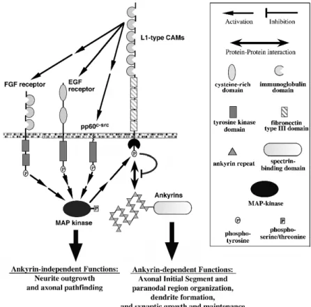
Abstract: L1-type cell adhesion molecules (CAMs) are important mediators of
neural differentiation, including axonal outgrowth and pathfinding and also of synapse formation and maintenance. In addition, their interactions with cytoskeletal components are highly conserved and regulated. How these different aspects of CAM functionality relate to each other is not well understood. Based on results from our and other laboratories we propose that ankyrin-binding to L1-type CAMs provides a master switch. The interaction with ankyrins directs L1-type adhesive proteins into different functional contexts, either ankyrin-independent functions, such as neurite outgrowth and axonal pathfinding or into ankyrin-dependent functions, such as L1’s role at axon initial segments (AIS), paranodal regions, synapses and in dendrites.
Key words: Cell adhesion, Ankyrins, Membrane skeleton, Tyrosine
INTRODUCTION
One of the major connections in non-erythroid cells between the actin-spectrin membrane skeleton and the plasma membrane is provided by the interaction between ankyrin linker proteins and the cytoplasmic domains of L1-type CAMs. L1-type proteins form a functionally and structurally conserved family of type 1 transmembrane proteins, which have been identified in the genomes of a wide range of species, from nematodes to humans [1]. Whereas invertebrate species usually contain only one L1-type gene, such as the Drosophilaneuroglian gene, most vertebrate genomes encode four L1-type proteins, named L1-CAM, CHL1, neurofascin and NrCAM. They share a common extracellular protein domain structure, typically consisting of six aminoterminal immunoglobulin domains that are followed by 5 fibronectin type III domains (Fig. 1). The size of their cytoplasmic domains varies from 85 to 148 amino acid residues and contains several highly conserved protein sequence motifs. L1-type proteins are highly expressed in neuronal and glial cells. However, they also have important functions in other tissues, e.g., for epithelial stability in Drosophila by
serving as essential components of septate junctions [2]. L1-type proteins normally perform these functions using their homophilic adhesive abilities. In addition, they also interact with a plethora of extracellular heterophilic binding partners, including other CAMs, Integrins, proteoglycans and RTKs.
It has been known for a considerable time that neuronally-expressed L1-type proteins promote neurite outgrowth and axonal pathfinding during nervous system development [3]. This important activity is in part mediated by the adhesion-dependent activation of neuronally-expressed RTKs, especially FGFR and EGFR. A relatively newly-identified functional role for L1-type CAMs is their involvement in the formation and maintenance of synaptic contacts in arthropod and chordate nervous systems [4-6]. They appear to be essential for the presynaptic organization of specific synapses in Drosophila, chicken and mice. L1-type proteins are also known for their ability to bind to members of the ankyrin family, which serve as cytoskeletal linker proteins. Almost all L1-type family members contain a highly conserved ankyrin-binding motif in their cytoplasmic domain [7] and this interaction is regulated at several levels.
In this mini-review, we will discuss two major aspects of L1-type function in the nervous system, neurite outgrowth and axonal pathfinding, as well as synaptogenesis and synapse maintenance. Based on the available published data, we will propose a hypothesis, how several functional aspects of L1-type proteins may be differentially regulated by the binding of ankyrin to the L1 cytoplasmic domain. As this interaction itself is subject to highly conserved regulatory processes, it may serve as a master switch for directing L1-type CAMs into different functional tasks.
THE REGULATON OF ANKYRIN-BINDING TO L1-TYPE CAMS
In 1993 the Bennett laboratory published the first report that a member of the L1 family has a specific binding activity for ankyrin proteins [8]. Using an ankyrin affinity chromatography approach they isolated rat neurofascin as an ankyrin-binding protein from a total protein homogenate of a rat brain. This result was extended by the demonstration that other vertebrate members of the L1-type family also have an ankyrin-binding activity [9] and that the Drosophila
member of the L1-type family, neuroglian, interacts with the Drosophila
ankyrin1 protein [10]. By expressing neuroglian in Drosophila S2 cells, Dubreuil et al. showed that in response to neuroglian protein that is engaged in a homophilic interaction ankyrin1 protein is selectively recruited to cell contact sites [10]. Similarly, human L1-CAM protein is also able to recruit Drosophila
ankyrin1 protein to cell contact sites and can bind Drosophila ankyrin1 in response to cis-interactions with homophilic human TAG-1 adhesion complexes [11]. This adhesion-dependent interaction between neuroglian/L1-CAM and
Drosophila ankyrin1 was subsequently mapped to a 36 amino acid
carboxyterminal region of the neuroglian167 cytoplasmic domain [12]. The amino
L1-type gene family, suggesting that all known L1-type family members are probably able to interact with ankyrin proteins (Fig. 2). It includes two tyrosine residues that are found in the sequences of almost all L1-type proteins (a LADY and an FIGQY motif). The second conserved tyrosine residue is a phospho-rylation target of a still unidentified tyrosine kinase [13]. Because fusion proteins containing all 24 aminoterminal Drosophila ankyrin1 and ankyrin2 repeats interact with the cytoplasmic neuroglian domain in a yeast two-hybrid assay, the dephosphorylated state appears to be the biological active form to bind ankyrins [14]. In contrast, phosphorylation of the FIGQY motif results in the complete inhibition of ankyrin-binding [15]. In its phosphorylated state, the FIGQY motif becomes the binding partner for the microtubule-associated
Fig. 2. Core sequences of the ankyrin-binding regions in the cytoplasmic domains of several L1-type CAMs. The P symbol indicates the conserved tyrosine residue that can be optionally phosphorylated and when modified abolishes ankyrin-binding.
doublecortin protein instead [16]. Antibodies that are specific for the phosphorylated form of the FIGQY peptide specifically recognize a subset of neuroglian protein at epidermal muscle attachment sites, developing NMJs and septate junctions in epithelia of Drosophila embryos and L1-type proteins in vertebrates at paranodal regions, NMJs and areas of neuronal migration and axon extension [17]. Several publications report that EGFR, FGFR and MAP kinase activation indirectly result in the phosphorylation of the FIGQY motif, placing the responsible tyrosine kinase downstream of the MAP kinase signaling pathway [13, 18-20]. A mutated form of the Drosophila neuroglian protein, in which the FIGQY motif had been changed to FIGQF and thereby preventing its phosphorylation, mediates a significantly lower level of homophilic adhesion, when expressed in S2 cells [12]. In a yeast two-hybrid assay the same mutant protein also exhibits an approximately 50% reduction in its ability to bind
Drosophila ankyrins, indicating that this conserved cytoplasmic tyrosine residue
regulated by at least two mechanisms, first by the adhesive activity of the L1-type protein and also by the phosphorylation state of the FIGQY motif.
THE ANKYRIN2 PROTEIN, A BINDING PARTNER OF THE
NEUROGLIAN CYTOPLASMIC DOMAIN IN Drosophila NEURONS
Genes encoding the principal components of the membrane skeleton, like actin, spectrins, band 4.1 and ankyrins, can be found throughout the metazoan kingdom [21]. Although initially identified and characterized in erythrocytes, these genes significantly predate the evolutionary appearance of red and white blood cells in vertebrates. Vertebrate genomes usually encode 3 paralogous ankyrin genes/proteins, designated ankyrin1, 2 and 3 or ankyrinR/B and G [22]. In contrast, the nematode C. elegans genome contains only a single gene
encoding an ankyrin-type protein (unc-44) [23]. An independent gene
duplication event in the arthropod lineage has resulted in two genes encoding ankyrin-type proteins in the fruit fly Drosophila melanogaster, which are identified as Drosophilaankyrin1 and 2 [14, 24]. The Drosophila ankyrin1 gene encodes only a single canonical ankyrin protein, containing 24 ankyrin protein repeats and a single spectrin binding domain [24]. In contrast, the Drosophila
ankyrin2 gene gives rise to a multitude of small, medium, large and extra large
protein products, ranging from 1159 to 11640 amino acid residues [25, 26]. Also the expression pattern of the two Drosophila ankyrin loci differs dramatically.
The ankyrin1 gene appears to be ubiquitously expressed. In contrast, expression
of the Drosophila ankyrin2 gene is completely restricted to neuronal cells throughout the life cycle of the fly [14, 26]. As neuroglian is co-expressed with ankyrin2 in most neurons, it probably constitutes the major membrane-associated binding partner for the different ankyrin2 isoforms. However, neuroglian is not expressed by all neurons and therefore other ankyrin2-binding membrane proteins must exist, e.g., in ventral unpaired median (VUM) neurons along the midline of the embryonic ventral cord [14].
All Drosophila neurons appear to express multiple different ankyrin2 isoforms,
which are specifically localized to different neuronal subcellular domains. The smallest Drosophila ankyrin2 isoform remains restricted to neuronal cell bodies, whereas the longer protein isoforms are localized to dendrites, axons and synaptic contacts [25-27]. A differential subcellular localization of ankyrin protein isoforms has also been reported for other invertebrate and some vertebrate ankyrins [28, 29]. With the exception of synaptically-localized ankyrins, it remains currently unknown whether these different ankyrin protein forms are involved in specific functions at their respective locations.
There is only limited genetic information about the biological role of ankyrins in
the Drosophila nervous system. The cellular localization of ankyrin proteins is
neuroglian expression affects the stability of ankyrin2 protein in Drosophila
neurons. When comparing neuroglian null mutants with wild type embryos, immunostaining for ankyrin2 protein is significantly lower in mutant cells [14]. Although the nervous system of fruit flies with mutations affecting or abolishing the longer ankyrin2 protein isoforms is somewhat less organized and causes a lethal phenotype, overall axonal growth and pathfinding appears to be normal [25-27]. This indicates that neuroglian’s interaction with ankyrin proteins is not essential for this L1-type function.
NEURITE OUTGROWTH AND AXONAL PATHFINDING, ANKYRIN-INDEPENDENT FUNCTIONS OF L1-TYPE CAMS
The ability of L1-type proteins to induce neurite outgrowth has been extensively studied in vitro [3]. In addition, the in vivo analysis of mutant L1-type proteins
in Drosophila, mice and humans uncovered a wide range of neurological
dysfunctions, including underdeveloped axonal tracts, axon guidance defects, mental retardation, spastic paraplegia and others. All these studies point to an important role of L1-type proteins during the extension and pathfinding of many axons during nervous system development. These effects are mediated by neuronally-expressed RTKs and/or non-receptor tyrosine kinases. The L1-dependent activation of these signaling molecules induces intracellular signaling pathways, specifically MAP kinase and/or phospholipase C activities, which ultimately result in the localized release of calcium from intracellular stores and the activation of a calmodulin-dependent signaling cascade.
Co-transfection experiments in Drosophila S2 cells demonstrated that
homophilic, L1-CAM-mediated cell adhesion induces human EGFR tyrosine kinase activity [30]. These in vitro findings have been confirmed in vivo using a genetic approach in Drosophila [31, 32]. The overexpression of L1-type CAMs induces a gain-of-function phenotype in the peripheral nervous system of
Drosophila [30]. Sensory neurons in developing wings respond with robust
This conclusion is also supported by in vitro studies using vertebrate primary neuronal and ND-7 neuroblastoma cells [19, 33]. Although ankyrinB binding to the L1-CAM cytoplasmic domain appears to be important for neurite initiation, it is not involved in coupling L1-CAM to actin flow in growth cones or in L1-induced neurite elongation [33]. Rather ankyrin binding results in a stationary behavior of L1-CAM molecules in the neuronal plasma membrane and interferes with the actin-dependent retrograde movement of L1-CAM [19]. In addition, inhibiting the interaction between ankyrins and L1-CAM promotes axonal growth of mouse cerebellar granule neurons in culture.
NEUROGLIAN’S ROLE IN Drosophila SYNAPTOGENESIS:
ANKYRIN-BINDING REQUIRED
First reports that L1-type proteins are involved in the formation and maintenance of synaptic contacts were published fairly recently. Ango et al. demonstrated that the vertebrate L1-type CAM, neurofascin, is involved in directing GAGAergic innervation to AIS of cerebellar Purkinje cells [5]. The ability of basket interneurons to target and establish these “pinceau synapses” depends on the interaction between the 186 kDa isoform of neurofascin with ankyrinG. A role for L1-CAM at synapses of the nicotinic pathway in chick ciliary ganglia was demonstrated by Triana-Baltzer et al. [6]. In this system L1-CAM is needed pre-and postsynaptically for the positioning of presynaptic structures and for facilitating nicotinic synaptic signal transduction. Two independent publications reported an interaction of NrCAM with the synapse-associated proteins SAP90/PSD95 and SAP97, as well as SAP102, which colocalizes with NrCAM in cerebellar granule cells [34, 35]. However, the potential functional significance of these interactions for synaptogenesis remains unknown.
In Drosophila, neuroglian’s synaptic function was uncovered by a point mutation
and neuroglianY1234F results in a disruptive phenotype, which affects the GF synapse function. As the nrg849 mutation also reduces the level of neuroglian Y1234 tyrosine phosphorylation, these results only demonstrate that the FIGQY tyrosine residue is important for neuroglian’s synaptic function. However, they do not answer the question, whether this function involves the binding of ankyrin to the neuroglian cytoplasmic domain or not.
Evidence for the importance of the membrane skeleton during synapse maintenance was recently provided by two publications demonstrating the requirement for synaptic ankyrin2 protein at Drosophila NMJs [25, 27]. Both publications report that several point mutations and a transposon insertion in the
ankyrin2 gene, which only affect the larger ankyrin2 protein isoforms,
destabilize NMJs in Drosophila larvae. Two additional larger ankyrin2 isoforms were identified, which are localized to the presynaptic membrane. One of these isoforms contains a carboxyterminal microtubule-binding activity. Accordingly, in mutant larvae the presynaptic microtubular network is disassembled, resulting in the disruption of general presynaptic organization and synaptic activity. However, it is currently unknown, whether neuroglian participates in the stabilization and maintenance of Drosophila NMJs. Nevertheless, it appears likely that neuroglian provides the plasma membrane target for ankyrin2 binding at the NMJ. Therefore, the published data suggest an important role for neuroglian in the organization of microtubules at the presynaptic terminus, as well as for the stability of a number of different Drosophila synapses in general. This function may also extend to the postsynaptic side, as the non-neuronal neuroglian167 isoform is expressed by larval muscles [36] and because ankyrin1
has been identified as a component of the postsynaptic spectrin-based membrane skeleton at NMJs [37].
This newly discovered function of the lone Drosophila member of the L1-type family is not the only neuroglian function that involves and requires an interaction with one of the two Drosophila ankyrins. Yamamoto et al. recently identified a role for neuroglian and ankyrin2 during dendritic branch formation in embryonic peripheral sensory neurons [38]. Neuronally-expressed neuroglian interacts with peripheral glial cells and thereby prevents abnormal axonal sprouting. This function also requires the expression of Drosophila ankyrin2 in these neuronal cells, thus providing a second function for neuroglian in the
Drosophila nervous system, which depends on its interaction with ankyrins.
Several publications using vertebrate model systems suggest that L1-type proteins have additional ankyrin-dependent functions. A co-localization of L1-type proteins (specifically L1-CAM, neurofascin and NrCAM) with ankyrinB and ankyrinG has been reported in myelinated nerves at AIS and at paranodal regions of nodes of Ranvier [39-41]. As axons don’t become properly myelinated in invertebrate species, these structures can’t be studied in the
Drosophila nervous system. In vertebrate neurons, AIS have a barrier function
neurofascin accumulate together with ankyrinG at AIS, where they exhibit slow diffusion and a low detergent extractability [43]. This suggests that these L1-type CAMs interact with ankyrinG and the membrane skeleton at AIS. In contrast, membrane mobility and detergent extractability for both L1-CAM and neurofascin are considerably higher in distal axon regions of cultured hippocampal neurons [43]. This does not exclude that L1-ankyrin interactions have some additional roles in axons, as Bennett and Lambert reported that L1-CAM and ankyrinB both mediate axon fasciculation and stabilize axon bundles in premyelinated axons of the optic nerve [39]. Otsuka et al. reported that mutations in the C. elegans ankyrin gene unc-44 induce an axonal guidance phenotype [23]. However, as L1-type proteins are not the only membrane-associated ankyrin binding partners, it is currently unknown, whether this axonal pathfinding phenotype is mediate by an interaction with the nematode L1-type protein LAD-1 or by a different unc-44 ankyrin ligand. All these reports indicate that L1-type proteins have other ankyrin-dependent functions than synapse formation and maintenance. However, more research is needed to determine how the interactions between L1-type proteins and ankyrins influence these L1 functions and how they are regulated, especially at AIS and at the paranodal regions of nodes of Ranvier.
ANKYRIN-DEPENDENT AND -INDEPENDENT FUNCTIONS – A MODEL FOR THE REGULATION OF L1-TYPE PROTEINS
the formation of the dendritic tree. The binding of other proteins, for example doublecortin [16], [44], shootin1 [45], synapse-associated proteins SAP102 [34] and SAP90/PSD95 and SAP97 [35] and the AP-2 adapter [46], to L1-type cytoplasmic domains indicates that other switches for regulating L1-type function might exist. So far, the interaction between ankyrins and L1-type cytoplasmic domains provides the most convincing example for connecting a link with a specific family of cytoskeletal proteins to profound changes in the functional engagement of L1-type CAMs.
Acknowledgements. T. A. G. is supported by RO1 HD050725-01A1 and M. H.
appreciates the support by a GLOBAL Reach Research, Education and Collaboration in Health faculty grant from the University of Michigan.
REFERENCES
1. Hortsch, M. Structural and functional evolution of the L1-family: Are four adhesion molecules better than one? Mol. Cell. Neurosci. 15 (2000) 1-10. 2. Hortsch, M. and Margolis, B. Septate and paranodal junctions: Kissing
cousins. Trends Cell. Biol. 13 (2003) 557-561.
3. Panicker, A.K., Buhusi, M., Thelen, K. and Maness, P.F. Cellular signalling mechanisms of neural cell adhesion molecules. Front. Biosci. 8 (2003) D900-911.
4. Godenschwege, T.A., Kristiansen, L.V., Uthaman, S.B., Hortsch, M. and Murphey, R.K. A conserved role for Drosophila Neuroglian and human L1-CAM in central-synapse formation. Curr. Biol. 16 (2006) 12-23.
5. Ango, F., di Cristo, G., Higashiyama, H., Bennett, V., Wu, P. and Huang, Z.J. Ankyrin-based subcellular gradient of neurofascin, an immunoglobulin family protein, directs GABAergic innervation at purkinje axon initial segment. Cell 119 (2004) 257-272.
6. Triana-Baltzer, G.B., Liu, Z. and Berg, D.K. Pre- and postsynaptic actions of L1-CAM in nicotinic pathways. Mol. Cell. Neurosci. 33 (2006) 214-226. 7. Hortsch, M. The L1 family of neural cell adhesion molecules: Old proteins
performing new tricks. Neuron 17 (1996) 587-593.
8. Davis, J.Q., McLaughlin, T. and Bennett, V. Ankyrin-binding proteins related to nervous system cell adhesion molecules: candidates to provide transmembrane and intercellular connections in adult brain. J. Cell. Biol.
121 (1993) 121-133.
9. Davis, J.Q. and Bennett, V. Ankyrin binding activity shared by the neurofascin/L1/NrCAM family of nervous system cell adhesion molecules.
J. Biol. Chem. 269 (1994) 27163-27166.
11. Malhotra, J.D., Tsiotra, P., Karagogeos, D. and Hortsch, M. Cis-activation of L1-mediated ankyrin recruitment by TAG-1 homophilic cell adhesion.
J. Biol. Chem. 273 (1998) 33354-33359.
12. Hortsch, M., Homer, D., Malhotra, J.D., Chang, S., Frankel, J., Jefford, G. and Dubreuil, R.R. Structural requirements for "outside-in" and "inside-out" signaling by Drosophila neuroglian, a member of the L1 family of cell adhesion molecules. J. Cell. Biol. 142 (1998) 251-261.
13. Garver, T.D., Ren, Q., Tuvia, S. and Bennett, V. Tyrosine phosphorylation at a site highly conserved in the L1 family of cell adhesion molecules abolishes ankyrin binding and increases lateral mobility of neurofascin.
J. Cell. Biol. 137 (1997) 703-714.
14. Bouley, M., Tian, M.-Z., Paisley, K., Shen, Y.-C., Malhotra, J.D. and Hortsch, M. The L1-type cell adhesion molecule neuroglian influences the stability of neural ankyrin in the Drosophila embryo but not its axonal localization. J. Neurosci. 20 (2000) 4515-4523.
15. Tuvia, S., Garver, T.D. and Bennett, V. The phosphorylation state of the FIGQY tyrosine of neurofascin determines ankyrin-binding activity and patterns of cell segregation. Proc. Natl. Acad. Sci. USA 94 (1997) 12957-12962.
16. Kizhatil, K., Wu, Y.X., Sen, A. and Bennett, V. A new activity of doublecortin in recognition of the phospho-FIGQY tyrosine in the cytoplasmic domain of neurofascin. J. Neurosci. 22 (2002) 7948-7958. 17. Jenkins, S.M., Kizhatil, K., Kramarcy, N.R., Sen, A., Sealock, R. and
Bennett, V. FIGQY phosphorylation defines discrete populations of L1 cell adhesion molecules at sites of cell-cell contact and in migrating neurons.
J. Cell Sci. 114 (2001) 3823-3835.
18. Whittard, J.D., Sakurai, T., Cassella, M.R., Gazdoiu, M. and Felsenfeld, D.P. MAP kinase pathway-dependent phosphorylation of the L1-CAM ankyrin-binding site regulates neuronal growth. Mol. Biol. Cell (2006). 19. Gil, O.D., Sakurai, T., Bradley, A.E., Fink, M.Y., Cassella, M.R., Kuo, J.A.
and Felsenfeld, D.P. Ankyrin binding mediates L1CAM interactions with static components of the cytoskeleton and inhibits retrograde movement of L1CAM on the cell surface. J. Cell. Biol. 162 (2003) 719-730.
20. Chen, L., Ong, B. and Bennett, V. LAD-1, the Caenorhabditis elegans
L1CAM homologue, participates in embryonic and gonadal morphogenesis and is a substrate for fibroblast growth factor receptor pathway-dependent phosphotyrosine-based signaling. J. Cell. Biol. 154 (2001) 841-856.
21. Bennett, V. and Baines, A.J. Spectrin and ankyrin-based pathways: metazoan inventions for integrating cells into tissues. Physiol. Rev. 81 (2001) 1353-1392.
22. Mohler, P.J., Gramolini, A.O. and Bennett, V. Ankyrins. J. Cell Sci. 115 (2002) 1565-1566.
and Sobery, A. An ankyrin-related gene (unc-44) is necessary for proper axonal guidance in Caenorhabditis elegans. J. Cell. Biol. 129 (1995) 1081-1092.
24. Dubreuil, R.R. and Yu, J.-Q. Ankyrin and beta-spectrin accumulate independently of alpha-spectrin in Drosophila. Proc. Natl. Acad. Sci. USA
91 (1994) 10285-10289.
25. Koch, I., Schwarz, H., Beuchle, D., Goellner, B., Langegger, M. and Aberle, H.
Drosophila ankyrin 2 is required for synaptic stability. Neuron 58 (2008)
210-222.
26. Hortsch, M., Paisley, K.L., Tian, M.Z., Qian, M., Bouley, M. and Chandler, R. The axonal localization of large Drosophila ankyrin2 protein isoforms is essential for neuronal functionality. Mol. Cell. Neurosci. 20 (2002) 43-55. 27. Pielage, J., Cheng, L., Fetter, R.D., Carlton, P.M., Sedat, J.W. and Davis,
G.W. A presynaptic giant ankyrin stabilizes the NMJ through regulation of presynaptic microtubules and transsynaptic cell adhesion. Neuron 58 (2008) 195-209.
28. Otsuka, A.J., Boontrakulpoontawee, P., Rebeiz, N., Domanus, M., Otsuka, D., Velamparampil, N., Chan, S., Vande Wyngaerde, M., Campagna, S. and Cox, A. Novel UNC-44 AO13 ankyrin is required for axonal guidance in C.
elegans, contains six highly repetitive STEP blocks separated by seven
potential transmembrane domains, and is localized to neuronal processes and the periphery of neural cell bodies. J. Neurobiol. 50 (2002) 333-349.
29. Kordeli, E., Davis, J., Trapp, B. and Bennett, V. An isoform of ankyrin is localized at nodes of Ranvier in myelinated axons of central and peripheral nerves. J. Cell. Biol. 110 (1990) 1341-1352.
30. Islam, R., Kristansen, L.V., Romani, S., Garcia-Alonso, L. and Hortsch, M. Activation of EGF receptor kinase by L1-mediated homophilic cell interactions. Mol. Biol. Cell 15 (2004) 1509-1518.
31. Garcia-Alonso, L., Romani, S. and Jimenez, F. The EGF and FGF receptors mediate neuroglian function to control growth cone decisions during sensory axon guidance in Drosophila. Neuron 28 (2000) 741-752.
32. Kristiansen, L.V., Velasquez, E., Romani, S., Baars, S., Berezin, V., Bock, E., Hortsch, M. and Garcia-Alonso, L. Genetic analysis of an overlapping functional requirement for L1- and NCAM-type proteins during sensory axon guidance in Drosophila. Mol. Cell. Neurosci. 28 (2005) 141-152. 33. Nishimura, K., Yoshihara, F., Tojima, T., Ooashi, N., Yoon, W., Mikoshiba,
K., Bennett, V. and Kamiguchi, H. L1-dependent neuritogenesis involves ankyrinB that mediates L1-CAM coupling with retrograde actin flow.
J. Cell. Biol. 163 (2003) 1077-1088.
35. Dirks, P., Thomas, U. and Montag, D. The cytoplasmic domain of NrCAM binds to PDZ domains of synapse-associated proteins SAP90/PSD95 and SAP97. Eur. J. Neurosci. 24 (2006) 25-31.
36. Bieber, A.J., Snow, P.M., Hortsch, M., Patel, N.H., Jacobs, J.R., Traquina, Z.R., Schilling, J. and Goodman, C.S. Drosophila neuroglian: a member of the immunoglobulin superfamily with extensive homology to the vertebrate neural adhesion molecule L1. Cell 59 (1989) 447-460.
37. Pielage, J., Fetter, R.D. and Davis, G.W. A postsynaptic spectrin scaffold defines active zone size, spacing, and efficacy at the Drosophila
neuromuscular junction. J. Cell. Biol. 175 (2006) 491-503.
38. Yamamoto, M., Ueda, R., Takahashi, K., Saigo, K. and Uemura, T. Control of axonal sprouting and dendrite branching by the nrg-ank complex at the neuron-glia interface. Curr. Biol. 16 (2006) 1678-1683.
39. Bennett, V. and Lambert, S. Physiological roles of axonal ankyrins in survival of premyelinated axons and localization of voltage-gated sodium channels. J. Neurocytol. 28 (1999) 303-318.
40. Lambert, S., Davis, J.Q. and Bennett, V. Morphogenesis of the node of Ranvier: co-clusters of ankyrin and ankyrin-binding integral proteins define early developmental intermediates. J. Neurosci. 17 (1997) 7025-7036. 41. Davis, J.Q., Lambert, S. and Bennett, V. Molecular composition of the node
of Ranvier: Identification of ankyrin-binding cell adhesion molecules neurofascin (mucin+/third FNIII domain-) and NrCAM at nodal axon segments. J. Cell. Biol. 135 (1996) 1355-1367.
42. Winckler, B., Forscher, P. and Mellman, I. A diffusion barrier maintains distribution of membrane proteins in polarized neurons. Nature 397 (1999) 698-701.
43. Boiko, T., Vakulenko, M., Ewers, H., Yap, C.C., Norden, C. and Winckler, B. Ankyrin-dependent and -independent mechanisms orchestrate axonal compartmentalization of L1 family members neurofascin and L1/neuron-glia cell adhesion molecule. J. Neurosci. 27 (2007) 590-603.
44. Dickson, T.C., Mintz, C.D., Benson, D.L. and Salton, S.R. Functional binding interaction identified between the axonal CAM L1 and members of the ERM family. J. Cell. Biol. 157 (2002) 1105-1112.
45. Shimada, T., Toriyama, M., Uemura, K., Kamiguchi, H., Sugiura, T., Watanabe, N. and Inagaki, N. Shootin1 interacts with actin retrograde flow and L1-CAM to promote axon outgrowth. J. Cell. Biol. 181 (2008) 817-829. 46. Kamiguchi, H., Long, K.E., Pendergast, M., Schaefer, A.W., Rapoport, I.,