DIGEORGE SYNDROME PHENOTYPES REFLECT DISRUPTED
INTERACTION BETWEEN INDUCTIVE SIGNALS AND 22Q11
GENES
Deepak Gopalakrishna
A dissertation submitted to the faculty of the University of North Carolina at Chapel Hill in partial fulfillment of the requirements for the degree of Doctor of
Philosophy in the Curriculum in Genetics and Molecular Biology
Chapel Hill 2010
Approved by:
Anthony-Samuel LaMantia, PhD: Jenny Ting, PhD:
ABSTRACT
DEEPAK GOPALAKRISHNA: DiGeorge Syndrome phenotypes reflect disrupted interaction between inductive signals and 22q11 genes
(Under the direction of Anthony LaMantia)
We asked whether similar phenotypes that result from diminished 22q11 gene dosage and altered Sonic Hedgehog (Shh), Fibroblast Growth Factor (Fgf), Retinoic Acid (RA) or Bone morphogenetic protein (Bmp) signaling reflect interactions between 22q11 genes and these cardinal morphogenetic signals. When Shh, RA, Fgf, or Bmp signaling is disrupted, expression levels, but not patterns, of several 22q11 genes change in mid-gestation mouse embryos, with most substantial changes associated with altered Shh signaling. When 22q11 gene expression is diminished in mouse embryos by a deletion similar to that in DiGeorge/22q11 Deletion Syndrome (22q11DS), expression of a subset of Shh-, RA-, and Bmp-, but not Fgf-related signaling molecules is altered, with several RA intermediates most substantially changed. Shh and RA signaling, quantified using reporter mice, is altered in the brain or heart of 22q11 deleted, but not
Tbx1
TABLE OF CONTENTS
LIST OF TABLES ... vi LIST OF FIGURES ... vii
Chapter
I. INTRODUCTION - I ... 1 22Q11DELETION SYNDROME: CONSISTENT GENETIC
CAUSE AND VARIABLE PHENOTYPES…...………...…1 M/E INDUCTIVE SIGNALING AND ITS ROLE IN 22Q11DS
PHENOTYPIC VARIATION…..………..3 22Q11DS PHENOTYPE: VARIABILITY AND PHENOCOPIES…..…………...6 22Q11 GENES: THE SEARCH FOR A CANDIDATE………..…………...11 INTERACTION OF SIGNALING NETWORKS AND 22Q11 GENES…..…...14 GENE DOSAGE: MORPHOGENETIC THRESHOLDS
AND VARIABILITY…..………...15 II. INTRODUCTION - II ... 22 III. RESULTS ... 25
SHH, BMP, FGF AND RA SIGNALING INFLUENCE
22Q11 GENE EXPRESSION………25 SHH REGULATION OF 22Q11 GENE EXPRESSION………...…….28 RA SIGNALING AND REGULATION OF 22Q11
GENE EXPRESSION. ………..………32 FGF AND BMP SIGNALING AND REGULATION
OF 22Q11 GENE EXPRESSION………..………...36 DIMINISHED DOSAGE OF 22Q11 GENES DISRUPTS
TBX1+/- AND BROADER 22Q11 DELETION DO NOT
RESULT IN SIMILAR GENE EXPRESSION CHANGE………...…..41
DIMINISHED 22Q11 DOSAGE RESULTS IN ALTERED SHH SIGNALING AT SITES OF M/E INTERACTION………...…45
DIMINISHED 22Q11 DOSAGE RESULTS IN ALTERED RA SIGNALING AT SITES OF M/E INTERACTION……..………48
22Q11 GENE DOSAGE AND SHH SIGNALING INTERACT DURING MORPHOGENESIS………51
22Q11 GENE DOSAGE AND RA SIGNALING INTERACT DURING MORPHOGENESIS………55
22Q11DEL SENSITIZES CRANIAL AND CARDIOVASCULAR MORPHOGENESIS TO RA AVAILABILITY...………59
IV. DISCUSSION ... 67
SHH MAINTAINS 22Q11 GENE EXPRESSION……….67
RA SIGNALING PATHWAYS ARE DISRUPTED BY 22Q11 DELETION…..69
DIMINISHED 22Q11 DOSAGE SENSITIZES THE EMBRYO TO ALTERED SIGNALING………..70
A DYNAMIC RANGE FOR SHH AND RA SIGNALING AND PHENOTYPIC VARIATION IN 22Q11DS……….73
V. MATERIAL AND METHODS ... 77
MICE………...…77
QUANTITATIVE PCR (QPCR). ………....77
IMMUNOHISTOCHEMISTRY AND IN SITU HYBRIDIZATION.. ………...78
!-GALACTOSIDASE STAINING AND ENZYMATIC ACTIVITY QUANTIFICATION………...……….…….79
PHARMACOLOGICAL TREATMENT OF EMBRYOS……….79
FUNDING.. ……….……..80
VI. CONCLUSION ... 81
THE HEART….……….………82
BRAIN………...……….85
LIST OF TABLES
Table
LIST OF FIGURES
Figure
1. 3Mb gene map of the 22q11.2 region………18 2. Mosaic of M/E signaling centers in the developing brain………19 3. Neural crest contributes to caudal pharynx and outflow tract…………20
4. Overlapping phenotypes of etiologically diverse genetic and
epigenetic disorders……….………….…..21
5. 22q11 genes are expressed primarily in the mesenchyme at sites of M/E interaction, and are diminished by 50% by
heterozygous deletion……….………...26 6. Sonic hedgehog (Shh) signaling maintains normal levels of
a substantial number of 22q11 genes……….………..30 7. Altered RA signaling influences expression levels of a
subset of 22q11 genes……….34 8. Fgf and Bmp signaling have limited impact on maintenance
of normal levels of 22q11 gene expression……….….………....38 9. Divergent expression levels of M/E signaling genes—ligands
or synthetic enzymes, receptors, transcriptional or metabolic
cofactors—in LgDel and Tbx1+/- embryos………...….43 10. Shh signaling is increased in the developing heart in LgDel,
but not Tbx1
E10.5 embryos…………...………....46 11. Diminished RA signaling in the head/brain and heart in
LgDel, but not Tbx1+/- E10.5 embryos………...………49 12. Diminished 22q11 gene dosage sensitizes embryos to altered
Shh signaling……….………..53 13. Diminished dosage of 22q11 genes sensitizes embryos to
sub-teratogenic RA exposure………57 14. Diminished dosage of 22q11 genes results in a higher frequency
of exencephaly, without detectable changes in cellular
15. RA exposure results in enhanced pharyngeal arch artery (PAA)
dysmorphology in LgDel embryos….………..………64 16. Full 22q11 gene dosage supports, and diminished dosage
CHAPTER 1
INTRODUCTION - I
Patients with a heterozygous deletion of 30-40 genes on chromosome 22
(22q11 deletion syndrome) exhibit significant variability of cardiovascular and
brain phenotypes. This dissertation will focus on the analysis of the genetic, cell
biological and molecular mechanisms of variability in the abnormal heart and
brain development associated with 22q11 deletion syndrome.
22q11Deletion Syndrome: consistent genetic cause and variable phenotypes
22q11 deletion syndrome [DiGeorge syndrome (1)/ Velo-cardio-facial
syndrome (2): 22q11DS] is the consequence of a hemizygous microdeletion on
the proximal portion of the long arm of human chromosome 22. 22q11DS is the
most common (1:2000) multiple anomaly syndrome resulting from copy number
variation in humans (3, 4). The resulting wide spectrum of anomalies is due to a
change in dosage of multiple syntenic genes as opposed to rare recessive single
gene mutations or larger scale chromosomal rearrangements. This makes
22q11DS an ideal model system to study the role of gene networks and gene
dosage in early morphogenesis of various embryonic structures. Approximately
~90% of 22q11DS patients have a similar 3 Mb (million base pair) deletion within
the 22q11.2 region (5, 6) which contains about 40 genes (between LCR-A and
deletion, spanning 30 genes (LCR-1 to LCR-B; Figure 1). Loss of the critical
(1.5Mb) region seems to be sufficient to recapitulate penetrance and severity of
the phenotypes resulting from the loss of the larger 3Mb region. Similar
phenotypes resulting from non-overlapping deletions within the 22q11 region
indicate that no single gene in the region is fully responsible (7, 8). In addition,
there have been no single heterozygous gene mutations within the 22q11 region
that have a similar phenotypic profile to the full deletion (Figure 1 – Indiv K/O).
Thus, studying a contiguous gene syndrome like 22q11DS, whose genomic lesion
and range of developmental phenotypes can be modeled in mice, will allow
mechanistic molecular insight into multigenic networks of gene interactions that
are disrupted in a variety of congenital developmental defects. I have used
mouse models of 22q11DS to elucidate the interactions between the multigenic
genomic lesion and M/E signaling that are disrupted in 22q11DS resulting in
altered tissue morphogenesis.
22q11DS is characterized by a spectrum of phenotypes in multiple organ
systems whose early embryonic development is driven by
mesenchymal-epithelial interactions (M/E interactions). Common—and clinically significant—
manifestations of the disorder include craniofacial (cleft palate, velo-pharyngeal
insufficiency), cardiovascular anomalies, immunodeficiency (thymic aplasia),
short stature and hypocalcaemia (4, 9, 10). The syndrome has also drawn
considerable attention because of its association with number of cognitive and
behavioral impairments including attention deficit disorder, autism,
schizophrenia, and bipolar disorder (11, 12). None of these individual features or
some form of cognitive and behavioral deficit. About 50% of these cardiovascular
defects are classified under Tetralogy of Fallot, which consists of 4 defects
(ventricular septal defects, narrowing of outflow tract, aorta that grows from
both ventricles and right ventricular hypertrophy). The remaining 50% of cardiac
defects are split between individuals who only have ventricular septal defects
and individuals who have more severe outflow tract defects (10). The psychiatric
defects range from mild ADHD or learning disabilities to severely disabling
autistic spectral disorders and schizophrenia (35%). It is likely that this
variability reflects a multilevel interaction with the signaling networks that
provide the specification and differentiation cues to these morphogenetic
domains. Thus, I have characterized molecular interactions between multiple
genes in the 22q11 region and cardinal M/E signaling mechanisms in patterning
the pharyngeal and forebrain morphogenetic domains.
M/E inductive signaling and its role in 22q11DS phenotypic variation
The most anterior portion of the neural tube, the telencephalon, gives rise
to the cerebral cortex and its substructures including the forebrain (disrupted in
22q11DS). The development of the forebrain is governed by critical M/E
interactions between signaling from the surface ectoderm, genes and signals
expressed in the mesenchyme (mainly neural crest derived) and the developing
neuroepithelium of the telencephalic vesicles (Figure 2). The major structural
defects in the heart and aorta/outflow tract are all derived from the pharyngeal
apparatus, a temporary embryological structure that is caudal and lateral to the
developing head (13). It consists of a series of segmented bulges on the lateral
aspect of the embryo. Each pharyngeal arch is covered externally by epithelial
large population of neural crest cells (which delaminate from the
ectodermally-derived neural tube) and endodermally-ectodermally-derived mesenchymal cells (Figure 3).
Similar to the forebrain, pharyngeal development and morphogenesis is driven
by molecular interactions between the mesenchymal and epithelial (M/E
interaction) structures of the pharyngeal pouches. Thus, M/E interactions are a
shared morphogenetic mechanism that might be compromised in parallel in the
brain and heart in 22q11DS. I will attempt to delineate any parallel or divergent
nature of these interactions between 22q11 genes and multiple signaling
networks.
Clinical cardiovascular pathogenesis in 22q11DS ranges from the most
frequent tetralogy of fallot (TF; 40%), truncus arteriosus (TA; 10%) and
interrupted aortic arch (IA; 14%) to the less frequent double outlet right ventricle
(DORV; 1%), transposition of the great arteries (1%) and patent ductus arteriosus
(1%) (10, 14, 15). The pharyngeal apparatus gives rise to all the structures that are
affected in 22q11DS. The pharyngeal endoderm forms the thyroid, parathyroid
and thymus (16, 17) while the neural crest and cells of mesodermal lineage form
the connective tissue, musculature, endothelial cells and skeletal tissue of the
arch as well as its associated arteries (18-22). Multiple 22q11 genes are highly
expressed in the neural crest cells as well as pharyngeal epithelium. Ablation of
neural crest cells that migrate into the developing forebrain and pharyngeal
region results in dysmorphology that is reminiscent of the defects seen in
22q11DS (23, 24). Additional studies show that development of the arches
depends on both crest-dependent as well as crest-independent cues (25, 26). The
(28-30). Thus, it is likely that diminished dosage of a large number of 22q11 genes
in 22q11DS patients may disrupt morphogenetic function and identity of either
the neural crest derived mesenchyme, the surface epithelium or both, altering the
mesenchymal-epithelial (M/E) communication between the pharyngeal
endoderm, mesenchyme and epithelium leading to disrupted pharyngeal
morphogenesis. Thus I analyzed 22q11 gene distribution within each pharyngeal
compartment and examined altered pharyngeal M/E signaling in a 22q11DS
mouse model.
Approximately 35% of 22q11DS patients are develop schizophrenia and a
much larger percentage display other cognitive and behavioral deficits, including
learning disabilities (67%), mental retardation (53%), autism (31%) and ADHD
(16%) (11, 31). Given that specific regions and circuits are associated with these
functions that are variably affected in 22q11DS, it is to be expected that patients
with the deletion may have aberrant development of certain CNS structures that
depend on M/E interactions. Indeed, children with 22q11DS seem to have
altered grey and white matter volumes and exhibit enlargement of the lateral
structures also shape the development of midline patterning centers, the basal
ganglia primordia and axonal inputs from the thalamus and brain stem. The
cortex develops from a morphologically uniform ventricular zone located in the
dorsocaudal part of the telencephalic vesicles. Cranial neural crest cells, derived
from the dorsal mesencephalon and rhombencephalon, constitute a large part of
mesenchymal tissue that lies adjacent to the developing brain (concentrated in
anterior and ventral regions during early brain development) and acts as a
signaling source. It is likely that diminished expression of 22q11 genes likely
interferes with the ability of signaling centers to appropriately drive cortical
morphogenesis. The range of phenotypes in 22q11DS indicates that gene dosage
may be critical in these morphogenetic networks that drive remodeling of the
brain. Thus, I will analyze the restricted expression of 22q11 genes in
mesenchymal or epithelial compartments in the brain and heart and quantify any
22q11-gene-dosage dependent M/E signaling with in these morphogenetic
domains. Together, this data will provide insight into the role of 22q11 gene
dosage in signaling networks that pattern and drive the development of the brain
and heart.
22q11DS phenotype: Variability and Phenocopies
Even though most 22q11DS patients have the same 3 Mb deletion, the
phenotype is highly variable (10). Early specification and patterning during the
development of structures affected in 22q11DS is dependent upon FGF, RA, SHH
and BMPs----signals whose activity is vulnerable to disruption by genetic and
epigenetic variations. The cardiovascular phenotypes in 22q11DS range from
autism, psychosis, and mental retardation. The variable severity and penetrance
of the cardiovascular, psychiatric and behavioral phenotypes in 22q11DS
suggests that stochastic, environmental and genetic factors outside of the deleted
22q11 locus likely modify the phenotypes. It has also been shown in mouse
models that penetrance of the defects is highly dependent upon genetic
background (37, 38). These findings suggest the presence of modifier genes
elsewhere in the genome, perhaps on other chromosomes. Thus it is likely that
signaling molecules are likely modifiers of the phenotypes.
One way to identify candidate genes is to examine mouse mutants that
carry null alleles that produce phenotypically similar defects to 22q11DS. SHH,
RA, FGFs, BMPs and related signaling molecules have been proposed as likely
candidates. Some phenotypes in mutant mice with disrupted Fgf8 (39) and RA
(Raldh2) (40) signaling have been identified as 22q11DS “phenocopies”, while Shh
(41) and Noggin/Chordin (Bmp signaling) anomalies (42) are more narrowly
interpreted as “parallel” to 22q11DS. It is unclear whether such similarities
reflect convergence of signals and 22q11 genes consistent with the strict
definition of “phenocopy”, or instead, the phenotypic similarities are due to the
influence of 22q11 genes and M/E signals on distinct aspects of development in
similar tissues. My comprehensive expression analysis in signaling mutants and
22q11DS mouse models will assess if the relationships between M/E signals and
22q11 genes constitutes a strict phenocopy dogma or it reflects a network of
interactions on multiple distinct levels.
Shh
Sonic hedgehog (SHH), a secreted, cholesterol-modified protein that binds
allowing them to translocate into the nucleus and induce transcription of
downstream targets (43). It is predominantly expressed in the pharyngeal
endoderm in the heart and the ventral telencephalon and hypothalumus in the
developing brain (44, 45). The Shh
mouse has a phenotype comparable to
Tetralogy of Fallot with complete pulmonary atresia (41), indicating deficits in
the secondary heart field, which gives rise to the outflow tract. Additional
studies have shown that SHH is needed for neural crest cell survival as well as
for the segmentation of the outflow tract (46). SHH-mediated proliferation in the
second heart field is required for the formation of the myocardium and smooth
muscle for the arterial pole and outflow tract (47). SHH is essential for the
regionalization of the subpallium and also regulates morphogenesis and
patterning of the pallium. Inactivation of Shh results in severe disruptions to
midline formation and in particular for the formation of ventral brain structures
(48) In addition to regulating cell fates during early neuronal specification, SHH
has also been implicated in the proliferation of precursor cells in the CNS (49, 50)
and granular cell precursors in the cerebellum during late embryogenesis (51-53).
Thus, SHH’s important roles in processes that appear to be disrupted in 22q11DS
(secondary heart field development and ventral pallial morphogenesis) indicate
that the signal and several 22q11 genes may be involved in the same
morphogenetic networks.
RA
During development Retinoic Acid (RA) serves as a ligand for nuclear
receptors that regulate developmentally important genes. It influences the
pharyngeal apparatus and the ventrolateral mesenchyme of the developing
forebrain (56, 57). Loss or gain of RA signaling by either genetic means or
exposure to exogenous RA has severe consequences to embryonic development.
Deletion of an RA synthetic enzyme Raldh2 results in a heart tube that fails to
loop or form any posterior compartments. Mice carrying one hypomorphic allele
and one null allele (Raldh2neo/−) display 22q11DS related thymic, parathyroid and
cardiovascular defects including a persistent truncus arteriosus (PTA),
ventricular septal defect (VSD) and aortic arch artery remodeling defects (40).
Ablation of the RA receptors (RARa and RARb) results in exencephaly,
underdeveloped telencephalic vesicles and abnormal frontonasal development
(58). Deletion of a RA metabolizing enzyme, Cyp26a1, results in severe
posteriorization of the embryo and disrupts rhombomere identity and in turn
neural crest cells that contribute to forebrain development (59). Given its role in
the development of the forebrain and heart structures that are also disrupted in
22q11DS, and its activity in 22q11-gene expressing tissue, it is likely that RA may
interact with 22q11 genes during cardiovascular and forebrain morphogenesis.
FGF’s
FGF8, a receptor-tyrosine-kinase signaling molecule, has a number of roles
throughout heart and brain development. In the heart it is expressed in the
pharyngeal endoderm and signals to the adjacent pharyngeal mesoderm and in
the developing brain it is expressed at the rostral margin of the telencephalon.
Fgf8-null mice have early heart looping abnormalities (60) while the Fgf8
hypomorphs develops double outlet right ventricle, persistent truncus arteriosus,
and has disrupted pharyngeal arch artery patterning (39). Thus, heart
expressing cardiac neural crest cells die en route to and within the pharyngeal
arches. Thus, FGF8 is necessary for 22q11-gene expressing neural crest cells to
reach the pharyngeal arches and function normally in the septation of outflow
tract (61). There is similar dose-dependency in the developing brain. FGF8
promotes telencephalic outgrowth and regulates its rostral regionalization. FGF8
dosage appears to mediate the size and nature of the neocortex as well as
telencephalic midline structures and basal ganglia (62-66). Together, these data
suggest that FGF signaling is important for processes that appear to be disrupted
in 22q11DS such as the addition of outflow tract myocardium by 22q11-gene
expressing cardiac neural crest cells from the secondary heart field and the
organization of the developing forebrain.
BMP’s
In both the heart and the brain, BMPs and WNTs seem to co-ordinate
proliferation and differentiation of progenitor cell populations (67, 68). Two
important mediators of BMP signaling, Chordin and Noggin, are expressed in
the dorsal endoderm and roofplate respectively (42, 69). Genetic ablation of
Chordin alone, or in conjunction with Noggin, induces severe cardiovascular as
well as cranial and spinal cord deficits in early gestation embryos (42). There is
early expansion of the ventral mesoderm indicating the need for appropriate
Bmp signaling for normal dorso-ventral patterning. The Chordin null lacks a
thymus and parathyroid and has a hypoplastic thyroid (all derivative of the 3rd
and 4rth pharyngeal pouch). In addition, the mutant exhibits persistent truncus
arteriosus and do not form any of the pharyngeal arch arteries (42). Chordin;
diencephalon are reduced to a small vesicle of thin neuroepithelium. In contrast,
more posterior brain structures are relatively normal (70). Thus, appropriate
antagonism and control of BMP signaling is required for the normal
development of the forebrain as well as the pharyngeal structures that are
disrupted in 22q11DS.
22q11 genes: the search for a candidate
Expression of 22q11 genes is spatially and temporally coincident with the
activity of the 4 signaling networks described above. In order to understand the
contribution of 22q11 genes to the complex network of cardinal-signal-driven
development events, several mouse models have been generated, with each
carrying deletions of varying lengths in the murine genome orthologous to the
22q11.2 region in the human (LgDel(71); Df1(72); Figure 1). The defects in these
mice closely recapitulate the cardiovascular phenotypes seen in human 22q11DS
patients. Some of these vascular defects are partially rescued by crossing the
LgDel mice with mice carrying 1-2 copies of artificial BAC’s containing 3-4 human genes from the 22q11 region. Complementing the deletion with one
particular BAC (BAC 316; Figure 1) containing Gnb1l, Tbx1, Septin5 and Cldn5 (2
structural proteins, 1 transcription factor and 1 protein of unknown function)
reduces the penetrance of vascular defects from ~45% to ~14%, indicating that
these 4 genes are at least partially responsible for the cardiovascular defects seen
in 22q11DS (71). Additional analysis of Tbx1 has shown localization to the
pharyngeal epithelium. The varied penetrance (19%-43%) of pharyngeal arch
defects in Tbx1
embryos has further implicated it in the development of
indicates that it is likely that additional genes within the 1.5Mb region contribute
to the cardiovascular defects.
To identify additional contributors to the phenotypes, several single-gene
as well as contiguous gene knockouts have been made. Knockdown of another
22q11 gene Dgcr6 results in cardiovascular defects in chicken (74) in addition to
altering expression of multiple other 22q11 genes (Tbx1, Ufd1l and Hira --- genes
potentially involved in cell cycle). Conditional ablation of a mitochondrial
metabolic gene, Txnrd2, disrupts cardiomyocyte proliferation and results in fatal
dilated cardiomyopathy (75). While several reports have claimed that Tbx1 alone
is responsible for the heart phenotypes (71, 76, 77), closer examination of the
above data and retrospective human genetic screens indicate that loss of Tbx1
only partially contributes to the cardiovascular phenotype. Dgcr6, Txnrd2 and
Ufd1L (and possibly Hira) potentially also contribute to the cardiovascular phenotype seen in 22q11DS by disrupting M/E signaling and associated cellular
processes that are critical for pharyngeal morphogenesis. It is likely that
disruption of many genes of varying function (cell cycle, transcription,
metabolism, nucleic acid processing, structural) within the region disrupt
signaling and morphogenesis to a greater degree than just one gene alone. Thus,
diminished dosage of multiple 22q11 genes, such as but not limited to, Dgcr6,
Txnrd2, Ufd1l, Tbx1 and Hira may disrupt cell-cell communication between the pharyngeal mesoderm and pharyngeal endoderm resulting in significant
variability in development of the musculature, connective tissue and endothelial
structures of the developing heart.
structural as well as functional CNS deficits seen in 22q11DS. The search for genes contributing to the psychiatric phenotypes in 22q11DS has lead to a
number of candidates, including Comt (a metabolic protein involved in the key
pathways for the degradation of catecholamines), Dgcr8 (part of the micro-RNA
processing complex), Prodh (proline metabolism), Rtn4r/NoGoR (a cell surface
protein potentially involved in axon pathfinding) and Arvcf (Catenin family
member involved in surface cell-cell interaction; potentially involved in Wnt
signaling). Comt is a critical component of the pathway that degrades
catecholamines including dopamine and epinephrine. Delayed or disrupted
metabolism of these catacholamines from synapses could impair brain function
due to elevated levels of these neurotransmitters (78). Dgcr8, is an integral part of
a microprocessor machinery that is required for miRNA production and
heterozygous loss leads to downregulation of a subset of mature miRNA’s (79).
Diminished expression of a microRNA gene contained within the minimal
deleted 22q11 region (mir-185) could have significant effects on the stability of
target transcripts. While specific targets of mir-185 have not been established it is
likely that altering miRNA levels during development due to deletion of Dgcr8
and mir-185 could disrupt expression of developmentally critical genes and lead
to disrupted cortical architecture and 22q11DS phenotypes. Diminished
expression of Prodh, responsible for metabolism of L-proline, an amino acid that
affects GABA-ergic neurotransmission, is responsible for increase levels of
L-proline found in about 50% of individuals with 22q11 deletion. Increased levels
of this amino acid has been linked to epilepsy (80) and schizoaffective disorder
significantly disrupt cortical circuitry and architecture leading to a high incidence of psychiatric disorders seen in 22q11DS patients. All these prior studies have taken the initial step of understanding the physiological contribution of some individual genes, but 22q11DS is the consequence of a multigenic deletion and must be studied as such. Understanding the developmental and physiological implications of a combined reduction in dosage of more than 30 genes will allow us to truly discern the molecular basis for 22q11DS. Hence I will undertake a comprehensive analysis of the interaction of the 22q11.2 region and M/E signaling mechanisms that drive early morphogenetic events of the heart and brain.
Interaction of signaling networks and 22q11 genes
If multiple 22q11 genes and the cardinal M/E signals that pattern the
forebrain and pharyngeal morphogenetic domains interact to facilitate
appropriate cell-cell communication and in turn normal morphogenesis, then
disrupting both should lead to new phenotypes not explained by additive effects
alone. Appropriate morphogenesis of embryonic structures is regulated by the
temporal and spatial integration of signals (BMP, SHH, RA and FGF) that
modulate cellular proliferation, survival and fate. The secreted signaling
molecules generated from a mosaic of patterning centers govern CNS as well as
cardiovascular development. Most BMPs are expressed in the dorsal
prosencephalon and dorsal endoderm (68), whereas SHH is ventromedially
restricted in the brain and in the pharyngeal endoderm (83, 84), RA is expressed
ventrolaterally in both regions (57) and FGF8 is expressed rostrally in the
anterioposterior axes. In addition, expression of inhibitors and receptors for each
signal create a more complex mosaic of signaling within each morphogenetic
domain. The existence of signaling centers in each structure where distinct
regions express BMP, SHH, RA and FGF sets up an interactive signaling
environment where the individual and combined actions of these cardinal
signals drives appropriate morphogenesis. Thus, temporally altering this
carefully controlled pattern of signaling in LgDel mice by ectopic administration
of signaling agonists and antagonists will provide insight into developmental
processes that rely on interactions between the deleted region and the altered
M/E signal.
Several studies in the developing brain, pharyngeal regions and limb (all
regions affected in 22q11DS) indicate that there is significant interaction between
these signaling pathways, (44, 86-90). In addition to interacting with each other,
these signaling morphogens pattern expression of homeobox genes and
important transcription factors in the developing brain and heart (44, 91).
Overlap of phenotypes in signaling mutants and 22q11DS mouse models
indicate that there might be interaction of 22q11 genes with in these signaling
networks. Indeed, some initial evidence shows that several signaling molecules
regulate expression levels and patterns of a 22q11 gene, Tbx1 (92) and that there
might be some interaction between multiple 22q11 genes and developmental
signals (93-96). Taken together, this data indicates that it is more appropriate to
generate a comprehensive and integrative model of signaling gene networks and
the role of 22q11 genes within them. My experiments will attempt to clarify the
(regulated by M/E signals) or ‘upstream’ (influencing M/E signaling) by
delineate the regulatory as well as interactive nature of this relationship.
Gene dosage: Morphogenetic thresholds and variability
Congential heart and brain defects have significant variability that is not
explained by total loss of one gene alone. 22q11DS accounts for 15% of patients
with tetralogy of Fallot (TF), and over 50% of children with interrupted aortic
arch (IAA) and truncus arteriosus (TA) (97). 22q11DS patients also account for
8% of children with cleft palate, submucous cleft palate and occult submucous
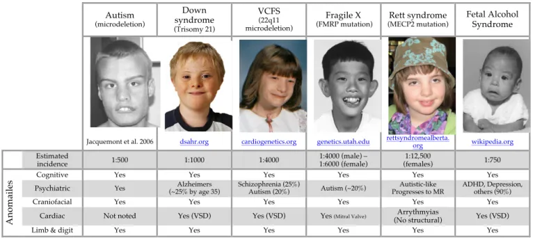
cleft palate (98, 99). There is also a high degree of overlap in phenotypes with
other etiologically diverse multiple-anomaly genetic and epigenetic disorders
(fetal alcohol syndrome (100), retinoic acid teratogenesis (101, 102), Downs
syndrome (103), Rett syndrome (104), Noonan syndrome (105), Fragile X (106);
Figure 4). While 22q11DS patients exhibit the full gamut of heart phenotypes,
patients suffering from Fetal Alcohol Syndrome and Downs Syndrome exhibit
TF and some septal defects while Noonan Syndrome patients have less severe
septal defects and Fragile-X patients do not exhibit any abnormal phenotypes of
the heart. Cognitive deficits in 22q11DS patients range from mild behavioral
deficits to high incidence of ADHD, autism, mood disorders, and schizophrenia.
None of the other single gene mutation or aneuploid diseases above have a high
incidence of schizophrenia but share increased incidence of ADHD, autism and
intellectual disability with 22q11DS. The partial penetrance and partial
recapitulation of the phenotype in single gene mutants underscore the
importance of studying 22q11DS and other multigenic diseases in a more
morphogenesis of the structures affected in 22q11DS will shed light on potential etiology of similar defects that occur more commonly in the general population.
Figure 1: Gene map of the 22q11.2 region and its orthologous region on the mouse chromosome 16. 3Mb deletion is between LCR-A and LCR-D. Nested
1.5MB deletion between LCR-A and LCR-B. Linkage of several individual genes
to schizophrenia. LgDel mice have deletion of 26 genes. Df1 mouse has a nested
21 gene deletion. BAC 316 used in complementation studies contains additional
copies of 4 genes (indicated with a +). Individual mouse knockouts that have
studied in the literature.
Serpind1
Zdhhc8
Ranbp1
Dgcr6 Prodh Dgcr5 Dgcr2 Stk22a Stk22b Dgcr1 Gscl Slc25a1 Cltcl Dvl1l Hira Mrpl40 Ufd1l Cdc45l Cldn5 Sept5 Gp1bb Tbx1 Gnb1L Txnrd2 Comt Arvcf T10 Dgcr8 Htf9c Rtn4r Dgcr6L Usp41 Znf74 Scarf2 KelchL Pcqap Pik4a Snap29 Crkl Aifm3 Lztr1 Thap7 P2rxl1 Slc7a4
ProdhL
LCR A hs#22q11 LCR B C D
mmChr16
LgDel Df1
Indiv#K/O BAC#316 Scz.#Linkage
Ranbp1
Dgcr6 Stk22a Dgcr2
Prodh Slc25a1 Gscl Dgcr1 Stk22b Car15 Scarf2 Khl22 Pcqap Smpd4 Slc7a4 P2rxl1 Lztr1 Aifm3 Crkl Snap29 Serpind1 Pik4a
Zdhhc8
Cdc45l
Ufd1l
Mrpl40
Hira Cldn5
Gnb1L
Tbx1
Gp1bb
Sept5 Txnrd2 Comt Arvcf T10 Dgcr8 Htf9c Ranbp1 Rtn4r
Smpd4
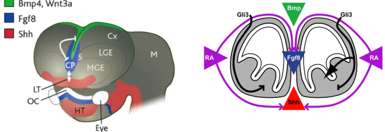
Figure 2: Mosaic of M/E signaling centers in the developing brain *Adapted from Ohkubo etal 2002 (44) and Tucker et al (107)
Shh
RA %%RA
Gli3 Gli3
Figure 3: Formation of the arterial pole (A) Cardiac neural crest (purple – from rhombomeres 5/6) populate the second heart field. (B) Origin of the secondary heart field cells (purple) in the ventral pharynx (C) Myocardium (green) and
smooth muscle (red) derived from the secondary heart field that form the arterial
pole.
*Adapted from Hutson and Kirby 2007 (108)
Autism
(microdeletion)
Down syndrome
(Trisomy 21)
VCFS
(22q11 microdeletion)
Fragile X
(FMRP mutation) (MECP2 mutation)Rett syndrome
Fetal Alcohol Syndrome
Jacquemont et al. 2006 dsahr.org cardiogenetics.org genetics.utah.edu rettsyndromealberta.org wikipedia.org
Estimated
incidence 1:500 1:1000 1:4000 1:4000 (male) – 1:6000 (female) (females) 1:12,500 1:750
An
o
m
ai
le
s Cognitive Yes Yes Yes Yes Yes Yes
Psychiatric Yes (~25% by age 35) Alzheimers Schizophrenia (25%) Autism (20%) Autism (~20%) Progresses to MR Autistic-like ADHD, Depression, others (90%)
Craniofacial Yes Yes Yes Yes Yes Yes
Cardiac Not noted Yes (VSD) Yes (VSD) Yes (Mitral Valve) Arrythmyias
(No structural) Yes (VSD)
Limb & digit Yes Yes Yes Yes Yes Yes
CHAPTER 2
INTRODUCTION - II
22q11 deletion syndrome (22q11DS), also known as DiGeorge or
Velocardiofacial Syndrome, is the consequence of a hemizygous loss of a
“critical” (1.5Mb) or larger “typical” (3MB) region of human Chr.22 (6, 109).
22q11DS phenotypes, seen with variable penetrance, include life-threatening
cardiovascular malformations (2, 10), craniofacial and limb abnormalities (110),
parathyroid and thymic hypoplasia (111), and increased susceptibility to
behavioral disorders and psychiatric diseases (112) suggesting altered brain
development or function. Initial differentiation of most affected structures,
including the brain and heart, depends upon mesenchymal/epithelial (M/E)
interactions, mediated by signaling via Sonic Hedgehog (Shh), Fibroblast Growth
Factors (Fgfs), Retinoic Acid (RA), and Bone Morphogenetic Proteins (Bmps)
(113, 114). Loss-of-function mouse models for these M/E signals have
phenotypes that overlap, to varying degrees, those in 22q11DS. Coincident
expression and activity of 22q11 genes, Shh, Fgfs, RA and Bmps at M/E sites—
including the limb, face, heart and brain—suggest that normal signaling may be
disrupted by altered 22q11 gene dosage (115). Nevertheless, it remains unknown
whether 22q11 genes interact significantly with the cardinal M/E signals. Thus,
we asked whether Shh, Fgf, RA, and Bmp signaling influences 22q11 gene
modifies these signaling pathways during mid-gestation, when M/E signaling
initiates significant morphogenesis at sites compromised in 22q11DS.
Variable severity and penetrance of 22q11DS phenotypes suggests modifiers
outside of the deleted 22q11 locus, and Shh, Fgfs, RA, Bmps, and related
signaling molecules are likely candidates. Some phenotypes in mutant mice with
disrupted Fgf8 (39) and RA (Raldh2)(40) signaling have been identified as
22q11DS “phenocopies”, while Shh (41) and Noggin/Chordin (Bmp signaling)
anomalies (42) are more narrowly interpreted as “parallel” to 22q11DS. It is
unclear whether such similarities reflect convergence of signals and 22q11 genes
consistent with “phenocopy”, or influence of 22q11 genes and M/E signals on
distinct aspects of development in similar tissues. Shh and RA apparently
regulate expression of at least one 22q11 gene, Tbx1 (92, 94) which whendeleted
in combination with Crkl (outside of the 1.5MB critical region, but within the
broader 3MB “typical” region;(6, 116)) can modulate RA and Fgf8 signaling (93),
while total loss of Tbx1 down-regulates Bmp4 (76). Moreover, recent observations
suggest that additional genes outside the 22q11 minimal region modulate Tbx1
mutant phenotypes (117, 118), some of which may be associated with RA (117)
and Bmp (119) signaling. These observations, however, do not address
interactions with 22q11 genes beyond Tbx1, which although critical, may not
explain the full range of 22q11DS phenotypes (93, 115, 120, 121). Thus, we asked
if M/E signals regulate 22q11 genes in the critical region; whether dosage of
these genes influences Shh, Fgf, RA or Bmp signaling, and whether phenotypes
in 22q11DS mouse models can be modified by altered inductive signaling.
We found clear quantitative evidence of significant interaction between 22q11
Shh and RA signaling in mouse embryos with a deletion parallel to that in
22q11DS results in significantly more severe phenotypes than altered signaling,
22q11 deletion or heterozygous Tbx1 deletion alone. The reciprocal regulation
and phenotypic modulation we have found does not support simple, linear,
uni-directional relationships between cardinal M/E signals and 22q11 genes; instead,
they are likely part of a broader network that modifies distinct aspects of
M/E-mediated morphogenesis in the heart, brain and other phenotypic sites. This
network may be an essential contributor to the phenotypic variability seen in
CHAPTER 3
RESULTS
Shh, BMP, FGF and RA signaling influence 22q11 gene expression
The 21 mouse orthologues of genes within the 22q11 critical region known to
be selectively or specifically expressed at sites of M/E interaction (122) are
selectively expressed in the mesenchymal compartment (Figure 5A; [21]).
Analysis of the developing forebrain and branchial arches show that with the
exception of 3 epithelium-restricted genes (3/21; Tbx1, Gnb1L and Slc25a1; Figure
5A) the majority of 22q11 genes (18/21) are exclusively or selectively expressed
in the mesenchyme. Heterozygous deletion of 28 contiguous murine 22q11
orthologues (including 7 not expressed in the embryonic or adult brain, and thus
not analyzed further here;(122)) in embryonic (E)10.5 LgDel embryos (71) results
in a 50% expression decrement for each corresponding mRNA (Figure 5B),
presumably primarily within the mesenchyme, where most of these genes are
highly expressed. Thus, we asked whether altered Shh, RA, Fgf8, or Bmp
signaling at E10.5, the peak age when each of these signals mediates M/E
interactions at 22q11DS phenotypic sites—limbs, heart, face and forebrain—
disrupts expression of any 22q11 gene, perhaps to levels that seen following
heterozygous deletion Dysmorphology and tissue loss complicates analysis at
mutant embryos. To circumvent this issue, we also manipulated each signal
using pharmacological antagonists or agonists between E9.5 and E10.5.
Slc25a1
RanBP1Dgcr2
Dgcr1
Dgcr6
Prodh
Zdhhc8
Htf9c
Dgcr8
T10
Arvcf
Comt
Txnrd2
Gnb1L
Tbx1
Septin5
Cldn5
Cdc45L
Ufd1L
Mrpl40
Hira
0.0 0.5 1.0 1.5 2.0
LgDel
A
C
Slc25a1 RanBP1Dgcr2 Dgcr1 Dgcr6 Prodh Zdhhc8 Htf9c Dgcr8 T10 Arvcf Comt Txnrd2 Gnb1L Tbx1 Septin5 Cldn5 Cdc45L Ufd1L Mrpl40 Hira Fgf8
0 2
1.5
1
0.5
n.d n.d n.d n.d n.d n.d n.d n.d n.d n.d n.d n.d 0 2 1.5 1 0.5
n.d n.d n.d n.d n.d n.d n.d n.d
n.d n.d n.d
n.d n.d n.d n.d
Figure 5: 22q11 genes are expressed primarily in the mesenchyme at sites of M/E
interaction, and are diminished by 50% by heterozygous deletion. (A) Tissue
specific mRNA expression of twenty-one 22q11 genes in the epithelium and
mesenchyme of the frontonasal mass (FnM; n=3). 11/17 detected genes are
restricted to mesenchyme while 3 (grey shaded box) are restricted to the
epithelium. The remaining 3 are expressed equally in mesenchyme and
epithelium. (B) Tissue specific expression of the same 22q11 genes in Branchial
Arch 1 (BA; n=3). Detection and tissue-specific expression mostly mirror that in
the FnM; however, Dgcr1 is detected only in the BA mesenchyme while Arvcf is
detected only in the FnM-mesenchyme. (C) Twenty-one 22q11 genes are
expressed at 50±10% of wild-type levels following genetic heterozygous loss of
22q11 genes in the LgDel embryo (n=3, p≤0.05; 2-tailed T-test). Mesenchymal or
dual expressed genes indicated in black boxes with white letters; epithelial
Shh regulation of 22q11 gene expression
Expression of ten of twenty-one highly expressed 22q11 genes was
significantly altered by constitutive loss of Shh signaling in E10.5 Shh-/- embryos.
Two of these ten are epithelially restricted (Figure 6A, thick outlined bars) while
the remaining 8 are preferentially expressed in the mesenchyme. In mRNA
samples from individual whole Shh-/- mutant embryos (n=4), seven of the ten
genes that have altered expression are reduced to 50% of wild type levels (Figure
6A, solid bars; comparisons based upon one way ANOVA for wild-type, LgDel,
and Shh-/- embryos, p
≤0.05), paralleling that in the LgDel embryo (LgDel-like).
Three additional genes are either reduced beyond 50% or significantly increased
(Figure 6A, hatched bars; p ≤ 0.05). Of the seven genes that have LgDel-like
expression changes, two are restricted to the epithelium (Figure 6A, thick
outlined bars) and the other five are mesenchymal. Analysis of the Gli3Xtj/Xtj
embryos (a presumed gain of Shh function;(45)) did not show reciprocal changes
of 22q11 genes, Fgf8, or P75 (Figure 6B).
mRNA levels for epithelial (Fgf8; (85)) and mesenchymal markers (P75; (123))
are also significantly diminished in Shh-/- embryos, complicating interpretation of
parallel diminished levels of 22q11 genes. Accordingly, to assess acute effects of
Shh signaling, independent of concatenated morphogenetic disruption at M/E
sites in Shh-/- embryos, we reduced Shh signaling between E9.5-E10.5 via
maternal exposure to cyclopamine, which blocks Shh signal transduction (124)
without the dysmorphogenesis seen in Shh-/- embryos. Brief, transient
cyclopamine exposure results in a 50% reduction of Ptch1, a known Shh target
Cyclopamine-dependent loss of Shh signaling also significantly alters expression
of ten 22q11 genes. Eight of these ten genes are diminished by 50%, thus
paralleling LgDel (Figure 6C; solid bars; p≤ 0.05), while an additional two are
either increased, or reduced by less than 50%. Three of these eight genes are
restricted to the epithelium (Figure 6C; solid yellow bars with thick outline),
overlapping Shh expression domains. Five genes: Slc25a1, Dgcr6, Arvcf, Tbx1, and
Septin5 are reduced to LgDel values in cyclopamine-treated as well as Shh
-/-embryos.
We next assessed whether Shh signaling regulates 22q11 genes by
maintaining normal expression levels or patterns using in situ hybridization
(ISH) to analyze genes that substantially decrease or increase in response to
genetic and pharmacological manipulation of Shh signaling. We evaluated Sept5,
which is significantly diminished by both Shh-/- and cyclopamine, as well as
Ranbp1, which is significantly increased by cyclopamine. For both genes, ISH
labeling in cyclopamine-exposed embryos indicates diminished (Sept5) or
enhanced (Ranbp1) levels, but not patterns of wild-type expression at sites of
M/E induction (Figure 6E; n=4 for each probe). Thus, Shh signaling likely
maintains normal levels, without apparent change in pattern, of a substantial
subset of 22q11 genes in the mesenchyme and epithelium at 22q11DS phenotypic
Slc25a1 RanBP1
Dgcr2 Dgcr6 Prodh Rtn4r Zdhhc8 Htf9c Dgcr8 T10 Arvcf Comt Txnrd2 Gnb1L Tbx1
Septin5 Cldn5 Cdc45L Ufd1L Mrpl40 Hira Fgf8 P75
Figure 6: Sonic hedgehog (Shh) signaling maintains normal levels of a
substantial number of 22q11 genes. Epithelium-restricted 22q11 genes are
indicated by thicker black outlines on bars. (A) Expression of ten 22q11 genes
(yellow bars, hatched or solid) changes significantly in E10.5 Shh
embryos (n=4;
p≤0.05; t-test); 7 of these changes are statistically equivalent to changes in the
LgDel (solid yellow bars; p≥0.05; 2-way ANOVA) embryo. Significantly
decreased expression of epithelial Fgf8 and mesenchymal P75 (black) suggests
altered integrity of the respective tissues. (B) Minimal expression changes of
22q11 genes in E10.5 Gli3-/- embryos (4/22 genes, hatched yellow bars); without
significant changes in Fgf8 or P75 (open bars). (C) Transient
cyclopamine-induced inhibition (24-hr exposure; see Methods) of Shh signaling decreases
expression of 10 22q11 genes (n=4, p≤ 0.05, t-test). Hatched bars indicate
significant changes not equivalent to LgDel; 8 solid yellow bars are equivalent to
LgDel (n=4; p≥0.05; 2-way ANOVA). Neither epithelial Fgf8 nor mesenchymal
P75 expression is altered. (D) Ptch1, a known Shh target gene is downregulated
to 50% of wild-type levels following 24-hr cyclopamine treatment (p≤0.05; n=4).
(E) Changes in apparent intensity, but not pattern, of expression of Septin5 and
Ranbp1 (largest magnitude changes) in response to 24-hr cyclopamine exposure
RA signaling and regulation of 22q11 gene expression
Disrupted RA signaling in Raldh2-/- embryos (54) is accompanied by
significantly decreased expression of sixteen 22q11 genes. Fifteen of these
expression changes are statistically equivalent to changes seen in LgDel embryos
(Figure 7A; solid bars; p ≤0.05). All but one gene is restricted to the mesenchyme,
overlapping with domains of RA activity. These changes, however, are paralleled
by significant expression changes of Fgf8 and P75 (50% and 200% respectively;
Figure 7A), and likely reflect severe morphogenetic consequences, especially for
mesenchyme, of constitutive loss of Raldh2 function at M/E sites (40). To
circumvent interpretative difficulties due to likely tissue loss versus altered
22q11 gene expression, we briefly enhanced or diminished RA signaling between
E9.5 and 10.5 using all trans RA ((126); Figure 7B) and DEAB (an RA synthesis
inhibitor; (127); Figure 7C), both of which alter expression of RA-regulated genes
including Rarα (126), but do not significantly change Fgf8 or P75 (Figure 7B,C).
RA significantly diminishes three genes—Ranbp1, Gnb1l, and Cdc45l—none,
however, by 50% or more (Figure 7B, p ≤0.05). In contrast, RA increases Prodh
and Septin5 (Figure 7B, 50-60%, p ≤0.05). These changes indicate that our sub-teratogenic RA dosing regimen is the least disruptive of all our pharmacological
manipulations. DEAB diminishes expression of six mesenchymal genes—Dgcr2,
Dgcr6, Dgcr8, Septin5, Cldn5 and Hira—to LgDel levels (Figure 7C, p ≤0.05). ISH for Ranbp1 and Dgcr8, which are significantly decreased by RA and DEAB treatments respectively, indicates these changes reflect local fluctuations in
modestly influences 22q11 gene expression levels at M/E inductive sites
compromised in 22q11DS; however, there is no clear relationship between
RA
0.0 0.5 1.0 1.5 2.00
B
Raldh2'/'
0.0 0.5 1.0 1.5 2.615
A
DEAB
0.0 0.5 1.0 1.5 2.06
C
0.0 0.5 1.0 1.5 2.0 RA DEAB RARD
Control +RADgcr8 +DEAB Control Septin56
E
Significant LgDel;Eqiv .;;
6
;;
16
;;
7
Slc25a1 RanBP1Dgcr2 Dgcr6 Prodh Rtn4r Zdhhc8 Htf9c Dgcr8 T10 Arvcf Comt Txnrd2 Gnb1L Tbx1
Septin5 Cldn5 Cdc45L Ufd1L Mrpl40 Hira Fgf8 P75
Figure 7: Altered RA signaling influences expression levels of a subset of 22q11
genes. Epithelium-restricted 22q11 genes are indicated by thicker black outlines
on bars. (A) Expression of 16 genes is significantly decreased in highly
dysmorphic E10.5 Raldh2
embryos (n=4; p≤0.05; t-test); 15 of these changes are
statistically equivalent to that in LgDel embryos (solid purple bars; p≥0.05; 2-way
ANOVA); however, significantly altered Fgf8 and P75 expression (solid black
bars) indicates disrupted epithelial and mesenchymal integrity. (B) 24-hr
sub-teratogenic RA exposure alters expression of 6 22q11 genes in E10.5 embryos
(hatched purple bars; p≤0.05; n=4; t-test); however, none are statistically
equivalent to that in LgDel embryos. (C) Transient inhibition of RA synthesis in
E10.5 embryos using DEAB leads to diminished expression of six 22q11 genes to
LgDel levels (solid purple bars). (D) Expression of the RA responsive gene, Rarα, is significantly increased following transient exposure of embryos to RA (50%)
and DEAB (75%; p≤0.05, n=4 for each condition). (E) Expression of Septin5 is
increased in the frontonasal mass, branchial arches and limb bud (solid white
arrows) in response to 24-hr RA treatment; Dgcr8 expression in branchial arches,
frontonasal mass and limb buds is decreased following DEAB mediated
Fgf and Bmp signaling and regulation of 22q11 gene expression
Fgf and Bmp signaling have less quantitatively detectable influence on
maintaining normal levels of 22q11 gene expression at midgestation than either
Shh or RA. In mildly dysmorphic Fgf8neo/neo embryos (approximately 40% of WT
level of Fgf8; Figure 8A) a modest (approx.15%) but significant increase of Ranbp1
is the only detectable 22q11 gene expression change (Figure 8A). PD173074 (128,
129), a small molecule inhibitor of FGF receptor-mediated signaling, disrupts Fgf
signaling at M/E sites, confirmed by the down-regulation of Mkp3 (130)). In
PD173074-exposed embryos expression of seven 22q11 genes is significantly
altered; five genes mirror LgDel changes, however, none parallel those in the
Fgf8neo/neo
embryos (Figure 8A,B). PD173074 also significantly diminishes Fgf8,
with a similar trend for P75 suggesting potential tissue loss at M/E sites. ISH in
PD173074-exposed embryos shows altered 22q11 gene expression levels at M/E
sites (Figure 8F); nevertheless, there is also hypoplasia at M/E sites suggesting
diminished tissue volumes not seen following 22q11 deletion (131). Thus,
PD173074-induced 22q11 gene expression changes may be secondary, due to
tissue loss. Together, this data indicates that the contribution of Fgf signaling to
normal 22q11 gene expression levels is less direct and substantial than that via
Shh or RA.
Enhanced Bmp signaling due to loss of Noggin mediated Bmp antagonism
(Nog
-/-(69)) significantly decreases one 22q11 gene (Txnrd2) by 50%, similar to
LgDel (p≤0.05). Expression of 6 additional 22q11 genes is modestly (10-30%) but
dorsomorphin (132) results in diminished expression of seven 22q11 genes, two
of which are shared with Nog
embryos. Only three of the dorsomorphin-induce
changes, however, are statistically similar to LgDel changes (Figure 8D), even
though Id1 a known Bmp target is enhanced by 30% (Figure 8G). ISH in
dorsomorphin-treated embryos is consistent with altered expression levels of
Prodh, the most substantially diminished 22q11 gene, without significant changes in pattern or morphology (Figure 8H). Thus, the contributions of Bmp signaling
to maintenance of normal 22q11 gene expression levels, judged by numbers of
genes whose expression is significantly altered by disrupted Bmp signaling,
Slc25a1 RanBP1
Dgcr2 Dgcr6 Prodh Rtn4r Zdhhc8 Htf9c Dgcr8 T10 Arvcf Comt Txnrd2 Gnb1L Tbx1
Septin5 Cldn5 Cdc45L Ufd1L Mrpl40 Hira Fgf8 P75
Fgf$I
0.0 0.5 1.0 1.5 2.0 ++5
B
Fgf8+hyp
0.00.51.0 1.5 2.0
A
+FGFRi Septin5 Control Significant LgDel+Eqiv .++
7
*NogginL/L
0.0 0.5 1.0 1.5 2.0 ++1
Dorsomorphin 0.0 0.5 1.0 1.5 2.0 ++3
D
C
E
Control +DorsomorphinProdh
F
++
7
++++++9
Dgcr1
Figure 8: Fgf and Bmp signaling have limited impact on maintenance of normal
levels of 22q11 gene expression. (A) Fgf8neo/neo
embryos express 40% levels of
wild-type Fgf8 (solid black bar), but have no LgDel-like changes in expression of
22q11 genes; Ranbp1 is modestly but significantly increased. (B) Transient
inhibition of FgfR mediated signaling reduces Fgf8 and P75 expression as well as
eight 22q11 genes; 6 gene expression changes are equivalent to LgDel (solid red
bars) while Prodh and Zdhhc8 display increased and greatly reduced expression,
respectively (hatched red bars). (C) Noggin-/- embryos have no measurable
changes in epithelial or mesenchymal markers, but display significant and
modest decrements in 7 genes that are not equivalent to LgDel embryo dosage
(hatched blue bars), with one exception, Txnrd2 (solid blue bar). (D) Transient
pharmacological inhibition of Bmp signaling by dorsomorphin induces LgDel
-like decrements in Dgcr1, Arvcf, Tbx1 and Ufd1l (solid blue bars) as well as
smaller magnitude changes in Prodh, Dgcr8, Comt, Septin5 and Hira (hatched blue
bars). (E) Inhibition of FgfR signaling results in 55% reduced expression of the
Fgf target gene Mkp3. (F) Septin5 expression is severely diminished in the
developing forebrain, branchial arches and limb buds; structures that are also
severely morphologically compromised. (G) Id1, a Bmp target gene is
upregulated following 24-hr dorsomorphin treatment. (H) Prodh, quantitatively
down regulated by dorsomorphin, is diminished in the developing forebrain and
Diminished dosage of 22q11 genes disrupts M/E signaling pathway gene
expression
There may be reciprocal regulation of 22q11 genes and cardinal M/E
inductive signals including Shh, RA, Fgfs, and Bmps. If this were the case,
diminished 22q11 gene dosage should alter ligand expression, availability,
abundance or activity of receptors and signaling co-factors. Accordingly, we first
asked whether diminished 22q11 gene dosage modifies Shh, Fgf, RA or Bmp
signaling pathways. To evaluate such interactions, we first analyzed mRNA
levels for Shh, Fgf, or Bmp ligands, RA synthetic enzymes, which are essential
for ligand production, and key receptors, signaling co-factors, and metabolic
regulators for each pathway in whole E10.5 LgDel embryos (71).
Three of the four M/E signaling pathways appear altered—all due to
decreased, rather than enhanced expression—by varying degrees as a result of
diminished 22q11 gene dosage in LgDel embryos. Two Shh transcriptional
mediators, Gli1 and Gli3, decline in LgDel embryos (25%; p≤ 0.05; Figure 9A, far
left). We detect no change in Shh ligands or receptors in whole embryo samples,
including Ptch1 and Ptch2 which are unchanged despite known regulation by
Shh-activated release of Gli transcriptional control (125, 133, 134). We next
evaluated genes associated with RA synthesis, as well as RA receptors and
co-factors. Raldh3, an RA synthetic enzyme highly enriched in the brain (135, 136) is
diminished by nearly 50% (p≤0.05; Figure 9A, left center). Two receptors, Rarα
as does Cyp26a1, an RA degrading enzyme whose expression is RA-dependent
(138) (p≤0.05, n=4). There were no changes in expression of two Fgf ligands, 2
receptors, 3 signaling regulators and an established Fgf-regulated gene
(Mkp3;(130)) in the LgDel embryo (Figure 9A, right center). Bmp ligands are not
significantly altered; however, Id1 (a Bmp target gene) is modestly but
significantly diminished (14%) as is the Bmp receptor BmpR1 (17%) and two
transcriptional cofactors, Smad2 (16%), and Smad4 (26%; p≤0.05, n=4; Figure 9A,
right). Thus, based upon analysis of representative molecular mediators of the
four signaling pathways, we do not detect changes in Fgf signaling molecules,
Shh and Bmp mediators change modestly, and the most frequent, substantial and
statistically significant expression changes for multiple elements (ligand-related,
receptors, and co-factors) in mid-gestation LgDel embryos are seen for RA
signaling.
Tbx1
and broader 22q11 deletion do not result in similar gene expression
changes
Tbx1 has been proposed as a key, if not singularly explanatory, gene for
several 22q11 phenotypes, especially those that reflect aberrant heart and brain
development (71, 77, 139). If Tbx1 alone is responsible for the majority of 22q11
phenotypes, and these phenotypes reflect altered Shh, RA, Bmp or Fgf signaling
as suggested in several reports (93, 95, 96, 140), one would expect significant
overlap between expression changes in Shh, RA or Bmp signaling molecules in
LgDel and Tbx1
+/-embryos. Of the 42 signaling intermediates we assessed, only 4
are significantly altered in Tbx1+/- embryos (Figure 9B): 2 Shh signaling mediators
Rara overlaps with changes in E10.5 LgDel embryos; however, the change is of
smaller magnitude. One Fgf signaling intermediate, Spry4 (141) is altered in
Tbx1+/- embryos, and no Bmp-related transcripts are changed (Figure 9B). Thus,
there are fewer, and mostly divergent expression changes for M/E signaling