Coupled Electron Proton Transfer Reactions in Biological Redox Active Substrates
Christine Fecenko Murphy
A dissertation submitted to the faculty of the University of North Carolina at Chapel Hill in partial fulfillment of the requirements for the degree of Doctor of Philosophy in the
Department of Chemistry
Chapel Hill 2009
Approved By
Advisor: Professor H. Holden Thorp
Advisor: Professor Thomas J. Meyer
Reader: Professor Joseph Templeton
ii ABSTRACT
Christine Fecenko Murphy: Coupled Electron Proton Transfer Reactions in Biological Redox Active Substrates
(Under the direction of H. Holden Thorp and Thomas J. Meyer)
The kinetics of oxidation of amino acids and nucleobases by the series of metal complex oxidants, M(bpy)33+ (M = Os, Fe, Ru) in aqueous solution at an ITO electrode, in the presence of added bases, was investigated utilizing catalytic cyclic voltammetry. A common mechanism involving initial formation of a hydrogen bond complex between the substrate and a base in solution was identified for all substrates. After association,
iii
Acknowledgements
Holden, first off, thank you for taking me on as a graduate student. When I first joined the group, I thought I wanted to be a biological chemist. In fact, I was shocked when I was handed the initial patent on tyrosine oxidation. You must have known what you were doing, because now I have found my passion for amino acid oxidation and how oxidoreductases function. You have always supported my ideas and interest both in the lab and out. I don’t know any other advisor would have given a fourth year graduate student a high-five when she announced her pregnancy. It is for this and many other reasons, I’m so glad that I chose to work in your group and have learned so much from you.
Dr. Meyer, thank you for taking such an interest in me and my work. Our daily meetings to discuss science always left me with a great grasp on my work and my plate overflowing with work. You’ve kept me busy, productive, and on task. As someone that is as scatterbrained as me, it was definitely the fire under me that I needed. You’ve allowed me to do so many things in graduate school that many students only dream of and have supported even my craziest ideas. I doubt you ever thought you would venture into protein design and protein electrochemistry. I’m very grateful to you for selecting me to be part of your group. I’ve learned so much and have laid the ground work for my future interests.
Dr. Feldberg, you’ve taught me how to let the electrochemistry speak for itself. I looked forward to your visits to UNC every year, and have learned so much for your patient instruction.
To all the past and present members of the Thorp group, you’ve made working in lab a pleasure. Dom, thanks for being my electrochemistry mentor and sharing the ins and outs of digisim. Stephanie, you’re thesis has been more useful than you would have ever imagined when writing it. Julie, thanks for putting up with my 3 month hiatus into biochemistry. I learned so much, even though I wasted so many chemicals. Chris, I couldn’t have found a better person to leave my project to, I know you’ll do great things with it. To the Meyer group, thanks for all your ideas and thoughts on my project. Javier, sometimes I think you thought about my kinetics more than I did. Kyle, thanks for always being patient with me and saving me in my orals with a lesson on spin orbit coupling.
iv
inspiration to always do my best. Karen, thanks for being my “best sister” and always answering you phone at 6am when I’m awake and want to chat. Alison and Chetna, you’re the best roommates I could ever ask for. Grant, thank you for putting up with my seemingly endless nights of work. You have let me bounce ideas off of you and listened to more practice talks than any husband should be expected to. Grant and Gavin, you’ve made me the luckiest mom and wife in the world.
v
Table of Contents
List of Figures…………..………...ix
List of Tables………...………..………….xii
List of Abbreviations………....………xiii
Chapter 1: Introduction to EPT in Biological Redox Active Substrates………....1
1.1 Biological Oxidation………...2
1.2 Proton Coupled Electron Transfer………...4
1.3 Metal Mediated Electrocatalysis on ITO………7
1.4 Electrocatalysis of Amino Acids and Nucleobases………...10
1.5 References……….12
Chapter 2: The Oxidation of Tyrosine through Parallel Rate Determining Proton Transfer and MS-EPT ...…………14
2.1 Abstract……….15
2.2 Introduction………..17
2.3 Methods and Materials………..18
2.3.1: General………..18
2.3.2: Buffer Preparation……….18
2.3.3: Isotope Studies………..18
2.3.4: Sample Preparation………19
2.3.5: Electrochemistry………19
2.3.6: Digital Simulation……….20
vi
2.3.8 Mixing Studies………...22
2.4 Results and Discussion………..23
2.4.1: Electrocatalytic Studies in Phosphate Buffer………...23
2.4.2: Kinetic Studies on Tyrosine………..25
2.5 Conclusions………29
2.6 References ………..…31
Chapter 3: Coupled Electron-Proton Transfer Pathways in Tyrosine Oxidation……...33
3.1 Abstract………..34
3.2 Introduction………....35
3.3 Methods and Materials………...38
3.3.1: General………..38
3.3.2: Isotope Studies……….38
3.3.3: Electrochemistry………....39
3.3.4: Rate Law………40
3.3.5: Digital Simulation………..44
3.3.6: Electron Transfer Tyrosine and Anion………..45
3.4 Results………47
3.4.1: Oxidation of Tyrosine and Anion………...47
3.4.2: Determination of Rate Law………...….48
3.4.3: Pre-Association Between TyrOH + B………...…50
3.4.4: Dependence on [TyrOH] and [B]………...51
3.4.5: KA by 31PNMR………...52
3.4.6: Pre-Association between Complex and Metal………...53
3.4.7: Rapid Pre-equilibrium Formation of TyrO-………...…54
vii
3.4.9: Complete Rate Expression……….57
3.4.10: H/D Isotope Effects……….58
3.4.11: General Buffer Base Catalysis……….61
3.5 Discussion………..62
3.5.1: Pathways for Tyrosine Oxidation………..63
3.5.2: Kinetic Parameters……….66
3.5.3: Free Energy Dependence MS-EPT………....73
3.5.4: Free Energy Dependence ET………...75
3.6 Conclusions………....78
3.7 References………..80
Chapter 4: pH Dependent or Base Assisted Tyrosine Oxidation...85
4.1 Abstract………..86
4.2 Introduction………85
4.3 Methods and Materials………...87
4.3.1 General………87
4.3.2: Kinetics-Spectrophotometric Monitoring………..89
4.3.3: Second Order Unequal Concentration Kinetics……….89
4.3.4: Kinetics Cyclic Voltommetric Studies………..89
4.3.5: Digital Simulation………..92
4.4 Results and Discussion………..92
4.4.1: pH Dependence in the Oxidation of Tyrosine by Os(bpy)33+…………92
4.4.2: Hydroxide as an EPT Base………94
4.4.3: Additional EPT Bases………96
4.4.4: General Role of OH-………100
viii
4.6 References ………106
Chapter 5: Coupled Electron Proton Transfer in Guanine Oxidation………...108
5.1 Abstract………...….109
5.2 Introduction………..110
5.3 Methods and Materials……….111
5.3.1: DNA………111
5.3.2: General………112
5.3.3: Isotope Studies………112
5.3.4: Electodes……….113
5.3.5: Electrochemistry……….113
5.3.6: Mixing Studies………...115
5.4 Results and Discussion………...….117
5.4.1: Oxidation of dGMP………117
5.4.2: EPT Oxidation of dGMP and G with dCMP and C………127
5.4.3: Oxidation of ssDNA……….….130
5.4.4: Oxidation of ssDNA with Base Complement………...132
5.4.5 Oxidation of dsDNA………..133
5.5 Conclusions………135
5.6 References ……….143
ix
List of Figures
Chapter 1: Introduction to EPT in Biological Redox Active Substrates
Figure 1: Electroactive amino acids……….2
Figure 2: Electroactive nucleobases……….3
Figure 3: Electron Transfer followed by Proton Transfer………5
Figure 4: Proton Transfer followed by Electron Transfer………6
Figure 5: Electron Proton Transfer………..6
Figure 6: Electrocatalytic cyclic voltammogram……….8
Figure 7: Digital simulation of electrocatalytic cyclic voltammogram………9
Chapter 2: The Oxidation of Tyrosine through Parallel Rate-Determining Proton Transfer and Multi-Site Electron Proton Transfer Figure 1: Driving force dependence of tyrosine and tryptophan……….24
Figure 2: Electrocatalysis of tyrosine at constant pH………..24
Figure 3: Cyclic Voltammogram at pH 8.0 showing loss of Os(bpy)33+……….28
Figure 4: Driving Force Dependence study at pH 7.5 and 6.0 phosphate…………...28
Chapter 3: Coupled Electron Proton Transfer Pathways in Tyrosine Oxidation Figure 1: Cyclic Voltammogram of Os(bpy)33+ with TyrOH A) no buffer, B) pH 6.0, and C) pH 8.5………...46
Figure 2: Saturation kinetics exhibited from plots of kobs vs [HPO42-]………...47
Figure 3: Kinetic plots from variation of A) Base and B) TyrOH………...48
Figure 4: Association constant calculation from 31PNMR……….……...49
Figure 5: Saturation kinetics observed from plots of kobs vs [Os(bpy)33+]……...……50
Figure 6: Linearization of Rate Law through plots of kobs vs [B]/[HB+]………...…..51
Figure 7: Cyclic Voltammogram at pH 8.0 showing loss of Os(bpy)33+...52
x
Figure 9: Cyclic voltammogram and digital simulation at pH 7.5………54
Figure 10: Mole Fraction study under limiting conditions………55
Figure 11: -RTLnk1 vs RTLnKHB+……….64
Figure 12: RTLNk1 vs RTLnKa………..65
Figure 13: Proton Transfer in the Inverted Region……….66
Figure 14: RTLnkred vs RTLnk1………..67
Figure 15: Variation of kobs by A) variation of oxidant or B) variation of base…….69
Figure 16: Free Energy Dependence for MS-EPT………..70
Figure 17: Driving force dependence study of anion………..72
Chapter 4: pH Dependent or Base Assisted Tyrosine Oxidation? Figure 1: pH dependent oxidation of Ru-Y adduct……….88
Figure 2: Absorbance over time studies-First order kinetics………..92
Figure 3: Plot of kobs/[TyrOH] vs pH………..97
Figure 4: Plot of kobs/[TyrOH] vs [HPO42-] pH 7.0 and 9.0………98
Figure 5: Absorbance over time studies-Second order kinetic………99
Figure 6: Plot of kobs/[TyrOH] vs [Base]………..………102
Figure 7: Plot of kobs/[TyrOH] vs [OH-]……….………..103
Figure 8: Plot of kobs/[TyrOH] vs pH……….………..104
Chapter 5: Coupled Electron Proton Transfer in Guanine Oxidation Figure 1: Absorbance over time studies-first order kinetics………..………...116
Figure 2: Saturation kinetics exhibited in plots of kobs vs [HPO42-]……….…120
Figure 3: Saturation kinetics exhibited in plots of kobs vs [Ru(bpy)33+]……….120
Figure 4: Cyclic voltammogram of dGMP in D2O and H2O………..……...121
Figure 5: Variation of kobs by A) variation of oxidant and b) variation of base..…..123
xi
Figure 7: Cyclic voltammogram of dGMP with dCMP in phosphate buffer……..128
Figure 8: Plot of kobs/[G] vs [C]………..129
Figure 9: Cyclic votammograms of ssDNA and dsDNA in phosphate buffer……131
Figure 10: Plot of kobs vs [HPO42-] obtained by stopped flow on ssDNA...………132
Figure 11: Cyclic voltammogram and digital simulation of ssDNA with 7mer….133
xii
List of Tables
Chapter 1: Introduction to EPT in Biological Redox Active Substrates
Chapter 2: The Oxidation of Tyrosine through Parallel Rate-Determining Proton
Transfer and Multi-Site Electron Proton Transfer
Table 1: Rate and equilibrium constants for tyrosine in phosphate buffer………….26
Table 2: Isotope effects for tyrosine in phosphate buffer………...26
Chapter 3: Coupled Electron Proton Transfer Pathways in Tyrosine Oxidation Table 1: Rate constants for all bases………..58
Table 2: Isotope effects for all bases………..60
Table 3: Buffer bases with pKa………..61
Table 4: Oxidant investigations with rate constants for k2 and kred………...62
Chapter 4: pH Dependent or Base Assisted Tyrosine Oxidation? Table 1: Slope and Intercept from plot of kobs/[TyrOH] vs [Base]………102
Chapter 5: Coupled Electron Proton Transfer in Guanine Oxidation Table 1: Rate and Equilibrium constants for dGMP and TyrOH……….123
xiii
List of Symbols and Abbreviations
[TyrOH]T total concentration of tyrosine
∆E change in reduction potential
∆G Free Energy
∆GEPT free energy change for electron proton transfer
∆GET free energy change for electron transfer
∆GPT free energy change for proton transfer
°C degrees celsius
1H NMR Proton Nuclear Magnetic Resonance
31P NMR phosphorus nuclear magnetic resonance
A Adenosine A Ampere
A electrode area
Å Angstrom Abs absorbance
Ag/AgCl Silver/Silver Chloride Reference
B base form of buffer
C Cytosine
CV cyclic voltammogram
CySH Cysteine d distance D+ deuteron
D2O deuterium oxide
DCl deuterochloric acid
dCMP deoxycytodine 5' monophosphate
DFT density functional theory
dGMP deoxyguanosine 5' monophosphate
dmb dimethylbipyridine
DNA Deoxyribonucleic Acid
dsDNA double stranded DNA
E electrode potential
ɛ molar absorbtivity
e- electron
E°´ Formal Potential
E1/2 Midpoint Potential
EIE equilibrium isotope effect
xiv
ET-PT Step-wise Electron Transfer followed by Proton Transfer
eV electron volt
F Faraday constant
Fe Iron
FT-IR fourier transfer infrared spectroscopy
G Guanine
G-H+/. guanine radical cation
H+ proton
H190 Histidine 190 in Photosystem II
H2PO4- monobasic hydrogen phosphate
H3O+ hydronium ion
HB+ acid form of buffer
HCl Hydrochloric Acid
His histidine
HPO42- dibasic hydrogen phosphate
i electrode current
io exchange current density
ITO Indium Tin Oxide
kD rate constant for diffusion
kEPT electron proton transfer rate constant
KIE kinetic isotope effect
kobs observed rate constant
ks heterogeneous electron transfer rate constant
ƛ reorganizational energy
ƛmax maximum absorbance
M Molar
M(bpy)33+/2+ Metal tris(2,2'-bipyridine) complex
MeCN acetonitrile
MLCT metal-ligand charge transfer
mM millimolar
MS-EPT Multi-Site Electron Proton Transfer
N1 Proton in guanine
NA Avogadros number
NaOD Sodium Deuroxide
NaOH Sodium Hydroxide
NHE Normal Hydrogen Electrode
xv
OH- hydroxide anion
Os Osmium P680 Pigment found in PSII acts as primary electron donar.
P680+ Oxidized form of P680
PCET Proton Coupled Electron Transfer
pD -Log[D+]
pH -Log[H+]
phos dibasic phosphate
pKa -Log[Ka]
PSII Photosystem II
PT-ET Step-wise Proton Transfer followed by Electron Transfer
R universal gas constant
RNR Ribonucleotide Reductase
Ru Ruthenium
ssDNA single stranded DNA
Succ succinate T Thiamine T Temperature
tris tris base
TrpH Tryptophan
TyrO- tyrosyl anion
TyrO. tyrosyl radical
TyrOH Tyrosine
TyrOH+/. tyrosine radical cation
TyrOH--B tyrosine adduct
UV Ultraviolet V volts
Yz Tyrosine 161 in Photosystem II
α symmetry factor
δ chemical shift
η difference between formal potential and electrode potential
μM micromolar
χD mole fraction deuterium
Ω resistance
Chapter 1
2
1.1 Biological Oxidation: Understanding oxidation and reduction reactions in biologically relevant systems is important to gain insight into how enzymes and DNA function.Amino acid oxidation acts as a redox mediator in vital life processes such as photosynethesis and respiration.1-5 In photosystem II, tyrosine (Yz) aids in electron movement in the production of atmospheric oxygen in the thylakoid membrane of green plants.5 Oxidation of the amino acids, tyrosine, tryptophan, and cysteine (Figure 1) is also of central importance to electron transfer reactions in oxidoreductases, a diverse family of redox active enzymes. Electroactive amino acids are involved in charge migration over long distances in the protein environment. Studies on ribonuceotide reductase (RNR), a DNA repair protein, have found that electron transfer occurs over a distance of ~35Å. EPR studies on RNR, isolate radical formation at tyrosines, tryptophans, and cysteines found within 10Å of one another in the protein framework.6
+
H3N CH C
CH2 O -O
OH
+
H3N CH C CH2
O -O
HN
+
H3N CH C
CH2 O -O
SH Tyrosine - TyrOH Tryptophan-TrpH Cysteine-CySH
Figure 1: Structures of electroactive amino acids, tyrosine, tryptophan, and cysteine.
Oxidation of deoxyribonucleic acids (DNA) leads to DNA damage and degredation, and has been associated with neurodegenerative disorders such as Alzheimer’s and
3
and thiamine (figure 2) that are found in hydrogen bond partners of A-T and G-C in the DNA phosphate backbone. Guanine acts as a radical trap due its low reduction potential of 1.1V vs NHE as compared to the other nucleobases and is often the site of oxidative damage. 6,8
N N N N NH2 O OH O P -O O-O NH N N O NH2 N O OH O P -O O O-N NH2 O N O OH O P -O O-O NH O O N O OH O P -O O-O Adenosine Thyamine Guanine Cytosine
Figure 2: Structures of the DNA nucleobases
4
hydrogen bond partners of histidine and aspartate respectively.21,22 As the electron hops from one electroactive residue to the next, the proton is thought to transfer to a neighboring base.
The coupled movement of protons and electrons is supported by experimental results. Studies on DNA and electroactive proteins have reported pH dependent oxidations in which the rate of electron transfer is directly proportional to the pH of solution.23 Proton movement is also supported by isotope effects reported in the literature for the electroactive amino acids and the guanine nucleobase. Isotope effects in oxidation reactions, indicate the reaction proceeds through a rate-determining step involving the transfer of a proton.24 It is commonly agreed that both electrons and protons are involved in these reaction mechanisms, the
mechanistic details of these reactions remains unclear.1-24
1.2 Proton Coupled Electron Transfer: Proton coupled electron transfer (PCET) describes any reaction that involves the net movement of protons and electrons.5 Oxidation of tyrosine, tryptophan, cysteine, and guanine all involve the net movement of electrons and protons, but the specific reaction mechanism is highly debated.23, 25
Oxidation can occur in one of three ways, electron transfer followed by proton transfer (ET-PT), proton transfer followed by electron transfer (PT-ET) and concerted electron proton transfer (EPT).5 Each mechanism will be addressed independently and in terms of a common reaction, tyrosine Yz in photosystem II, but the general mechanism can be applied to all four substrates.
5
the P680+ is 0.9V vs NHE making the transfer of an electron a thermodynically unfavorable reaction. The second step, proton transfer, however, occurs rapidly due to the acidity (-2) of the protonated radical. The initial electron transfer to the P680 is uphill by +0.08eV due to the formation of a highly charged intermediate.
Figure 3: Mechanistic outline of ET-PT in PSII
Proton Transfer followed by Electron Transfer (PT-ET): Oxidation of tyrosine can also occur through another step-wise process involving initial deprotonation to the deprotonated tyrosyl anion shown in figure 4. The pKa of the hydroxyl proton is 10.1 making initial
6 Figure 4: Mechanistic outline of the PT-ET reaction.
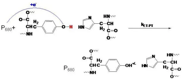
Concerted Electron Proton Transfer (EPT): A third type of oxidation mechanism has been proposed. As shown in figure 5, the reaction of tyrosine involves the simultaneous transfer of a proton to His190 and an electron to the P680+. Microscopically more complex then the step-wise reactions, the concerted mechanism avoids the thermodynamic cost of forming a charge intermediate. The simultaneous movement of the electron and the proton is actually thermodynamically favored over the step-wise reactions by 0.1eV.
7
Electron proton transfer occurs through simultaneous transfer of protons and electrons. EPT results from a redox process that allows for transfer of the electron and proton from different orbital sites on the same molecule to different acceptor sites on the same molecule.5 Multi-site electron proton transfer is a subset of EPT and is defined by transfer of the proton and the acceptor to different electron and proton acceptor sites.5 The fundamental principles for EPT have been addressed by Hammes-Schiffer, Cukier, and others where EPT theory is based on traditional Marcus electron transfer theory.3,5,12,15,16
1.3 Metal Mediated Electrocatalysis on ITO Electrodes: Model systems are routinely used to investigate specific interactions inside biological species to avoid the added complications of the cellular environment.12 Models systems have helped elucidate the fundamental aspects of PCET reactions with regard to the oxidation of tyrosine, cysteine, tryptophan, and guanine.23-25 Experiments have been designed to study PCET reactions chemically, photochemically, and electrochemically in solution.6, 18-25 Specifically,
electrochemistry has the advantage because the reaction is controlled by the potential of the electrode and reaction kinetics can be monitored directly from current over-potentials.15,23
Electrochemical techniques have been used to investigate the oxidation mechanisms of guanine and DNA and have recently applied this technique to amino acids through the use of electrocatalytic oxidation26-28. In electrocatalytic oxidation, metal complexes M(bpy)32+ (M=Ru, Fe, Os) are placed in the presence of organic donors and can be studied on indium tin oxide doped electrodes in aqueous solution in a near neutral pH window.26,27
8
6a). When an organic donor, such as tyrosine is present in solution and positive potentials are applied to the electrode, M(bpy)33+ gains an electron from the electron donor to reform the M(bpy)32+ in solution instead of at the electrode surface. The electrode only detects the presence of the M(bpy)32+ species, and the cyclic voltammograms show significant
enhancement of the oxidative wave (Figure 6b).
Figure 6: Cyclic voltammograms of Os(bpy)33+ a) alone in phosphate buffered solution and b) in the presence of tyrosine. When an electron donor is present as in (b), electron transfer occurs form the tyrosine to the Os(bpy)33+ in solution. The electode only detects the
conversion from Os2+ to Os3+ which results in enhancement of the catalytic oxidative wave. Kinetic information can be obtained from cyclic voltammetric studies exhibiting current enhancement of the oxidative wave through digital simulation. Current and potential information is related through the Butler Volmer equation 29,30 shown in eq 1. Digital simulation of the oxidative wave results in rate constants consistent with the homogeneous electron transfer rate constant for the exchange between the M(bpy)33+ and the electron donor in solution using equation 2, which relates reduction potential to the observed rate constant (kobs). Digital simulations are compared to experimental data as shown in figure 7.
[ ]
[ ]
(
)
i
nFA
k
A
exp
F
RT
E
B
exp 1
F
RT
E
s x 0 x 0
=
⎛
−
⎝⎜
⎞
⎠⎟
−
⎛
⎝⎜
−
⎞
⎠⎟
⎡
⎣⎢
⎤
⎦⎥
=
α
Δ
=α
Δ
(1)-4 10-6 -3 10-6 -2 10-6 -1 10-6 0 1 10-6 2 10-6 3 10-6
0 200 400 600 800 Potential (mV)
-2 10-5 -1.5 10-5 -1 10-5 -5 10-6 0 5 10-6
-200 0 200 400 600 800 1000 Potential (mV)
Potential vs Ag/AgCl Potential vs Ag/AgCl
C u rr en t (A ) C urr en t (A )
-4 10-6 -3 10-6 -2 10-6 -1 10-6 0 1 10-6 2 10-6 3 10-6
0 200 400 600 800 Potential (mV)
-2 10-5 -1.5 10-5 -1 10-5 -5 10-6 0 5 10-6
-200 0 200 400 600 800 1000 Potential (mV)
Potential vs Ag/AgCl Potential vs Ag/AgCl
9
(
)
Δ
G
=
nF E
oxo−
E
redo (2a)k
Aexp
G
RT
obs
=
−
Δ
(2b)
Figure 7: Electrocatalytic cyclic voltammograms of M(bpy)33+ in the presence of an organic donor (solid line) and a digitally simulated cyclic voltammogram (dashed line). Digital simulations are fit to experimental data to obtain rate constants for the homogeneous electron transfer between the metal complex
1.4 Electrocatalysis of Amino Acids and Nucleobases: We report here, the application of this electrochemical technique to the investigation of the oxidation mechanism of the biologically relevant substrates, tyrosine, tryptophan, cysteine and guanine.
Initial application of this technique to tyrosine resulted in isolation of a new mechanism for tyrosine oxidation through competitive PT-ET and EPT. The key mechanistic finding was the formation of an association complex with the basic form of the buffer (HPO42-),
-3 10-5 -2.5 10-5 -2 10-5 -1.5 10-5 -1 10-5 -5 10-6 0 5 10-6
0 200 400 600 800 1000 1200 experimental simulation Potential (mV)
-Experiment
--Simulation
-3 10-5 -2.5 10-5 -2 10-5 -1.5 10-5 -1 10-5 -5 10-6 0 5 10-6
10
before oxidation through either pathway. The pathway of oxidation was determined by solution conditions with PT-ET dominating at high concentrations of base and low concentrations of acid and MS-EPT dominating at low concentrations of base and high concentrations of acid. Rate constants and isotope effects were isolated that supported the oxidation mechanism. Further studies on the tyrosine system have found that previous reported pH dependent oxidation occurs through prior association with the basic form of the buffer (in the limit of pKa= 4.7-8.1 investigated) followed by either deprotonation or electron proton transfer depending on solution conditions.
The same experimental technique was applied to amino acids, cysteine, and tryptophan and the nucleobase guanine. Guanine, was studied in several environments, including dGMP, single stranded (ssDNA), and double stranded (dsDNA). We have found that the EPT reaction mechanism is a general mechanism for tyrosine, tryptophan, cysteine, and guanine. We have found that base association and redox activity through competitive pathways of deprotonation and electron proton transfer are common in biological redox systems, and play a role in tuning the reduction potentials of these substrates.
11 1.5 References:
1) Cukier, R. I; Nocera, D. G. Proton Coupled Electron Transfer, Annu. Rev.Phys. Chem.199849 337-69
2) Mayer, J. M. Proton Coupled Electron Transfer: A Reaction Chemist’s Point of View.Annu. Rev. Phys. Chem.200455 363-90
3) Hammes-Schiffer, S. Theoretical Perspectives on Proton Coupled Electron Transfer Reactions Acc. Chem. Res. 2001 34 273-81
4) Brudvig, G. W.; Thorp, H. H.; Crabtree, R. H. Probing the Mechanism of Water Oxidation.Acc. Chem. Res.199124, 311-316
5) Alstrum-Acevedo, J. H.; Brennaman, M. K.; Meyer, T. J. Inorg. Chem.2005,
3446802-27. Meyer, T. J.; Huynh, M-H. V.; Thorp, H. H. The Possible Role of Proton Coupled Electron Transfer (PCET) in Water Oxidation by Photosystem IIAngew. Chem., Int. Ed. 2007,46, 5284-5304
6) Stubbe, J.; Nocera, D. G.; Yee, C. S.; Chang, M. C. Y. Radical initiation in the Class I ribonucleotide reductase: long-rangeproton-coupled electron transfer?
Chem Rev2003, 103 2167-2202
7) Baraj, G. Free Radicals and Aging J. Neurosci.2004, 27, 595-600
8) Chan, P. H. Reactive Oxgyen Radicals in Signaling and Damage in the Ischemic Brain. J. Cereb. Blood Flow Metals.2001, 21, 2-14
9) Fancchinetti, F.; Dawson, V. L.; Dawson, T. M. Free Radicals as mediators of neuronal injury. Cell, Mol. Neurobiol.1998, 18, 667-82
10)Mattson, M. P.; Pederson, W. A.; Duan, W.; Culmsee, C.; Camanola, S. Cellular and molecular mechanisms underlying perturbed energy metabolism and neuronal degeneration in Alzheimer's and Parkinson's diseases.Ann. NY Acad. Sci.1999, 893, 154-75
11)Steenken, S.; Javanovic, S.V How Easily Oxidizable Is DNA? One-Electron Reduction Potentials of Adenosine and Guanosine Radicals in Aqueous Solution
J. Am. Chem. Soc. 1997, 119, 617-618
12
13)Rhile, I. J.; Markle, T. F.; Nagao, H.; DiPasquale, A. G.; Lam, O. P.; Lockwood, M. A.; Rotter, K.; Mayer, J. M. Concerted Proton-Electron Transfer in the Oxidation of Hydrogen-Bonded PhenolsJ. Am. Chem. Soc. 2006, 128, 6075-6088.
14)Chang, C. J.; Chang, M. C. Y.; Damrauer, N. H.; Nocera, D. G. Models for Proton Coupled Electron Transfer in Photosystem IIBiochim. Biophys. Acta-Bioenerg.
2004, 1655,13
15)Hammes-Schiffer, S; Iordanova, N. Theoretical Perspectives on Proton Coupled Electron Transfer Reactions Biochim Biophys Acta, 2004, 1655, 29-36
16)Cukier, R. I. Proton Coupled Electron TransferJ. Phys. Chem. 1996, 100, 15428-15443.
17)Abo-Riziq, A.; Grace, L.; Nir, E.; Kabelac, M.; Hobza, P.; deVries, M.S. Vibrational spectroscopy of the G...C base pair: experiment, harmonic and anharmonic calculations, and the nature of the anharmonic couplings. Proc. Natl. Acad. Sci.
U.S.A.2005, 102, 20-23
18)Sobolewski, A.L.; Domcke, W; Hattig, C. Tautomeric selectivity of the excited-state lifetime of guanine/cytosine base pairs: the role of electron-driven proton-transfer processes. Proc. Natl. Acad. Sci. U.S.A. 2005, 102, 17903-06
19)Sun, Lixiang; Bu, Yuxiang Marked variations of dissociation energy and H-bond character of the guanine-cytosine base pair induced by one-electron oxidation and Li+ cation coupling. J. Phys. Chem. B.2005109, 593-600
20)Ghosh, A. K.; Schuster, G. B. Role of the guanine N1 imino proton in the migration and reaction of radical cations in DNA oligomers.J. Am. Chem. Soc.
2006, 128, 4172-73
21)Ekberg, M.; Potsch, S.; Sadin, E.; Thunnissen, M.; Nordlund, P.; Sahlin, M.; Sjoberg, B.-M. Preserved Catalytic Activity in an Engineered Ribonucleotide Reductase R2 Protein with a Nonphysiological Radical Transfer Pathway. The Importance of Hydrogen Bond Connections between the Participating Residues. J. Biol.
Chem. 1998, 273, 21003-21008
22)Persson, A. L.; Eriksson, M.; Katterle, B.; Potsch, S.; Sahlin, A.; Sjoberg, B.-M. A New Mechanism-based Radical Intermediate in a Mutant R1 Protein Affecting the Catalytically Essential Glu441 in Escherichia coli Ribonucleotide Reductase
J. Biol. Chem. 1997272, 31533-31541
13
24)Weatherly, S. C.; Yang, I. V.; Thorp, H. H. Proton-coupled electron transfer in duplex DNA: driving force dependence and isotope effects on electrocatalytic oxidation of guanine. J. Am. Chem. Soc.2001, 123, 1236-37
25)a) Sjodin, M.; Styring, S.; Akermark, B.; Sun, L.; Hammerstrom, L. Proton-Coupled Electron Transfer from Tyrosine in a Tyrosine-Ruthenium-tris-Bipyridine Complex Comparison with TyrosineZ Oxidation in Photosystem II: J. Am. Chem.
Soc.2000 122, 3932b) Sjödin,M;,Styring,S;Wolpher,H.;Xu,Y.; Sun, L.;
Hammarström, L. Switching the Redox Mechanism: Models for Proton-Coupled Electron Transfer from Tyrosine and TryptophanJ. Am. Chem. Soc.2005, 127, 3855-3863. c) Costenin, C.; Robert, M.; Saveant, J. Electrochemical and
Homogeneous Proton-Coupled Electron Transfers: Concerted Pathways in the One-Electron Oxidation of a Phenol Coupled with an Intramolecular Amine-Driven Proton Transfer. J. Am. Chem.. Soc.2006, 128, 4552-53.
26)Johnston, D. H.; Glasgow, K. C.; Thorp, H. H. Electrochemical Measurement of the Solvent Accessibility of Nucleobases Using Electron Transfer between DNA and Metal Complexes. J. Phys. Chem. B.2000, 117, 8933-3938
27)Armistead, P. M.; Thorp, H. H. Oxidation Kinetics of Guanine in DNA Molecules Adsorbed onto Indium Tin Oxide Electrodes Anal. Chem.2001, 73, 558-564 28) a) Fecenko, C. J.; Meyer, T. J.; Thorp, H. H. Electrocatalytic Oxidation of
Tyrosine by Parallel Rate-Limiting Proton Transfer and Multisite Electron-Proton Transfer.J. Am. Chem. Soc. 2006, 128, 11020-11021. b) Fecenko, C. J.; Thorp, H. H.; Meyer, T. J. The Role of Free Energy Change in Concerted Electron Proton TransferJ. Am. Chem. Soc., 2007, 129, 15098-15099
29)Rudolph, M.; Reddy, D. P.; Feldberg, S. W. A Simulator for Cyclic Voltammetric Responses Anal. Chem.1994, 66, 589A-600A.
Chapter 2
The Oxidation of Tyrosine through Parallel Rate-Determining Deprotonation and MS-EPT
Reproduced with permission from the American Chemical Society
15
2.1Abstract:
The oxidation of the amino acid tyrosine and tryptophan by complexes based on M(bpy)33+
(M = Ru, Os) was studied by monitoring the cyclic voltammetry of the metal complex in the
presence of electroactive amino acids. Addition of both amino acids to aqueous solutions of
the metal complexes in phosphate buffer produced electrocatalytic enhancement in the
oxidative wave observed at indium tin oxide electrodes. The kinetics for the oxidation by the
Ru(III) and Os(III) forms was determined by digital simulation. The oxidation kinetics for
tryptophan were consistent with outer-sphere electron transfer, giving an expected
dependence of the oxidation rate constant on the reduction potential of the metal complex. In
contrast, oxidation of tyrosine at pH 7.5 did not give an appreciable dependence on the metal
complex potential. These results were explained by a mechanism involving preassociation of
tyrosine with the base form of the buffer (HPO42-). After association with the base, the
oxidation of tyrosine can occur through three competitive oxidation pathways depending on
solution conditions: electron transfer followed by proton transfer (ET-PT), rate-limiting
proton transfer followed by electron transfer (PT-ET), and concerted electron proton transfer
(EPT). In the absence of base, the ET-PT pathway dominates and kinetics are slow.
Significant rate enhancement occurs in the presence of base. Kinetic isolate methods through
alteration of acid and base concentrations in solution allow for distinction between the PT-ET
and EPT pathway with the PT-ET pathway dominating at high base concentrations and low
acid concentrations and the EPT pathway dominating at low concentrations of base and high
concentrations of acid. Rate constants and isotope effects have been isolated under limiting
conditions and support the presence of competitive kinetics in phosphate buffered solutions.
16
(PT-ET and EPT) depending on the solvent accessibility of the oxidized residue and the
17
2.2 Introduction:
Oxidation of the tyrosine phenol to its deprotonated, neutral radical is a critical step in
numerous enzymatic processes.1-5 In enzymes, loss of an electron is believed to accompany
proton transfer to an available proton acceptor residue.6 In photosystem II, for example, oxidation of Yz is thought to occur through a proton coupled electron transfer mechanism
with transfer of a proton to His 190.5, 7 Proton coupled electron transfer (PCET) reactions involve the net movement of a proton and an electron.7 Knowledge of whether this PCET occurs via concerted electron proton transfer (EPT) or sequential electron-proton transfers is
of importance in understanding how these reactions occur.2 A concerted EPT avoids build-up of charged species in over the course of the reaction. However, the microscopic demands
of the concerted reaction are greater because of the requirement for moving the proton and
the electron simultaneously. A reaction that proceeds through a step-wise reaction involving
initial deprotonation of the tyrosine would be controlled by the availability of a suitable base
with less demand on the precise nature of the oxidant. Like-wise, a reaction that proceeds
through a step-wise electron transfer followed by proton transfer would be controlled by the
strength of the oxidant and not by the availability of a proton acceptor.
The oxidation of complex organic redox donors, such as guanine, is conveniently
studied via electrocatalysis utilizing metal complexes, such as M(bpy)32+ (M=Ru, Fe, Os),
(bpy = 2,2’-bipyridine) at indium tin oxide (ITO) electrodes in neutral solution. Eo’ values for M(bpy)33+ oxidants range from 0.85 V(vs NHE) (M = Os) to 1.26 V (M = Ru).10,11 Rate
constants for the homogeneous electron transfer between the organic donor and oxidant can
18
(tyrosine and tryptophan), oxidant (M(bpy)33+, and added base on cyclic voltammetric
waveforms was analyzed by digital simulation to yield kinetic parameters for each amino
acid.12. The electrochemical results provide new insights into the intimate details of the
coupling of electron and proton transfer in amino acid oxidation.
2.3 Methods and Materials:
2.3.1 General: Deionized water was purified by passing in-house distilled water through a
MilliQ (18Ω) deionizing system and was used to prepare all aqueous solutions. All buffers
and tyrosine were purchased from Sigma-Aldrich as well. Metal complexes were prepared
previously or purchased from Sigma Aldrich and purified by recrystallization. The purity of
the sample was verified by UV-visible measurements and 1H NMR measurements.
2.3.2 Preparation of buffers:Buffers were prepared as a stock solution of 0.5M buffer and
diluted to the appropriate concentration with MilliQ water. The concentration of buffer
components was calculated based on the Henderson Hasselbach equation. The pKa values
used in the calculations are standard values for each buffer in aqueous solution. The pH was
verified by using a digital pH meter (Fischer Scientific Accumet AB 15 plus) with pH
adjusted by adding HCl or NaOH. Studies with no buffer were prepared by using MilliQ
water alone. The pH of stock solutions was verified by using a digital pH monitor with
adjustments made by adding HCl or NaOH and then diluting to the appropriate base
concentration by using MilliQwater.
2.3.3 Isotope Studies: Deuteriated tyrosine was prepared by dissolving protonated tyrosine in
D2O and deprotonating by adding NaOD. The pD (pH + 0.4) of the solution was decreased
to pD 7.0 by adding DCl (5M in D2O). The d-tyrosine was isolated by a distillation method
19
1H-NMR. Deuteriated buffers were prepared similarly and isolated through distillation. The
deuteriated components were subsequently re-dissolved in D2O and used in the voltammetric
studies.
2.3.4 Sample Preparation. After establishing the rate law dependence on tyrosine and metal
complex concentrations, the tyrosine concentration was held constant at 100µM, and the
concentration of complex at 20µM. Ionic strength was held constant by use of 800 mM
added NaCl throughout the study. The absolute concentration, base to acid ratio, was held
constant in all pH studies, while the actual concentration in solution was varied from 10-500
mM.
2.3.5 Electrochemistry: Electrochemical experiments were performed by using a
BAS100B/W series potentiostat. Electrochemical experiments were performed in a three
electrode cell. The working electrode was indium tin oxide (ITO) coated glass electrode
with a reaction area of 0.32cm2 was purchased from Delta Technologies (Stillwater, MN).
The reference electrode was a teflon coated Ag/AgCl micro-electrode purchased from
Cypress Systems, Inc (Lawrence, KS). The auxiliary electrode was platinum wire purchased
from Sigma Aldrich (St. Louis, MO). The auxiliary electrode was wrapped around the base
of the Teflon on the reference electrode. ITO electrodes were treated before use by
sonication in MilliQ water for 15 minutes, isopropanol for 15 minutes, and two washes with
MilliQ water for 15 minutes each. ITO electrodes were laid flat and allowed to dry
overnight. Experimental volumes were typically 50µL. The potential was swept in a
positive potential direction from 0 to up to1.3V. The ITO electrode was conditioned for 6
consecutive scans in phosphate buffer solution before the first measurement. A final
20
scan of buffer and 20µM metal complex with 100µM reductant (tyrosine). After a scan of
buffer with oxidant and reductant was taken, the ITO electrode was discarded and a new
electrode was used for the next sample. Cyclic voltammograms (CV) were background
corrected by subtracting buffer alone scans from CV’s of metal and metal + reductant.
2.3.6 Digital Simulation: Digital simulations were performed by using the DigiSim software
package purchased from BioAnalytical Systems (West Layfayette, IN). The diffusion
coefficients used were 6.0x10⁻⁶ cm2/s for Ru(bpy)32+/3+, 3.0x10⁻⁵cm2/s2 for tyrosine. The
reduction potential of the metal, complex (Eo’ = 0.85 V vs NHE in 0.05M phosphate and
0.8M NaCl at 23±2°C) and the heterogeneous electron transfer rate constant (ks = 0.01cm/s)
were obtained by fitting cyclic voltammograms of the metal complex alone in solution.
The general rate law for the mechanism in Scheme 1 in the text. The observed rate
constant (kobs) is given by equation 1,
[
−] [ ] [
+ −]
− ⎟⎟⎠ ⎞ ⎜⎜ ⎝ ⎛ + + ′ = 2 4 3 2 4 2 1 2 1 A red A A obs HPO Os k PO H k k k K k K K ] TyrOH [ k (1)Limiting forms of the rate law allow for kinetic isolation of each base catalyzed pathway.
Under conditions of high acid and low base, the equilibrium that dictates deprotonation is
pushed to the left, and reactivity through the deprotonation pathway is thermodynamically
unfavored. This means the k-1<<<k2 and KA[HPO42-<1 and results in isolation of the
MS-EPT pathway independently. In this limit the MS-MS-EPT pathway simplifies to eq 2.
[
−]
′ = 2 4 red A Aobs K K k HPO
] TyrOH [
k
21
In the limit where k2<<<k-1[H2PO4-], the rate law simplifies to eq 4 and the PT-ET pathway
dominates reactivity.
k [HPO ]
] TyrOH [
k 2
4 1
obs = − (3)
The cyclic voltammetric data were fit to DigiSim by assuming the electrochemical
mechanism in Scheme 1. Oxidation of tyrosine occurs by 1 e- followed by dimerization
consistent with the value of n = 1 used in the fits for the MS-EPT pathway of:
Scheme 1
Os(II) Æ Os(III) + e-
Os(III) + TyrOH ⎯k⎯→obs Os(II) +TyrOH’
ox
rate = kobs [TyrOH]
and the electrochemical mechanism expressed in Scheme 2 for the deprotonation pathway.
Scheme 2
Os(II) Æ Os(III) + e- TyrOH Æ TyrO- + H+ HB+Æ H+ + B (5c)
TyrOH + B Æ TyrO- + HB+ Os(III) + TyrO-Æ Os(II) +TyrOox
rate = kobs [TyrOH]
Limiting conditions were achieved in in the electrocatalytic analysisthrough alteration of acid
(H2PO4-) and base (HPO4-) . For the MS-EPT pathway, with H2PO4-/HPO42- as buffer and
with TyrOH in pseudo first order excess, this was achieved under conditions where the buffer
acid concentration was in a 10:1 excess over the base form. Under these conditions kobs from
the simulations is given by kobs = kredKAKA’[TyrOH] with the product kredKAKA’ obtained
from the slopes of plots of kobs vs. [adduct]. For the PT-ET pathway, isolation was achieved
at a 15:1 excess of HPO42-. Under these conditions, kobs from the simulations is given by
kobs=KAk1. At higher concentrations of TyrOH, > 0.001M, saturation kinetics are observed
22
Os(bpy)32+, onset of a second region of saturation kinetics allows for the separation of kred
and KA'.
2.3.7 31P NMR: 31P NMR was used to independently verify the existence of the proposed
association complex between TyrOH and HPO42- in Scheme 1. Solutions containing the
HPO42-/ H2PO4- buffer (1.0x10⁻⁴-1.0x10⁻³M) and tyrosine in pseudo first order excess
(2.0x10⁻²M) were prepared in D2O. A background 31P NMRspectrum was taken with
phosphate alone in solution and another with added tyrosine. A single 31P chemical shift was observed whose chemical shift was concentration dependence consistent with the rapid
exchange limit. Spectra were recorded and an small increase in chemical shift observed for
solutions of increasing tyrosine concentration with measurements made up to a 1:1 ratio of
tyrosine to dibasic phosphate. The association constant was calculated based on literature
sources outlining the calculation of association constants from NMR data (Equation 4).
TyrOH complex
2 4 TyrOH
complex A
obs
1 ]
HPO [
1 (
K
1 1
δ − δ
+ •
δ − δ
= δ
Δ − (4)
2.3.8 Mixing studies: Os(bpy)33+ in water was prepared by bubbling Cl2 gas through the
solution. The solution changed from dark green to red. Chlorine gas was bubbled through
the reaction vessel to maintain Os(bpy)33+ and avoid re-reduction to Os(bpy)32+, before use in
UV/visible experiments, the reaction cell is purged with argon gas to ensure removal of
excess Cl2. Os(bpy)33+ decomposition was monitored by UV/visible analysis and was not
found to be on the time scale of the experiment under all solution conditions. The reaction
was monitored by UV/vis measurements and disappearance of the absorption at λmax = 490
nm for Os(bpy)32+. Once oxidation was complete, the solutions containing Os(bpy)33+ were
23
measurements and the known molar extinction coefficient at 490 nm (ε=12,400M-1cm-1). Excess chlorine gas was removed from solution through purging the solution with argon gas.
In the absence of added bases, oxidation of TyrOH by Os(bpy)33+ is too slow for
electrochemical monitoring. Under these conditions rate constants and rate laws for TyrOH
oxidation were monitoring spectrophotometrically following conventional mixing by using
pseudo first order conditions in TyrOH as in the electrochemical experiments. Solutions of
1.0x10⁻³M-10-4M TyrOH were mixed with varying amounts of Os(bpy)33+ (1.0x10⁻⁵
M-1.0x10⁻⁴M) and the time of conversion from Os(III) to Os(II) monitored
spectrophotometrically at 490 nm. Kinetic parameters were determined through using
Equation 5. Values of kET’ = kETKA were obtained from the slopes of plots of kobs vs
[TyrOH].
(
)
k t) Abs (Abs
Abs Abs
Ln obs
0 t =
⎥ ⎦ ⎤ ⎢
⎣ ⎡
− −
∞
∞ (5)
2.4 Results and Discussion:
2.4.1 Electrocatalysis Studies in Phosphate: The addition of tyrosine and tryptophan to
aqueous solutions of the M(bpy)32+ complexes Os(bpy)32+, Fe(bpy)32+,
Ru(4,4’-dimethyl-bpy)32+, Ru(bpy)2(4,4’-dimethyl-bpy)2+, and Ru(bpy)32+ resulted in enhancement of the
oxidative wave. For tryptophan, the dependence of kon the redox potential of the metal
complex oxidant over the range 0.83 to 1.25 V (vs. NHE) displayed a predicted Marcus
dependence with RTlnkobs increasing with Eo’ with a slope of 0.42 (Figure 1), consistent with
outer-sphere, one-electron oxidation.13, 14 By contrast, oxidation of tyrosine at pH 7.5 proceeded with no detectable dependence on the redox potential of the oxidant (Figure 1),
24
increases in the phosphate buffer concentration increased the catalytic current (Figure 2) up
to 50 mM with limiting currents observed at phosphate concentrations around 80 mM.
Figure 1: Plot of RTLn(kobs) vs midpoint potential of the oxidant at pH 6.0 (open circles) and
25
Figure 2: Cyclic voltammograms of Os(bpy)32+ (0.02mM) in the presence of 0.1mM tyrosine
and increasing concentrations of phosphate buffer (10-50mM) at pH 7.5 at 23±2°C in 0.8 NaCl
2.4.2 Kinetic Studies on Tyrosine: The reaction of tyrosine with Os(bpy)32+ was investigated
over a wide range of tyrosine and complex concentrations and buffer ratios. The results of
this study revealed the rate law in eq 6a in which [TyrOH]T is the total concentration of
tyrosine. This rate law is consistent with the mechanism in Scheme 1 with the observed rate
described by eq 6b. In Scheme 3 oxidation of a hydrogen-bonded tyrosine intermediate
occurs by parallel pathways, one involving EPT (KA’kred) and the other, initial deprotonation
26 Scheme 3
[ ]
[
]
[
]
[
] [ ]
[
] [ ]
⎟⎟ ⎠ ⎞ ⎜⎜ ⎝ ⎛ + + ′ ⎥ ⎦ ⎤ ⎢ ⎣ ⎡ + = − + − + − − + 3 2 4 2 1 2 1 red A 3 2 A 2 4 T A 2 Os k PO H k k k k K Os PO H K 1 HPO TyrOH K dt Os d (6a)[ ]
+ =[
]
[ ]
3+ obs 2 Os TyrOH k dt Os d (6b)Base pKA KA (M-1)
k1 (s-1)
k-1 (M-1s-1)
k2 (M-1s-1)
KA’ (M-2)
kred (s-1) HPO42- 7.2 30.0±0.1 3.3±0.1x10⁵ 7.8±0.4x10⁹ 1.7±0.3x10⁷ 22.2±0.1 9.6±0.5x10⁴
Table 1: Table of rate and equilibrium constants in phosphate buffer with 0.8M NaCl at 23±2°C
The rate law in eq 6 was verified through several observations. Saturation kinetics
were observed at concentrations of base greater than 50mM indicating the formation of the
association complex with HPO42-. In the limit with k-1<<k2, and the term k1 dominates, the
27
KA'kred dominates the rate law, and the dependence of the rate constant (kobs) on the reduction
potential (Eo') reappears. This observation is reflected in plots of RTLn(kobs) vs Eo' shows an
increasing trend with a slope of m=0.25 (Figure 4). When the reaction with added HPO4
2-was performed in D2O a quadratic dependence on the mole fraction of D2O was found
indicating the involvement of 2 protons in the reaction. Under these kinetic limits, H2O/D2O
isotope effects of 1.7±0.1 and 1.0±0.1 for KA and KA’ were observed and 2.1±0.6 and
1.2±0.4 for kred and k1 summarized in table 2.Bulk electrolysis in HPO42-/H2PO4- occurs with
n = 1 consistent with radical coupling following one-electron oxidation. The catalytic effect
of added base is considerable and studies in the absence of buffer were too slow to study on
the electrochemical time scale. Oxidation of TyrOH by Os(bpy)32+ followed by
spectrophotometric monitoring in 0.8 M NaCl at room temperature (pH = 7) occurs with k ~
1.7x10² M-1s-1, which is slower by ~ 104 than KAKA'kred in Scheme 1 with added HPO42- at
neutral pH.
Base pKA EIE KA KIE k1 KIE kred
HPO42- 7.2 1.7±0.4 1.2±0.4 2.1±0.6
28
Figure 3: Cyclic voltammograms of 20μM (solid) and 40μM Os(bpy)32+ in the presence of
0.1mM tyrosine in 50mM phosphate buffer (pH 8.5, [HPO42-/H2PO4-] = 15/1) at 23±2°C in
0.8M NaCl
Figure 4: Plot of RTLn(kobs) vs reduction potential (Eo') of the oxidant at pH 6.0 (open
29
These results demonstrate that catalyzed oxidation of tyrosine in water occurs at a
significant rate following association with the base form of the H2PO4-/HPO42- buffer,
presumably forming H-bonded association complexes. Once formed, the association
complexes can react either via concerted loss of electrons and protons (EPT), kred, or by
rate-limiting proton transfer followed by electron transfer (PT-ET) oxidation of the phenoxide
anion, Scheme 1.
2.5 Conclusions:
In the EPT pathway, electron and proton transfers occur to separate acceptors,
(Os(bpy)33+ and HPO42- in Scheme 1), and the reaction can be described as occurring by
Multi Site-Electron Proton Transfer (MS-EPT).5 It is important as a possible model for the tyrosine-histidine pair in photosystem II.5, 7,15
Recent results on the oxidation of hydrogen-bonded phenols in acetonitrile also support a
concerted reaction16 as do voltammetric results on hydrogen bonded phenols in nonpolar solvent.17 By contrast, in closely related studies,intramolecular oxidation of a phenol linked
to a Ru(bpy)32+ derivative has been found to respond to changes in the external pH at low
concentrations of added buffer.18, 19 Recent results, however, by Hammarstrom, Hammes-Schiffer and others have resulted in a rate law consistent with MS-EPT through a base
acceptor in solution at high concentrations of buffer.19
Recent work on tryptophan supports an outersphere mechanism in phosphate buffer
solution due to the high pKa of tryptophan (pKa =16.4). The free energy of EPT is
thermodynically unfavorable under the solution conditions described here-in due to the lack
of a proton acceptor with a high enough pKa to accept a proton from the imidozole group on
30
The observation of competing pathways suggests that enzymes may have the ability to tune
kinetic pathways based on solvent accessibility of the oxidized tyrosine.12 Such tunability may be critical in regulating enzyme kinetics and in allowing enzyme mechanisms to respond
31
2.6 References:
1. Cukier, R. I.; Nocera, D. G. Proton Coupled Electron Transfer Annu. Rev. Phys.
Chem. 1998,49, 337-369.
2. Mayer, J. M. Proton Coupled Electron Transfer: A Reaction Chemist's Perspective. Annu. Rev. Phys. Chem. 2004,55, 363-390.
3. Hammes-Schiffer, S. Theoretical Perspectives on Proton Coupled Electron Transfer Reactions. Acc. Chem. Res. 2001,34, 273-281.
4. Brudvig, G. W.; Thorp, H. H.; Crabtree, R. H. Probing the Mechanism of Water Oxidation in Photosystem II.Acc. Chem. Res. 1991,24, 311.
5. Alstrum-Acevedo, J.H.; Brennaman, M.K.; Meyer, T.J.; Chemical Approaches to Artificial Photosynthesis. Inorg. Chem; 2005, 44, 6802-6827; Meyer, T. J.; Huynh,
M.-H. V.; Thorp, H. H.The Possible Role of Proton Coupled Electron Transfer in Water Oxidation by Photosystem II Angrew. Chem. Int. Ed.2007, 46, 5284-5304
6. Stubbe, J.; Nocera, D. G.; Yee, C. S.; Chang, M. C. Y. Radical initiation in the Class I ribonucleotide reductase: long-range proton-coupled electron transfer?
Chem. Rev. 2003,103, 2167-2202.
7. Tommos, C.; Babcock, G. T.The protein environment appears to regulate the biological function of tyrosyl radicals Acc. Chem. Res. 1998,31, 18-25.
8. Johnston, D. H.; Glasgow, K. C.; Thorp, H. H. Electrochemical Measurement of the Solvent Accessibility of Nucleobases Using Electron Transfer between DNA and Metal Complexes. J. Am. Chem. Soc. 1995,117, 8933-8938.
9. Napier, M. E.; Hull, D. O.; Thorp, H. H. Electrocatalytic Oxidation of DNA-Wrapped Carbon Nanotubes. J. Am. Chem. Soc. 2005,127, 11952-11953.
10.Sistare, M. F.; Holmberg, R. C.; Thorp, H. H. Electrochemical Studies of Polynucleotide Binding and Oxidation by Metal Complexes: Effects of Scan Rate, Concentration, and Sequence. J. Phys. Chem. B 1999,103, 10718-10728.
11.Armistead, P. M.; Thorp, H. H. Oxidation Kinetics of Guanine in DNA Molecules Adsorbed onto Indium Tin Oxide Electrodes Anal. Chem. 2000,72, 3764-3770.
12.Di Bilio, A. J.; Crane, B. R.; Wehbi, W. A.; Kiser, C. N.; Abu-Omar, M. M.; Carlos, R. M.; Richards, J. H.; Winkler, J. R.; Gray, H. B. Properties of Photogenerated Tryptophan and Tyrosyl Radicals in Structurally Characterized Proteins
Containing Rhenium(I) Tricarbonyl Diimines. J. Am. Chem. Soc. 2001,123,
32
13.Bock, C. R.; Connor, J. A.; Gutierrez, A. R.; Meyer, T. J.; Whitten, D. G.; Sullivan, B. P.; Nagle, J. K. Estimation of excited-state redox potentials by electron-transfer quenching. Application of electron-electron-transfer theory to excited-state redox processes. J. Am. Chem. Soc. 1979,101, 4815-4824.
14. Marcus, R. A. Chemical and Electrochemical Electron Transfer Theory Annu. Rev. Phys. Chem.1964, 15, 155-196.
15.L. Biczok, L; Gupta, N; Linschitz,H. Coupled Electron-Proton Transfer in
Interactions of Triplet C60 with Hydrogen-Bonded Phenols: Effects of Solvation,
Deuteration, and Redox Potentials. J. Am. Chem. Soc.1997, 119, 12601-12609
16.Rhile, I. J.; Mayer, J. M. One-Electron Oxidation of a Hydrogen-Bonded Phenol Occurs by Concerted Proton-Coupled Electron TransferJ. Am. Chem. Soc. 2004, 126, 12718-12719.
17.Costenin, C.; Robert, M.; Saveant, J. Electrochemical and Homogeneous Proton-Coupled Electron Transfers: Concerted Pathways in the One-Electron
Oxidation of a Phenol Coupled with an Intramolecular Amine-Driven Proton Transfer. J. Am. Chem.. Soc.2006, 128, 4552-53.
18.Sjödin,M;,Styring,S;Wolpher,H.;Xu,Y.; Sun, L.; Hammarström, L. Switching the Redox Mechanism: Models for Proton-Coupled Electron Transfer from Tyrosine and TryptophanJ. Am. Chem. Soc.2005, 127, 3855-3863.
19.a) Irebo, T.; Reece, S. Y.; Sjödin, M.; Nocera, D. G.; Hammarström, L. Proton-Coupled Electron Transfer of Tyrosine Oxidation: Buffer Dependence and Parallel Mechanisms J. Am. Chem. Soc.2007, 129, 15462-15464. b) Costentin, C.;
Robert, M.; Saveant, J. M. Concerted Proton-Electron Transfer Reactions in Acts as Proton Donor or Acceptor? J. Am. Chem. Soc. 2007, 129, 5870-5879. c) Ishikita,
H.; Soudackov, A. V.; Hammes-Schiffer, S. Buffer-Assisted Proton-Coupled Electron Transfer in a Model Rhenium-Tyrosine Complex. J. Am. Chem. Soc.
2007, 129, 11146-11152.
20.a) Gagliardi, C.; Thorp, H. H.; Meyer, T. J. 2008, unpublished results. b) Fecenko, C.
Chapter 3
Coupled Electron-Proton Transfer Pathways in Tyrosine Oxidation
Reproduced with permission of the American Chemical Society
Christine F. Murphy, H. Holden Thorp, Thomas J. Meyer Coupled Electron-Proton Transfer Pathways in Tyrosine Oxidation J. Am. Chem. Soc. 2009, submitted. © American
33
3.1 Abstract
Catalytic enhancements occur in cyclic voltammograms for the oxidative waves for,
[M(bpy)3]2+Æ[M(bpy)3]3+ (bpy is 2,2’-bipyridine; M = Fe, Ru, Os), in the presence of
tyrosine with added aqueous buffers at ITO electrodes. Analysis of these data over a wide
range of tyrosine, metal complex, and buffer concentrations and buffer ratios reveal a
complex catalytic mechanism involving prior association between the base form of the buffer
(B) and tyrosine, TyrOH---B. Complex formation is followed by: 1) Initial loss of a proton to
give TyrO- followed by its oxidation by electron transfer (PT-ET); 2) Further association with
oxidant [M(bpy)3]3+ followed by simultaneous transfer of a proton to the base and electron to
the oxidant in a Multiple Site-Electron Proton Transfer (MS-EPT) step. All of the rate and
equilibrium constants in this mechanism have been resolved for the series of oxidants and a
family of buffer bases ranging from from acetic acid (pKa = 4.7) to Tris (pKa = 8.1). The
kinetics of oxidation of both tyrosine and tyrosyl anion by [M(bpy)3]3+ in the absence of
buffer have also been investigated.
The results of these studies reveal the existence of multiple competing pathways for
tyrosine oxidation: electron transfer followed by proton transfer (ET-PT); proton transfer
followed by electron transfer (PT-ET), concerted electron-proton transfer (EPT) with an
added base or OH- as proton acceptor. Depending on reaction conditions all of these
pathways can be competitive.
Because of the high reduction potential for the tyrosine radical cation, TyrOH+., mechanisms
involving initial oxidation in the absence of an EPT base are inhibited and addition of added
34
dependences, at the microscopic level EPT is dominated by the quantum nature of the
35
3.2 Introduction
Tyrosine oxidation is a critical pathway in many enzymatic reactions . and been
studied extensively in model reactions.1-16 In some reactions it has been suggested that loss
of an electron from tyrosine is accompanied by proton transfer to a neighboring base by
coupled or concerted electron-proton transfer (EPT).3-21 This is the suggested pathway, for
example, in Photosystem II with oxidation of Tyr161 by oxidized chlorophyll P680+ thought
to be accompanied by proton transfer to His190. In this Multiple Site-Electron Proton
Transfer (MS-EPT) pathway an electron is transferred to P680+ in concert with proton transfer
to His190 (Eq 1).
Although EPT pathways are mechanistically more complex than either electron
transfer or proton transfer, they offer the advantage of avoiding high-energy intermediates in
the prevailing medium. For example, ΔGo’~ –0.36eV eV for EPT in eq 1 as opposed to ΔGo’
36
recovered in the proton transfer step following electron transfer with ΔGo’(25 ºC in eV) =
-0.059(pH -pKa(TyrOH+.) with pKa(TyrOH+.) = -2.22
There is growing recognition of the importance of EPT pathways in both chemistry
and biology and current interest in identifying the full scope of this reactivity and the factors
at the microscopic level that dictate reaction rates and barriers. Theoretical insight is
available largely from the work of Cukier and Hammes-Schiffer and their coworkers.4,6,17,19
There are controversies. One is the origin of pH dependences in Proton Coupled Electron
Transfer (PCET) reactions with added buffers and whether it arises from the pH dependence
of the driving force for electron transfer5,7,8,10 or from coupled proton transfer to the basic
form of the buffer.15,16,23-25
We investigated tyrosine oxidation by metal complex oxidants M(bpy)33+ (M = Fe,
Ru, Os) with added phosphate buffers (H2PO4-/ HPO42-) by a catalytic cyclic voltammetric
technique and established the mechanism in Scheme 1.15 In this mechanism prior association
with the basic form of the buffer is followed by competitive pathways for tyrosine oxidation
involving proton transfer followed by electron transfer (PT-ET) or Multiple Site-Electron
Proton Transfer (MS-EPT) in which electron transfer occurs to the oxidant and proton
transfer to the base.
In this and the following paper we document a detailed study of tyrosine oxidation
with a family of added buffer bases by application of the electrochemical approach. We
present experimental evidence that Scheme 1 is a common mechanism for tyrosine oxidation
by a series of metal complex oxidants and variety of bases. We also discuss implications and
37
an extensive range of ΔG values including experimental evidence for a quantum effect in the
38
Scheme 1
3.3 Methods and Materials:
3.3.1 General: All chemicals were purchased from Sigma Aldrich (St. Louis, MO) and used
without further modification. Solutions for kinetic studies were prepared by using MilliQ
water. Buffers (acetate, succinate, histidine, phosphate, and tris) were purchased from Sigma
Aldrich. Buffers were prepared as stock solutions at a concentration of 0.5M and were
brought to the appropriate pH by adding appropriate amounts of stock NaOH or HCl
solutions. Ionic strength was maintained with added 0.8M NaCl as supporting electrolyte.
Background voltammetric scans were limited to potentials where there was no contribution
from the NaCl electrolyte.
3.3.2 Isotope Studies: Deuterated solutions were prepared using deuterium oxide (99 % D)
purchased from Sigma Aldrich (St. Louis, MO). Tyrosine and buffer compounds were
dissolved in D2O and protons were removed by the addition of NaOD (99% D Sigma


![Figure 2: Plot of k obs /[TyrOH] vs [HPO 4 2- ] in a H 2 PO 4 - -HPO 4 2- buffer at pH 7.5 ([H 2 PO 4 -](https://thumb-us.123doks.com/thumbv2/123dok_us/8230355.2181801/67.918.308.690.108.480/figure-plot-obs-tyroh-hpo-po-hpo-buffer.webp)
![Figure 3: A) Plot of k obs vs [HPO 4 2- ] at pH 6.5 {H 2 PO 4 - /HPO 4 2- =3/1} at 1.0x10 -3 M TyrOH in 0.8M NaCl at 23±2ºC](https://thumb-us.123doks.com/thumbv2/123dok_us/8230355.2181801/68.918.150.824.112.434/figure-plot-obs-hpo-po-hpo-tyroh-nacl.webp)
![Figure 5: Plot of k obs vs [Os(bpy) 3 3+ ] in oxidation of TyrOH by Os(bpy) 3 3+ exhibiting saturation kinetics in complex at [TyrOH] > 5.0x10 -4 M , 0.5 M in H 2 PO 4 - -HPO 4 2- at pH 6.0 ([HPO 4 2- ] = 0.07M under conditions of complete TyrOH- H](https://thumb-us.123doks.com/thumbv2/123dok_us/8230355.2181801/70.918.264.676.107.480/figure-oxidation-exhibiting-saturation-kinetics-complex-conditions-complete.webp)
