The FASEB Journal
•
Research Communication
Duplicated zebrafish insulin-like growth factor binding
protein-5 genes with split functional domains:
evidence for evolutionarily conserved IGF binding,
nuclear localization, and transactivation activity
Wei Dai, Hiroyasu Kamei, Yang Zhao, Jun Ding, Zhou Du, and Cunming Duan1
Department of Molecular, Cellular, and Developmental Biology, University of Michigan, Ann Arbor, Michigan, USA
ABSTRACT Insulin-like growth factor binding pro-tein (IGFBP)-5 is a secreted propro-tein that binds to IGF and modulates IGF actions. IGFBP-5 is also found in the nucleus of mammalian cells and has transactivation activity. The structural basis of this transactivation activity and its role in mediating IGF-independent actions are not clear. Here we report that there are 2 igfbp-5 genes in zebrafish and other teleost fish. In zebrafish, igfbp-5a and -5b are expressed in spatially restricted, mostly nonoverlapping domains during early development. The IGF binding site is conserved in both zebrafish IGFBP-5s, and they are both secreted and capable of IGF binding. Both proteins contain a con-sensus bipartite nuclear localization signal and were found in the nucleus when introduced into cultured cells. Although zebrafish IGFBP-5b possesses transacti-vation activity, zebrafish IGFBP-5a lacks this activity. Mutational analysis demonstrated that 2 unique amino acids in positions 22 and 56 of IGFBP-5a are responsible for its lack of transactivation activity. These findings suggest that the duplicated zebrafish IGFBP-5s have evolved divergent regulatory mechanisms and distinct biological properties by partitioning of ancestral struc-tural domains and provide new evidence for a conserved role of the IGF binding, nuclear localization, and transac-tivation domain of this multifunctional IGFBP.—Dai, W., Kamei, H., Zhao, Y., Ding, J., Du, Z., Duan, C. Duplicated zebrafish insulin-like growth factor binding protein-5 genes with split functional domains: evidence for evolu-tionarily conserved IGF binding, nuclear localization, and transactivation activity. FASEB J. 24, 2020 –2029 (2010). www.fasebj.org
Key Words: IGFBP䡠IGF-independent action䡠 gene expression
䡠subfunctionalization
Regulation of peptide growth factor actions by se-creted binding proteins has emerged as a common mechanism in cellular signaling. Among the most ex-tensively studied examples are the insulin-like growth factor binding proteins (IGFBPs). Six distinct IGFBPs, designated as IGFBP-1 to -6, have been characterized in
humans and other mammals (1, 2). These IGFBPs bind to IGFs with equal or even greater affinities than do the IGF-1 receptors (IGF-1Rs) and modulate the distribu-tion, stability, and biological activities of IGFs.
IGFBP-5 is the most conserved member of the IGFBP family. Mammalian IGFBP-5 has a highly conserved N domain where the primary ligand binding domain (LBD) is located (3) and a conserved C domain containing a nuclear localization signal (NLS) (1, 4). The central variable linker (L) domain contains several post-translational modi-fication sites. Studies have suggested that IGFBP-5 is a multifunctional protein. In the blood, IGFBP-5 can form a ternary complex with IGF and the acid labile subunit (ALS). This ternary complex controls the efflux of IGFs from the vascular space and prolongs the half-lives of IGFs (1). In cultured cells, IGFBP-5 has been shown to inhibit IGF activities by binding to IGF and inhibiting IGF binding to the cell surface IGF-1 receptor (IGF-1R) (5). IGFBP-5 has also been shown to potentiate IGF actionsvia
its interactions with extracellular matrix (ECM) compo-nents (6 – 8). Recent studies suggest that IGFBP-5 itself can act as a growth factor with cellular effects that are not dependent on its IGF-binding ability (9 –12). In addition to these findings based on studies in various mammalian cell culture systems, recent mouse genetic studies have begun to shed light on the IGF-dependent and -indepen-dent actionsin vivo(13–15).
Despite this progress, the molecular and biochemical mechanisms underlying the IGF-independent actions of IGFBP-5 are still poorly understood. Recent studies suggest that mammalian IGFBP-5 is not only secreted but also can be found in the nucleus and has the ability to interact with nuclear proteins (4, 16 –20). Further-more, the IGFBP-5 N domain has been shown to have a functional transactivation (TA) domain that is separa-ble from its IGF binding site (19, 21). Thein vivoroles of the nuclear IGFBP-5 and its TA domain in mediating the IGF-independent actions, however, are not clear. 1Correspondence: Department of Molecular, Cellular, and Developmental Biology, University of Michigan, Ann Arbor, MI 48109, USA. E-mail: [email protected]
Addressing this issue using the mouse model is difficult because of the redundancy issues inherited with the mammalian systems and because of the multiple func-tionality nature of IGFBP-5.
Like in mammals, the IGF signaling systems in te-leosts are composed of IGF ligands, receptors, and IGFBPs (22). Recent studies have suggested that many teleost fish, including zebrafish, experienced an addi-tional genome wide duplication event (23, 24). For instance, there are 2 functional genes for IGF-1R, IGFBP-1, IGFBP-2, and IGFBP-6 in zebrafish (25–29). In this study we present evidence that zebrafish and several other fishes possess 2 functional igfbp-5 genes. Exploiting the availability of 2 zebrafish IGFBP-5s, we show that the duplicated zebrafish IGFBP-5s have evolved distinct biological properties by partitioning of ancestral structural domains, such as the TA domain.
MATERIALS AND METHODS
All chemicals and reagents were purchased from Fisher Scientific (Pittsburgh, PA, USA) unless stated otherwise. Restriction enzymes were purchased from Promega (Madi-son, WI, USA).TaqDNA polymerase and Vent DNA polymer-ase were purchpolymer-ased from New England Biolabs (Ipswich, MA, USA). Oligonucleotide primers and cell culture media were purchased from Invitrogen (Carlsbad, CA, USA).
Animals
Wild-type zebrafish (Danio rerio) were maintained on a 14/10 h light-dark cycle at 28°C and fed twice daily. Fertilized eggs were raised in embryo medium at 28.5°C and staged accord-ing to the standard method (30). To inhibit pigmentation, embryo medium was supplemented with 0.003% (w/v) N -phenylthiourea. All experiments were carried out in accor-dance with the guidelines established by the University Com-mittee on the Use and Care of Animals at the University of Michigan (http://www.ucuca.umich.edu).
Molecular cloning and molecular evolutionary analyses
Two zebrafish cDNAs encoding IGFBP-5-like sequences were found by database search and by screening a cDNA library. Their full-length cDNA sequences were determined by 5⬘and 3⬘ rapid amplification of cDNA ends (RACE) using the SMART RACE kit (Clontech, Mountain View, CA, USA). Amino acid sequences of IGFBP-5s were aligned by ClustalX (31). Phyloge-netic analyses were conducted using full-length amino acid sequences by the minimum evolution method in MEGA4 (32). The GenBank accession numbers of various IGFBPs are listed in Supplemental Table 1. The structures of the 2 zebrafishigfbp-5
genes were determined by comparing full-length cDNAs and the zebrafish genome sequence (http://www.genome.ucsc.edu/ cgi-bin/hgBlat). Synteny analysis was carried out based onHomo sapiens Build 36.3, Mus musculus Build 37.1, Danio rerio Zv7,
Takifugu rubripes FUGU 4.0, andGasterosteus aculeatus BROAD S1. Genes used for this study are summarized in Supplemental Table 2.
Reverse transcription (RT)-PCR and whole-mountin situ hybridization
Total RNA was isolated from embryos and adult zebrafish tissues using TRIzol reagent (Invitrogen). One microgram of total RNA
was reverse transcribed to single-strand cDNA using M-MLV reverse transcriptase (Invitrogen) according to the manufacturer’s instructions. RT-PCR was performed with 3 sets of primers (igfbp-5a: 5⬘-GGGTACATGTGGACGAGGA-3⬘ and 5⬘ -GAAAGAGCCATCACTCTGGAA-3⬘;igfbp-5b:5⬘ -GGGAGTGTG-TACGAACGAGAA-3⬘ and 5⬘ -TCCTGTCACAGTTAGGCAG-GTA-3⬘;-actin:5⬘-GCCGGTTTTGCTGGAGATGAT-3⬘ and 5⬘ -ATGGCAGGGGTGTTGAAGGTC-3⬘) using TaqDNA polymerase.
For whole-mountin situhybridization analysis, plasmids con-taining complete CDS (igfbp-5a: 807 bp; igfbp-5b: 798 bp) or partial 3⬘ UTRs (igfbp-5a: 501 bp; igfbp-5b: 485 bp after stop codon) were linearized by restriction enzyme digestion, followed byin vitrotranscription reactions with either T7 or SP6 RNA polymerase (Promega), to generate antisense or sense ribo-probes using DIG RNA labeling mix (Roche, Indianapolis, IN, USA). The specificity of the riboprobes was verified by dot-blot assay, and they did not cross-react with each other’s target. Hybridization was carried out as described previously (25).
Construction of plasmids
To produce purified recombinant proteins for biochemical assays, the open reading frames (ORFs) (with the stop codon deleted) of zebrafish IGFBP-5a and IGFBP-5b and human IGFBP-5 were amplified by PCR and subcloned into pcDNA3.1(⫺)/myc-His A expression vector (Invitrogen) atXhoI andHindIII sites. To determine the subcellular localization of the 2 zebrafish IGFBP-5s, their ORFs (with the stop codon deleted) were amplified by PCR and subcloned into pCS2⫹/ enhanced green fluorescent protein (EGFP) expression vector as reported previously (33). The construction of human IGFBP-4:EGFP and IGFBP-5:EGFP constructs was already reported (19). To produce the GAL4 DNA-binding domain (DBD) and IGFBP-5a N-domain fusion protein, a DNA fragment corre-sponding to the N domain of zebrafish IGFBP-5a was generated by PCR and subcloned into pBIND vector (Promega). The pBIND constructs containing human IGFBP-5 or zebrafish IGFBP-5b N domain were already reported (19, 21). The IGFBP-5 N-domain mutants were generated by PCR using Pfu Turbo DNA polymerase (Stratagene, La Jolla, CA, USA) as described previously (21). Primers used for constructing these plasmids are listed in Supplemental Table 3. All con-structs were sequenced at the University of Michigan DNA Sequencing Core Facility.
Expression and purification of recombinant proteins
Myc- and 6xHistidine-tagged human and zebrafish IGFBPs were produced and purified following previously reported proce-dures (27). The purified proteins were quantified using a BCA protein assay kit (Pierce Biotechnology, Rockford, IL, USA). The purity was confirmed by silver staining and Western immu-noblot using an anti-c-myc (9E10) antibody (Santa Cruz Biotech-nology, Santa Cruz, CA, USA). Their IGF binding abilities were determined by ligand blot using digoxigenin-labeled human IGF-1 following published procedure (34).
Cell-growth assay
The biological activities of various IGFBP-5s were studied using the MTS assay (Promega) in cultured human embryonic kidney (HEK) 293 cells and human osteosarcoma (U2OS) cells. Cells were cultured in DMEM (HEK 293) or McCoy’s 5A (U2OS) supplemented with 10% FBS, penicillin, and streptomycin in a humidified-air atmosphere incubator containing 5% CO2. After
20 h serum starvation, 25 nM purified IGFBP was added in the presence or absence of 25 nM IGF-1 (Novozymes GroPep,
Adelaide, SA, Australia). The assays were terminated after 48 h, following the manufacturer’s instructions.
Subcellular localization of zebrafish IGFBP-5a and -5b
U2OS cells were transiently transfected with an IGFBP:EGFP expression vector using Lipofectamine 2000 (Invitrogen), following the manufacturer’s instructions. Twenty-four hours after transfection, cells were washed with 1⫻PBS, fixed by 4% paraformaldehyde for 1 h, and stained with 0.5 g/ml 4⬘,6-diamidino-2-phenylindole (DAPI) for 5 min. The cells were washed, mounted, and examined under a fluorescence microscope (Nikon Eclipse E600; Nikon, Tokyo, Japan). Images were acquired using Leica TCS SP5 confocal micro-scope with the Leica LAS AF software (Leica Microsystems, Wetzlar, Germany).
Transcription activation assay
Mammalian one-hybrid transcription activation assay was per-formed as described previously (21). The TA activities of various IGFBPs were also examined in a zebrafish embryonic cell line (ZF4). ZF4 cells were cultured in DMEM/F12 supplemented with 10% FBS, penicillin, and streptomycin in a humidified-air atmosphere incubator containing 5% CO2at
28°C. FuGENE 6 (Roche) was used for transfection in ZF4 cells. Twenty-four hours after transfection, cells were washed and lysed. TA activity was quantified using the Dual-Lucif-erase Reporter Assay System (Promega). To detect the expres-sion level of GAL4 (DBD):IGFBP N-domain fuexpres-sion proteins, equal amounts of cell lysates were separated by 12.5% SDS-PAGE and transferred to Immobilon P membranes (Milli-pore, Billerica, MA, USA), followed by Western immunoblot using an anti-GAL4 (DBD) (RK5C1) antibody (Santa Cruz Biotechnology) and an anti-tubulin antibody (Sigma-Aldrich, St. Louis, MO, USA).
Statistics
All values are represented as means ⫾sd. Statistical differ-ences among experimental groups were analyzed by 1-way analysis of variance (ANOVA), followed by the Newman-Keuls multiple comparison test using GraphPad Prism 5 (GraphPad Software, La Jolla, CA, USA).
RESULTS
Identification of 2igfbp-5genes in zebrafish and other teleost fish species
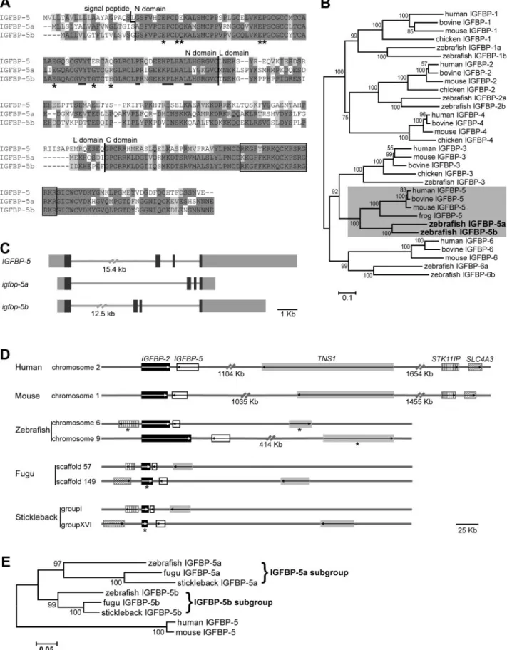
By searching public databases, screening a cDNA library, and performing 5⬘- and 3⬘-RACE experiments, we identi-fied and cloned 2 distinct zebrafish genes (GenBank accession numbers GQ892882 and AY100478). For rea-sons evident thereafter, we termed them asigfbp-5aand-5b and the encoded proteins as IGFBP-5a and -5b. As shown inFig. 1A,zebrafish IGFBP-5a has a putative signal pep-tide of 19 amino acids (aa) and a mature protein of 249 aa. IGFBP-5b has a putative signal peptide of 17 aa and a mature protein of 248 aa. Comparison of the 2 zebrafish IGFBP-5 sequences with 6 human IGFBPs revealed that they share the highest sequence identities with that of human IGFBP-5 (47–52%; see Supplemental Table 4). Their sequence identities to human IGFBP-3 are 36 and
37%, and below 30% to other human IGFBPs. There is a typical IGFBP motif in the N domain and a thyroglobulin type-1 repeat in the C domain in both zebrafish IGFBP-5s. Both proteins contain a consensus LBD motif (3) in the N domain and an NLS motif (4) in the C domain (Fig. 1A). Phylogenetic analysis grouped both proteins into the IGFBP-5 subgroup (Fig. 1B). The 2 zebrafish igfbp-5 genes also share similar exon/intron organization with the humanIGFBP-5gene: they all contain 4 exons and 3 introns (Fig. 1C). Although zebrafish igfbp-5a is lo-cated on LG6,igfbp-5bis on LG9. The 2 zebrafishigfbp-5 genes are adjacent to the 2 previously reportedigfbp-2 genes (28), arranged in a tail-to-tail fashion (Fig. 1D). This is very similar to the situations in the human and mouse genomes (35).
To determine whether there are 2 igfbp-5 genes in other teleost fish, we searched the fugu, stickleback, medaka, and tetraodon genome databases and found that they all contain 2igfbp-5genes. Again, the 2igfbp-5 genes are adjacent to 2 igfbp-2 genes in a tail-to-tail fashion in all these teleost genomes (Fig. 1D). Likewise, there are several other syntenic genes (TNS1, STK11IP, SLC4A3) (Fig. 1D). Phylogenetic analysis indicated that the duplication of the igfbp-5a/b subfamily likely origi-nated from a genome duplication event that occurred early during ray-fin fish evolution (Fig. 1E).
The duplicatedigfbp-5genes exhibit distinct expression patterns
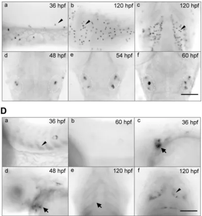
As shown inFig. 2A,in adult tissues,igfbp-5amRNA was detected in brain and gill at high levels. It was also detected in eye, heart, gut, kidney, and gonad, but not in liver and muscle by RT-PCR (Fig. 2A). In comparison, igfbp-5bmRNA was expressed in all adult tissues examined. There were no obvious gender differences (Fig. 2A). During early development,igfbp-5amRNA was not detect-able until 14 h postfertilization (hpf). It gradually in-creased from 14 to 72 hpf and was maintained at high levels thereafter (Fig. 2B). In 8-hpf embryos, igfbp-5b mRNA was detected at low levels. Starting from 12 hpf, igfbp-5bmRNA levels maintained at high levels thereafter (Fig. 2B). The results of whole-mountin situhybridization analysis are shown in Fig. 2C,D.We first detectedigfbp-5a mRNA at 20 hpf in a small number of cells on the surface of the yolk sac and yolk tube, and this became more evident at 36 hpf (Fig. 2Ca). The number of igfbp-5a mRNA-expressing cells increased as the embryos grew (Fig. 2Cb). At 96 and 120 hpf, igfbp-5a mRNA was also highly expressed in cells spreading in the gill filament regions (Fig. 2Cc). We also detected igfbp-5amRNA in a small number of cells located within the inner ear (Fig. 2Cd–f). In contrast,igfbp-5bmRNA was primarily detected in differentiating somites, gill arches, pectoral fin, and some neural tissues (Fig. 2D). Its expression in the somites disappeared at 60 hpf (Fig. 2Db). At 36 and 48 hpf,igfbp-5b mRNA was also highly expressed in the epithelial cells in the otic vesicles (Fig. 2Dc,d). In larvae (120 hpf),igfbp-5b mRNA was detected in the layers of cells surrounding the gill cartilage (Fig. 2De) and some cells in the brain (Fig.
Figure 1.There are 2igfbp-5genes in zebrafish and other teleost fish.A) Sequence alignment of human IGFBP-5 and zebrafish IGFBP-5a and -5b. Identical and similar amino acid residues are darkly and lightly shaded, respectively. Vertical lines indicate the boundaries between signal peptide, N, L, and C domains. Residues in the N domain known to be critical for IGF binding (LBD) are underlined. NLS is shown in box. Asterisks indicate residues that are critical for the TA activity of human IGFBP-5.B) Phylogenetic tree of the IGFBP family. Values on branches are percentages of replicate trees in which the genes clustered together in the bootstrap test (1000 replicates). Branch lengths are drawn in units of 0.1 aa substitutions/site.C) Structure of humanIGFBP-5and zebrafish
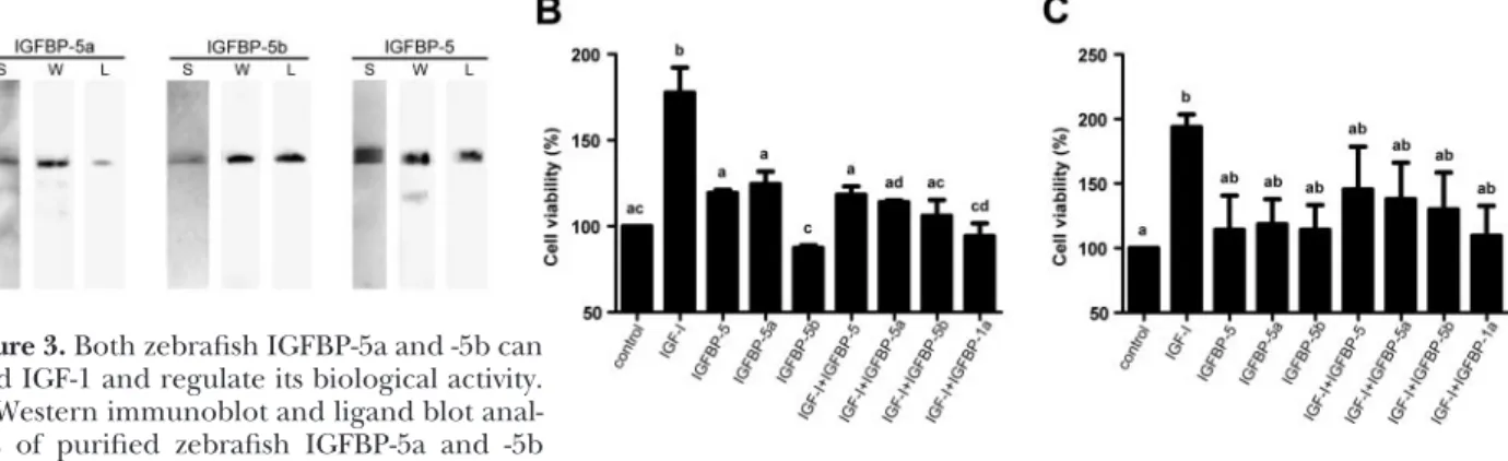
2Df). These results suggest that the 2 igfb-5 genes are ex-pressed in nonoverlapping domains during development. Both IGFBP-5a and -5b are secreted proteins, and they both bind to IGF and modulate IGF actions To test whether zebrafishigfbp-5a and -5b encode func-tional IGFBPs, recombinant zebrafish IGFBP-5a and -5b and human IGFBP-5 were produced in HEK 293 cells and purified from the culture medium. HEK 293 cells were used because of their high transfection efficiency. Human and zebrafish IGFBP-5s had apparent sizes of⬃36 kDa on SDS-PAGE (Fig. 3A), and they were all able to bind IGF-1, as shown by ligand blot (Fig. 3A).
We next determined the biological activities of ze-brafish IGFBP-5a and -5b and compared them to that of
human IGFBP-5. As shown in Fig. 3B,addition of IGF-1 to cultured U2OS cells caused a significant increase in cell growth. When human IGFBP-5, zebrafish IGFBP-5a, or zebrafish IGFBP-5b was added together with IGF-1 at a 1:1 M ratio, they abolished the IGF-1-induced increase. Addi-tion of any one of these IGFBP-5s alone had little effect. Similar results were also obtained in HEK 293 cells (Fig. 3C). These results suggest that zebrafishigfbp-5a and -5b encode secreted proteins that bind IGF-1 and modulate IGF-1 actions.
Both IGFBP-5a and -5b are localized in the nucleus, but only IGFBP-5b has TA activity
Previous studies have shown that human IGFBP-5 is not only secreted but can also be found in the nucleus of
UTRs and ORF, respectively.D) Chromosomal loci ofIGFBP-5genes in various vertebrate genomes. Arrows indicate the transcript orientation.IGFBP-2, IGFBP-5, TNS1, STK11IP,andSLC4A3are shown as black box, open box, gray box, vertical-line filled box, and wave-line filled box, respectively. Asterisk indicates partial transcript information from gene annotation. (Chromosomal locus of
SLC4A3in zebrafish is unknown.)E) Phylogenetic analysis of several teleost IGFBP-5a and -5b. Values on branches are percentages of replicate trees in which genes clustered together in the bootstrap test (1000 replicates). Branch lengths are drawn in units of 0.05 aa substitutions/site.
Figure 2.Zebrafishigfbp-5aand-5bexhibit distinct spatial and temporal expression patterns.A) RT-PCR analysis ofigfbp-5aand-5bmRNAs in female and male adult zebrafish tissues.B) RT-PCR
anal-ysis ofigfbp-5aand-5bmRNAs in zebrafish embryos at the indicated stages. Developmental stages are shown at top. hpf, hours postfertilization.C)In situhybridization analysis ofigfbp-5amRNA in whole-mount zebrafish embryos. Developmen-tal stage is at top right in each panel.a,b) Lateral view of yolk tube/sac region with anterior at left.c) Ventral view of gill arch region with anterior at top. Arrowheads indicate one representative epidermal cell.d–f) Dorsal view of head region with anterior at top. Dotted line indicates position of otic vesicles.D)In situhybridization analysis ofigfbp-5bmRNA.a,b) Lateral view of trunk region with anterior at left. Arrowhead indicates signals in somites.c,d) Lateral view of head region with anterior at left. Dotted line indicates position of otic vesicles.e) Ventral view of gill arch region with anterior at top. Arrows indicate signals in gill arch.f) Dorsal view of head region with anterior at top. Arrowhead indicates signal in midbrain. Scale bars⫽100m.
cultured mammalian cells and mouse embryos (4, 17–19, 21). Furthermore, the IGFBP-5 N domain has TA activity (19, 21). Because both zebrafish IGFBP-5s contain a consensus NLS in their C domains (Fig. 1A, open box), we investigated the possible nuclear local-ization of zebrafish IGFBP-5a and -5b. U2OS cells were chosen for subcellular localization analysis because of their large and flattened cell bodies. When cells were transfected with human IGFBP-4:EGFP, the EGFP sig-nal was detected only in the cytoplasm (Fig. 4A). But when cells were transfected with human IGFBP-5:EGFP, the EGFP signal was seen in the nucleus. Like human
IGFBP-5:EGFP, zebrafish IGFBP-5a:EGFP, and -5b: EGFP were also found in the nucleus (Fig. 4A). Similar results were also observed in HEK 293 cells (data not shown).
We next investigated whether zebrafish IGFBP-5a and/or -5b have any TA activity. HEK 293 cells were used because of the high transfection efficiency (21). The zebrafish IGFBP-5b N domain caused a GAL4-dependent TA 4-fold greater than the pBIND control group when tested in HEK 293 cells (Fig. 4B). In contrast, the zebrafish IG-FBP-5a N domain did not cause any significant increase. As reported previously (19, 21), the human IGFBP-5 N Figure 3.Both zebrafish IGFBP-5a and -5b can
bind IGF-1 and regulate its biological activity.
A) Western immunoblot and ligand blot anal-yses of purified zebrafish IGFBP-5a and -5b
and human IGFBP-5 proteins. L, ligand blot with DIG-labeled IGF-1; S, silver staining; W, Western immunoblot with an anti-myc antibody.B, C) Effects of various IGFBP-5s on IGF-1-stimulated cell growth in U2OS cells (B) and HEK 293 cells (C). Values are means ⫾sd of 2 separate experiments, each performed in triplicate. Groups with different letters are significantly different from each other (P⬍0.05).
Figure 4. Nuclear localization and TA activities of zebrafish IGFBP-5a and -5b.
A) Subcellular localization of IGFBP:EGFP. U2OS cells were transfected with human IGFBP-4:EGFP (IGFBP-4), human IGFBP-5:EGFP (IGFBP-5), zebrafish IGFBP-5a: EGFP (IGFBP-5a), and zebrafish IGFBP-5b:EGFP (IGFBP-5b) expression plasmid. EGFP signal was visualized (left panels) 24 h after transfection. Corresponding DAPI staining is shown in the middle panels and merged views in the right panels. Scale bar⫽25m.B, C) N domain of zebrafish IGFBP-5b but not that of IGFBP-5a has TA activity in HEK 293 cells (B) and ZF4 cells (C). TA activity is expressed as fold over the pBIND control group. Values are means⫾sd(n⫽3–5). Groups with different letters are significantly different from each other (P⬍0.05).D) Expression levels of fusion proteins were analyzed by Western immunoblot using an anti-GAL4 (DBD) antibody and an anti-tubulin antibody.
domain caused a 20-fold increase in activating the re-porter gene expression. To rule out the possibility of species-specific effect, the TA activities of these fusion proteins were also tested in ZF4 cells, a cell line derived from zebrafish embryos. In these zebrafish cells, human IGFBP-5 still had the strongest activity (9-fold increase over the pBIND control, P⬍0.001) and the zebrafish IGFBP-5b N domain had significant activity (3-fold in-crease,P⬍0.05). Again, the zebrafish IGFBP-5a N domain had no activity (Fig. 4C). Western immunoblot analysis revealed that expression levels of these fusion proteins were similar (Fig. 4D), thus excluding the possibility that the difference was due to different levels of protein expression and/or degradation.
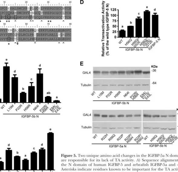
Several unique residues in the IGFBP-5 N domain are critical for the TA activity
The 2 zebrafish IGFBP-5s share the same domain arrangement, high sequence identity, nuclear
localiza-tion, and the ability to bind IGFs, but only IGFBP-5b has TA activity. Taking advantage of this finding, we compared the amino acid sequences of these 2 highly homologous proteins with that of human IGFBP-5. Among the 8 residues in the human IGFBP-5 N domain that are known to be critical for its TA ability (21), 5 are conserved in zebrafish IGFBP-5a and -5b (Fig. 5A, asterisk). When 3 of these conserved residues in ze-brafish IGFBP-5b were substituted with their corre-sponding residues from human IGFBP-1 (E8A/D11S/ E43L), the TA activity was abolished (Fig. 5B). Because zebrafish IGFBP-5b, but not IGFBP-5a, has TA activity, we focused on the 12 residues that differed between these 2 zebrafish IGFBP-5s in this region (Fig. 5A, caret). Among the 4 tested, we found that changing P22 or H56 in IGFBP-5b to the corresponding residue in IGFBP-5a reduced the TA activity by ⬎50%, and the P22R/H56R double mutant had essentially no TA activity (Fig. 5B). In comparison, the L15M and N64I
Figure 5.Two unique amino acid changes in the IGFBP-5a N domain are responsible for its lack of TA activity.A) Sequence alignment of the N domain of human IGFBP-5 and zebrafish IGFBP-5a and -5b. Asterisks indicate residues known to be important for the TA activity of human IGFBP-5; carets indicate residues that differ between zebrafish IGFBP-5a and -5b; open squares indicate residues tested in this study. B–D) TA activities of wild-type and mutant zebrafish IGFBP-5b and -5a. Indicated constructs were introduced into HEK 293 cells together with a GAL4 reporter plasmid by transient transfection. Values are expressed as percentages of the wild-type zebrafish IGFBP-5b N-domain group (B, C) or the human IGFBP-5 N-domain group (D). Values are means⫾sd
(n⫽3). Groups with the same letters are not significantly different from each other (P⬍0.05).E) Expression levels of mutant fusion proteins were analyzed by Western immunoblot using an anti-GAL4 (DBD) antibody and an anti-tubulin antibody.
mutants had only moderate effects. These results sug-gest that R22 and R56 in the IGFBP-5a N domain are largely responsible for its lack of TA activity. To further test whether these 2 positions are sufficient to establish the TA activity, we changed R22 and R56 in the zebrafish IGFBP-5a N domain to the corresponding residues of IGFBP-5b in individual and double mutants. The R22P mutant had significant TA activity. The R22P/R56H dou-ble mutant had TA activity comparadou-ble to that of zebrafish IGFBP-5b (Fig. 5C). Taken together, our results suggest that the 2 different amino acids in positions 22 and 56 are responsible for the different TA activity observed in the 2 zebrafish IGFBP-5s.
We also investigated the structural determinants ac-counting for the different activities observed between human IGFBP-5 and zebrafish IGFBP-5b. Among the 8 residues in the human IGFBP-5 N domain that are known to be critical for its TA ability (21), 3 of them differ between zebrafish IGFBP-5b and human IGFBP-5 (Fig. 5A, asterisk). Changing the zebrafish IGFBP-5 resi-due at position 56 into the corresponding resiresi-due from the human sequence resulted in a significant increase in its activity (R56Q and R22P/R56Q mutants in Fig. 5Cand H56Q mutant in Fig. 5D). We further generated double and triple zebrafish IGFBP-5b mutants, H56Q/G52E and H56Q/G52E/Q12E, by changing residues from the ze-brafish IGFBP-5b into the corresponding ones from the human IGFBP-5 sequence. The H56Q/G52E mutant had 80% activity compared to the human IGFBP-5 (Fig. 5D), and the H56Q/G52E/Q12E mutant fully achieved the same high TA activity as human IGFBP-5. All mutants were expressed at comparable levels as shown by immu-noblot (Fig. 5E). These results indicate that the difference in TA activities observed between human IGFBP-5 and zebrafish IGFBP-5b is due to their different amino acid residues in positions 12, 52, and 56.
DISCUSSION
In this study we identified 2igfbp-5 genes in zebrafish. Several lines of evidence indicated that they are co-orthologs of humanIGFBP-5: 1) sequence comparison at the protein level showed that they share the highest identity with human IGFBP-5,2) phylogenetic analysis grouped the 2 zebrafish IGFBPs in the IGFBP-5 cluster, 3) the gene structure and exon/intron size are most similar to humanIGFBP-5,and4) the chromosome loci indicated conserved synteny with human IGFBP-5.We also found 2igfbp-5genes in other teleost fish. Both the conserved synteny and phylogenetic analyses indicate that the duplication event that produced these 2 genes happened early in teleost evolution (Fig. 6).
In humans and mice, IGFBP-5 mRNA has been de-tected in a wide range of tissues and cell types. Analyzing the expression patterns in zebrafish, we found that the 2 duplicateigfbp-5genes diverge in expression pattern both spatially and temporally. In adult tissues, althoughigfbp-5b is expressed in all the tissues examined at relatively high levels,igfbp-5ais most strongly expressed in brain and gill.
During embryogenesis,igfbp-5bis expressed earlier than igfbp-5a.More interestingly, they are expressed in dis-tinct tissues and cells. Although igfbp-5a expression is restricted in the epidermal cells and in the inner ear, igfbp-5b is expressed in somites, branchial arches, pec-toral fin, and several domains in the brain. The diver-gent temporal and spatial expression patterns indicate that the cis-regulatory elements in these 2 genes may have diverged after the duplication event. Studies of duplicated genes in zebrafish indicated that this may be a common mechanism for diversification (36, 37). Further studies are needed to determine the conserved and divergent cis-regulatory elements in these 2 ze-brafishigfbp-5genes.
Mammalian IGFBP-5 is a multifunctional protein that contains several structural modules/domains. We performed molecular, biochemical and cell biological approaches to identify structural components that have diversified in the duplicate zebrafish IGFBP-5s. We found that although some of the domains are preserved in both genes (LBD and NLS), others (TA) diverged during evolution (Fig. 6). Protein function usually requires intra- and intermolecular domain interactions. Therefore, the related domains usually coevolve (38). It has been shown that TA activity and nuclear localiza-tion are well correlated in the 6 IGFBPs, with IGFBP-3 and -5 showing nuclear localization and possessing the highest TA activity (21). It is intriguing to ask why IGFBP-5a preserves the NLS while losing the TA do-main. One possible explanation from the structural point of view is that the conserved stretch of basic residues in the C domain has multiple functional roles. In addition to being a functional NLS (4, 19), this region is also involved in IGFBP-5 interaction with ALS, heparin, and ECM components (39, 40). Therefore, selection force may conserve these resi-dues even if one of their functions become unneces-Figure 6. Proposed model for the gene expression and functional divergence of igfbp-5 genes in teleost fish. The ancestralIGFBP-5gene was duplicated early in teleost evolu-tion as the result of a genome duplicaevolu-tion event. The dupli-catedigfbp-5genes may have undergone subfunctionalization at the levels of expression pattern, through divergent expres-sion regulation (indicated by different arrows), and protein functionality, through the loss of critical structural motifs, such as the TA domain in the case of zebrafishigfbp-5a.
sary. The LBD motif and the TA domain have been shown to be structurally separable and functionally independent (19). It is possible that IGFBP-5a may have lost the TA activity while retaining its IGF-binding function.
The 2 zebrafish IGFBP-5s share the same domain arrangement, high sequence identity, and the ability to bind IGFs, but only zebrafish IGFBP-5b has TA activity. Taking advantage of this finding, we were able to identify the key residues critical for the TA activity. By swapping their different residues, we discovered that 2 residues at positions 22 and 56 are both necessary and sufficient for the TA activity in zebrafish IGFBP-5s. These results indicate that IGFBP-5a lacks TA activity because of its unique residues in these critical positions. This conclusion is also consistent with our current understanding of gene evolution. After gene duplica-tion, it is thought that the duplicated genes are likely to be retained if they acquire nonredundant functions (41). The high divergence of the TA domains observed in the duplicated zebrafishigfbp-5paralogs may account for an adaptation or specialization of function of these 2 genes. We speculate that this may not be unique to zebrafish, as aligning the IGFBP-5 N domain sequences of zebrafish, fugu, and stickleback together with that of human and mouse suggests that position 56 showed clear divergence in the 2 branches. Specifically, it is an R in all members of the IGFBP-5a group, whereas Q or H in the IGFBP-5b group (Supplemental Fig. 1). The other position, 22, identified in this study does not seem to be conserved in the 2 groups, suggesting that this residue change is a more recent event during evolution in zebrafish. When we tested the TA activity in ancestral IGFBPs in amphioxus (unpublished re-sults), we found that they do exhibit high TA activity like the human IGFBP-5, suggesting that this activity has an ancient origin.
It has been shown that zebrafish contain 2igf-1genes, 2 igf-2 genes, and 2 igf-1rgenes (26, 42). In addition, zebrafish have 2 igfbp-1 genes, 2 igfbp-2 genes, and 2 igfbp-6genes (27–29). In this study, we provide evidence that there are 2 functionaligfbp-5genes in zebrafish and other teleost fish,igfbp-5aandigfbp-5b.We show that the duplicated igfbp-5s exhibited nonoverlapping expres-sion patterns during zebrafish embryogenesis. We also determined the structural changes accounting for func-tional divergence by mapping critical amino acid changes in the TA domain. These findings provide insight into the evolution of the IGFBP gene family and lay the foundations for further elucidation of the physiological functions of the nuclear IGFBP-5 and its TA activityin vivo.
The authors thank John Allard for critical reading of this manuscript. This study was supported by U.S. National Insti-tutes of Health grant 2RO1HL60679 and National Science Foundation research grant IOB 0110864 to C.D. H.K. is supported in part by a postdoctoral fellowship from the Japan Society for the Promotion of Science.
REFERENCES
1. Firth, S. M., and Baxter, R. C. (2002) Cellular actions of the insulin-like growth factor binding proteins. Endocr. Rev. 23,
824 – 854
2. Duan, C., and Xu, Q. (2005) Roles of insulin-like growth factor (IGF) binding proteins in regulating IGF actions.Gen. Comp. Endocrinol.142,44 –52
3. Kalus, W., Zweckstetter, M., Renner, C., Sanchez, Y., Georgescu, J., Grol, M., Demuth, D., Schumacher, R., Dony, C., Lang, K., and Holak, T. A. (1998) Structure of the IGF-binding domain of the insulin-like growth factor-binding protein-5 (IGFBP-5): im-plications for IGF and IGF-I receptor interactions.EMBO J.17,
6558 – 6572
4. Schedlich, L. J., Le Page, S. L., Firth, S. M., Briggs, L. J., Jans, D. A., and Baxter, R. C. (2000) Nuclear import of insulin-like growth factor-binding protein-3 and -5 is mediated by the importin beta subunit.J. Biol. Chem.275,23462–23470 5. Rozen, F., Yang, X. F., Huynh, H., and Pollak, M. (1997)
Antiproliferative action of vitamin D-related compounds and insulin-like growth factor-binding protein 5 accumulation. J. Natl. Cancer Inst.89,652– 656
6. Mohan, S., Nakao, Y., Honda, Y., Landale, E., Leser, U., Dony, C., Lang, K., and Baylink, D. J. (1995) Studies on the mecha-nisms by which insulin-like growth factor (IGF) binding pro-tein-4 (IGFBP-4) and IGFBP-5 modulate IGF actions in bone cells.J. Biol. Chem.270,20424 –20431
7. Parker, A., Rees, C., Clarke, J., Busby, W. H., Jr., and Clemmons, D. R. (1998) Binding of insulin-like growth factor (IGF) -bind-ing protein-5 to smooth-muscle cell extracellular matrix is a major determinant of the cellular response to IGF-I.Mol. Biol. Cell9,2383–2392
8. Ren, H., Yin, P., and Duan, C. (2008) IGFBP-5 regulates muscle cell differentiation by binding to IGF-II and switching on the IGF-II auto-regulation loop.J. Cell Biol.182,979 –991
9. Abrass, C. K., Berfield, A. K., and Andress, D. L. (1997) Heparin binding domain of insulin-like growth factor binding protein-5 stimulates mesangial cell migration.Am. J. Physiol.273,F899 – F906
10. Berfield, A. K., Andress, D. L., and Abrass, C. K. (2000) IGFBP-5(201–218) stimulates Cdc42GAP aggregation and filop-odia formation in migrating mesangial cells. Kidney Int. 57,
1991–2003
11. Hsieh, T., Gordon, R. E., Clemmons, D. R., Busby, W. H., Jr., and Duan, C. (2003) Regulation of vascular smooth muscle cell responses to insulin-like growth factor (IGF) -I by local IGF-binding proteins.J. Biol. Chem.278,42886 – 42892
12. Miyakoshi, N., Richman, C., Kasukawa, Y., Linkhart, T. A., Baylink, D. J., and Mohan, S. (2001) Evidence that IGF-binding protein-5 functions as a growth factor.J. Clin. Invest.107,73– 81 13. Salih, D. A., Tripathi, G., Holding, C., Szestak, T. A., Gonzalez, M. I., Carter, E. J., Cobb, L. J., Eisemann, J. E., and Pell, J. M. (2004) Insulin-like growth factor-binding protein 5 (Igfbp5) compromises survival, growth, muscle development, and fertility in mice.Proc. Natl. Acad. Sci. U. S. A.101,4314 – 4319 14. Ning, Y., Hoang, B., Schuller, A. G., Cominski, T. P., Hsu, M. S.,
Wood, T. L., and Pintar, J. E. (2007) Delayed mammary gland involution in mice with mutation of the insulin-like growth factor binding protein 5 gene.Endocrinology148,2138 –2147 15. Tripathi, G., Salih, D. A., Drozd, A. C., Cosgrove, R. A., Cobb,
L. J., and Pell, J. M. (2009) IGF-independent effects of insulin-like growth factor binding protein-5 (Igfbp5) in vivo.FASEB J. 23,2616 –2626
16. Jaques, G., Noll, K., Wegmann, B., Witten, S., Kogan, E., Radulescu, R. T., and Havemann, K. (1997) Nuclear localization of insulin-like growth factor binding protein 3 in a lung cancer cell line.Endocrinology138,1767–1770
17. Schedlich, L. J., Young, T. F., Firth, S. M., and Baxter, R. C. (1998) Insulin-like growth factor-binding protein (IGFBP)-3 and IGFBP-5 share a common nuclear transport pathway in T47D human breast carcinoma cells.J. Biol. Chem.273,18347– 18352
18. Amaar, Y. G., Thompson, G. R., Linkhart, T. A., Chen, S. T., Baylink, D. J., and Mohan, S. (2002) Insulin-like growth factor-binding protein 5 (IGFBP-5) interacts with a four and a half LIM protein 2 (FHL2).J. Biol. Chem.277,12053–12060
19. Xu, Q., Li, S., Zhao, Y., Maures, T. J., Yin, P., and Duan, C. (2004) Evidence that IGF binding protein-5 functions as a ligand-independent transcriptional regulator in vascular smooth muscle cells.Circ. Res.94,E46 –E54
20. Li, W., Fawcett, J., Widmer, H. R., Fielder, P. J., Rabkin, R., and Keller, G. A. (1997) Nuclear transport of insulin-like growth factor-I and insulin-like growth factor binding protein-3 in opossum kidney cells.Endocrinology138,1763–1766
21. Zhao, Y., Yin, P., Bach, L. A., and Duan, C. (2006) Several acidic amino acids in the N-domain of insulin-like growth factor-binding protein-5 are important for its transactivation activity. J. Biol. Chem.281,14184 –14191
22. Wood, A. W., Duan, C., and Bern, H. A. (2005) Insulin-like growth factor signaling in fish.Int. Rev. Cytol.243,215–285 23. Taylor, J. S., Braasch, I., Frickey, T., Meyer, A., and Van de Peer,
Y. (2003) Genome duplication, a trait shared by 22000 species of ray-finned fish.Genome Res.13,382–390
24. Postlethwait, J., Amores, A., Cresko, W., Singer, A., and Yan, Y. L. (2004) Subfunction partitioning, the teleost radiation and the annotation of the human genome.Trends Genet.20,481– 490 25. Maures, T., Chan, S. J., Xu, B., Sun, H., Ding, J., and Duan, C.
(2002) Structural, biochemical, and expression analysis of two distinct insulin-like growth factor I receptors and their ligands in zebrafish.Endocrinology143,1858 –1871
26. Schlueter, P. J., Royer, T., Farah, M. H., Laser, B., Chan, S. J., Steiner, D. F., and Duan, C. (2006) Gene duplication and functional divergence of the zebrafish insulin-like growth factor 1 receptors.FASEB J.20,1230 –1232
27. Kamei, H., Lu, L., Jiao, S., Li, Y., Gyrup, C., Laursen, L. S., Oxvig, C., Zhou, J., and Duan, C. (2008) Duplication and diversification of the hypoxia-inducible IGFBP-1 gene in ze-brafish.PLoS ONE3,e3091
28. Zhou, J., Li, W., Kamei, H., and Duan, C. (2008) Duplication of the IGFBP-2 gene in teleost fish: protein structure and function-ality conservation and gene expression divergence.PLoS ONE3,
e3926
29. Wang, X., Lu, L., Li, Y., Li, M., Chen, C., Feng, Q., Zhang, C., and Duan, C. (2009) Molecular and functional characterization of two distinct IGF binding protein-6 genes in zebrafish.Am. J. Physiol.296,R1348 –R1357
30. Kimmel, C. B., Ballard, W. W., Kimmel, S. R., Ullmann, B., and Schilling, T. F. (1995) Stages of embryonic development of the zebrafish.Dev. Dyn.203,253–310
31. Larkin, M. A., Blackshields, G., Brown, N. P., Chenna, R., McGettigan, P. A., McWilliam, H., Valentin, F., Wallace, I. M., Wilm, A., Lopez, R., Thompson, J. D., Gibson, T. J., and Higgins,
D. G. (2007) Clustal W and Clustal X version 2.0.Bioinformatics 23,2947–2948
32. Tamura, K., Dudley, J., Nei, M., and Kumar, S. (2007) MEGA4: Molecular Evolutionary Genetics Analysis (MEGA) software version 4.0.Mol. Biol. Evol.24,1596 –1599
33. Li, Y., Xiang, J., and Duan, C. (2005) Insulin-like growth factor-binding protein-3 plays an important role in regulating pharyngeal skeleton and inner ear formation and differentia-tion.J. Biol. Chem.280,3613–3620
34. Shimizu, M., Swanson, P., Fukada, H., Hara, A., and Dickhoff, W. W. (2000) Comparison of extraction methods and assay validation for salmon insulin-like growth factor-I using commer-cially available components.Gen. Comp. Endocrinol.119,26 –36 35. Beattie, J., Allan, G. J., Lochrie, J. D., and Flint, D. J. (2006)
Insulin-like growth factor-binding protein-5 (IGFBP-5): a critical member of the IGF axis.Biochem. J.395,1–19
36. McClintock, J. M., Kheirbek, M. A., and Prince, V. E. (2002) Knockdown of duplicated zebrafish hoxb1 genes reveals distinct roles in hindbrain patterning and a novel mechanism of dupli-cate gene retention.Development129,2339 –2354
37. Kleinjan, D. A., Bancewicz, R. M., Gautier, P., Dahm, R., Schonthaler, H. B., Damante, G., Seawright, A., Hever, A. M., Yeyati, P. L., van Heyningen, V., and Coutinho, P. (2008) Subfunctionalization of duplicated zebrafish pax6 genes by cis-regulatory divergence.PLoS Genet.4,e29
38. Chothia, C., Gough, J., Vogel, C., and Teichmann, S. A. (2003) Evolution of the protein repertoire.Science300,1701–1703 39. Firth, S. M., Clemmons, D. R., and Baxter, R. C. (2001)
Mutagenesis of basic amino acids in the carboxyl-terminal region of insulin-like growth factor binding protein-5 affects acid-labile subunit binding.Endocrinology142,2147
40. Arai, T., Clarke, J., Parker, A., Busby, W., Jr., Nam, T., and Clemmons, D. R. (1996) Substitution of specific amino acids in insulin-like growth factor (IGF) binding protein 5 alters heparin binding and its change in affinity for IGF-I response to heparin. J. Biol. Chem.271,6099 – 6106
41. Force, A., Lynch, M., Pickett, F. B., Amores, A., Yan, Y. L., and Postlethwait, J. (1999) Preservation of duplicate genes by com-plementary, degenerative mutations.Genetics151,1531–1545 42. Zou, S., Kamei, H., Modi, Z., and Duan, C. (2009) Zebrafish IGF
genes: gene duplication, conservation and divergence, and novel roles in midline and notochord development.PLoS ONE 4,e7026
Received for publication October 29, 2009. Accepted for publication December 17, 2009.