East Tennessee State University
Digital Commons @ East
Tennessee State University
Electronic Theses and Dissertations Student Works
5-2018
A Survey of the Implementation and Usage of
Electronic Dental Records and Digital Radiographs
in Private Dental Practices in Mississippi
Barbara K. Brent
East Tennessee State Universtiy
Follow this and additional works at:https://dc.etsu.edu/etd
Part of theDental Public Health and Education Commons, and theOther Dentistry Commons
This Thesis - Open Access is brought to you for free and open access by the Student Works at Digital Commons @ East Tennessee State University. It has been accepted for inclusion in Electronic Theses and Dissertations by an authorized administrator of Digital Commons @ East Tennessee State University. For more information, please contactdigilib@etsu.edu.
Recommended Citation
Brent, Barbara K., "A Survey of the Implementation and Usage of Electronic Dental Records and Digital Radiographs in Private Dental Practices in Mississippi" (2018).Electronic Theses and Dissertations.Paper 3365. https://dc.etsu.edu/etd/3365
A Survey of the Implementation and Usage of Electronic Dental Records and Digital Radiographs in Private Dental Practices in Mississippi
_______________________________ A thesis
presented to
the faculty of the Department of Allied Health Sciences East Tennessee State University
In partial fulfillment of the requirements for the degree Master of Science in Allied Health
____________________ by
Barbara Brent May 2018
____________________
Dr. Ester L. Verhovsek, EdD, Chair Dr. Randy Byington, EdD Dr. Deborah Dotson, PhD
Dr. Susan Epps, EdD
Keywords: electronic health records, digital radiography, conventional radiography, dental, private practice, dental health records, electronic dental records
2
ABSTRACT
A Survey of the Implementation and Usage of Electronic Dental Records and Digital Radiographs in Private Dental Practices in Mississippi
by Barbara Brent
Implementation of electronic health records by the Health Information Technology for Economic and Clinical Health has led to the implementation of electronic dental records
(EDRs) and digital radiography in dental offices. The purpose of this study was to determine the state of the implementation and usage of EDRs and digital radiographs by the private general and pediatric dental practices in Mississippi as well as reasons why the dental practices are not moving forward with the advanced technology.
A survey was emailed to 712 dental practices: 116 responded (16% response rate), and 104 consented to participate (89.66%). Results indicated dental practices in Mississippi using EDRs was 46.07%, EDRs with paper records was 42.70%, and only paper records was 11.24%. Results indicated dental practices using digital radiography was 76.40%, conventional
radiography was 13.48%, and both was 10.11%. Common reasons for not advancing were cost, insufficient training, computer/software issues, and “too old.”
3
DEDICATION
I dedicate this thesis to my husband, children, and parents. You are the most important aspect in my life, second only to my relationship with Jesus Christ. Without your constant support, understanding, and love during this process, I would not have been able to accomplish this milestone in my life. Thank you for your belief in me through this process.
4
ACKNOWLEDGEMENTS
I would like to express gratitude and appreciation to my thesis committee: Dr. Ester Verhovsek, Dr. Randy Byington, Dr. Susan Epps, and Dr. Deborah Dotson. Your knowledge, guidance, and assistance in the development and writing of this thesis was invaluable. Words cannot express my gratitude for the support you provided during my family’s crisis. A special thanks to my chair, Dr. Verhovsek, for your constant encouragement. Our conversations came at the time I needed a boost of confidence. Thank you!
A special thank you to my coworkers at the University of Mississippi Dental Hygiene Program. Thank you for listening, encouraging, and providing guidance during the writing of this thesis. Thank you to Dr. Kim Tolbert and Lisa Owen, R.D.H. for your support and encouragement.
5 TABLE OF CONTENTS Page ABSTRACT……… 2 DEDICATION……… 3 ACKNOWLEDGEMENTS………... 4 LIST OF FIGURES...………. 9 Chapter 1. INTRODUCTION……….. 10 Statement of Problem………. 14
Purpose of the Study……….. 15
Research Questions……… 15 Significance of Study………. 15 Delimitations……….. 16 Limitations………. 16 Assumptions……….. 16 Operational Definitions………. 16 2. LITERATURE REVIEW……….. 18
Electronic Health Records……… 18
Electronic Medical Records……… 21
Electronic Dental Records……….. 21
EDRs and Medically Complex Conditions……… 23
EDRs and Digital Radiography……….………. 24
Digital Radiography……… 25
6
Types of Digital Images………. 27
Direct Digital Images ……… 27
Indirect Digital Images ………. 28
Conventional Film-based Radiography……….. 29
Digital Radiography versus Conventional Radiography……… 29
Advantages……….. 30 Disadvantages………. 31 Summary………. 32 3. METHODOLOGY………. 33 Overview……… 33 Research Questions……… 33 Research Design………. 33
Strengths and Limitations of the Design……… 34
Population……….. 34
Informed Consent……….. 35
Survey Instrument Development……… 35
Instrument Validity……….……… 36
Data Collection Procedure………. 37
Data Analysis……….. 39
Summary………. 39
4. RESULTS……… 40
Introduction………. 40
7
Practices’ Demographics………. 41
Analysis of Data……….. 43
Research Question 1: Are private dental practices in Mississippi currently using electronic dental records?... 43
Research Question 2: What factors influence the selection of paper dental records or electronic dental records by private dental practices in Mississippi?... 44
Research Question 3: Are private dental practices in Mississippi currently using digital radiography?... 45
Research Question 4: What factors influence the selection of radiography exposure technique, digital or conventional, by private dental practices in Mississippi?... 46
Research Question 5: What factors influence the selection of the type of digital radiography, direct or indirect, by private dental practices in Mississippi?... 47
Electronic Dental Records Conversion Plans Data………. 47
Digital Radiography Conversion Plans Data……….. 49
Summary………. 50
5. SUMMARY OF FINDINGS, CONCLUSIONS, AND RECOMMENDATIONS……… 51
Overview………. 51
Summary of Findings……….. 51
Dental Records……… 52
8
Conclusions………. 56
Recommendations for Change to Electronic Dental Records……….…… 58
Future Research……… 58
REFERENCES……… 60
APPENDICES………. 67
Appendix A: Core Objectives and Menu Objectives of the Meaningful Use Incentive Plan………. 67
Appendix B: Survey Instrument……… 69
Appendix C: Invitation Email……… 71
Appendix D: Email for Pilot Study……… 73
Appendix E: Email Reminder ………... 75
9
LIST OF FIGURES
Figure Page
1. Percentage of Informed Consent Agreements Received ………... 41
2. Number of Years Current Practice Established……….. 42
3. Location of Respondents’ Practices……… 42
4. Type of Dental Records……….. 43
5. Type of Radiography Technique……… 45
6. Type of Digital Radiography Technique……… 46
7. Converting to Electronic Records: Yes or No……… 48
10
CHAPTER 1 INTRODUCTION
An electronic health record (EHR) is an electronic version of every health-related event encountered by individuals over their lifetimes (HealthIT.gov., 2016; Nguyen, Bellucci, & Nguyen, 2014). EHRs provide different healthcare professionals instant access to
documentation of a patient’s medical history including diagnoses, treatment, surgeries, radiographs, allergies, immunization records, lab tests and results, and vital signs (Center for Medicare and Medicaid Services, 2016; HealthIT.gov. 2016). Usage of EHRs improves management of patient care, promotes evidence-based decision-making, and increases the efficiency of health care services by promoting effective communication and coordination between the different health care specialists. A decrease in medical errors exists with the implementation of EHRs due to the accuracy and transparency of the patient’s record (Center for Medicare and Medicaid, 2012; Ghazisaeedi, Mohammadzadeh, & Safdari, 2014;
HealthIT.gov., 2016).
In 1991, the Institute of Medicine issued a report advocating within ten years the
implementation of EHRs in health systems (Dick, Steen, & Detmer, 1997). The implementation of EHRs by physicians increased from 18% in 2001 to 57% in 2010 (Hsiao, Hing, Socey, & Cai, 2011). On February 17, 2009, the Health Information Technology for Economic and Clinical Health (HITECH) was signed into law as part of the American Recovery and Reinvestment Act (ARRA). A primary objective of the HITECH was the nationwide
implementation and usage of EHRs. The HITECH provided financial incentives to encourage providers to implement and use a certified EHR (Jamoom, Yang, & Hing, 2016; Kempfert & Reed, 2011; Thurston, 2014). The certified EHR must meet standards of the Meaningful Use Incentive Program established by the Centers for Medicare and Medicaid Services and the
11
Office of the National Coordinator for Health Information Technology (ONC) in order for providers to qualify for the incentive payments (Center for Medicare and Medicaid Services, 2016; HealthIt.gov, 2016; Jamoom et al., 2016; Thurston, 2014). The standards of the
Meaningful Use Incentive Plan required the certified EHRs to:
1) Improve quality, safety, efficiency and reduce health disparities; 2) Engage patients and family
3) Improve care coordination, and population and public health
4) Maintain privacy and security of patient health information (Thurston, 2014, p. 511). In January 2016, the National Center for Health Statistics reported results from the 2014 National Electronic Health Records Survey. Data from the survey indicated the percentage of office-based physicians with a certified EHR system increased from 67.5% in 2013 to 74.1% in 2014 (Jamoom et al., 2016).
In the dental profession, chair-side computers function as the computer-based patient records (CPRs) which store and manage digital imaging and clinical data (Schleyer et al., 2006). Features of practice management systems (PMS) on chair-side computers include scheduling, billing, insurance processing, treatment planning, periodontal charting, and hard tissue charting (Schleyer et al., 2006). Being a paperless dental office indicates “all patient records are kept electronically with no paper backup” (Schleyer et al., 2013, p. 50). Data in the CPRs are backed up daily to avoid loss of patients’ records in a fire, flood, tornado, or any other natural disaster. For security, off-site backup systems for CPRs encrypt the data during transmission. Protection of paper records requires scanning the documents or converting into microfilm or microfiches (American Dental Association, 2010; Leeuw, 2014; Shagam & Kleiman, 2011).
12
The Center for Dental Informatics conducted a survey in 2004 through 2005 to
determine the percentage of general dentists in America using chair-side computers as well as the percentage of offices which are totally paperless. The results indicated that 25% of the general dentists had implemented chair-side computers in the office and 1.8% offices were entirely paperless (Schleyer et al., 2006). In 2006, data collected from a survey conducted by the American Dental Association (ADA) revealed that the implementation of chair-side computers had increased to 55.5% and the percentage of offices that were entirely paperless increased to 9.5% (American Dental Association Survey, 2007). On June 15, 2010, the Government and Public Affairs Division of the ADA posted a summary of the HITECH provisions providing information of interest to dentists. The summary acknowledged that the implementation and use of electronic dental records (EDRs) by the HITECH pertained to private practice dentists, the dental profession, and dental schools (American Dental Association, 2010). In 2013, Schleyer et al. conducted a survey to determine the use of EDRs by members of The Dental Practice-Based Research Network (DPBRN). The DPBRN members were “a consortium of dental practices with a broad representation of practice types, practitioners, and treatment philosophies” (Schleyer et al., 2013, p. 50). The data from the survey indicated that 73.8% of the solo practitioners used EDRs to store patients’ information and 14.3% were entirely paperless. The results indicated that 78.7% of the group practitioners used EDRs and 15.9% were paperless (Schleyer et al., 2013). Over a period of eight years, the number of general dentists and dental groups using EDRs has grown from 25% to 75% which signifies the importance of
incorporating the EDRs by every dental practice in Mississippi and throughout the United States.
13
According to the United States Surgeon General’s report on Oral Health in America, “oral health is essential to the general health and well-being of all Americans” (U. S.
Department of Health & Human Services, 2000, p. 1). The report included findings from studies that indicated an association between periodontal disease and systemic diseases such as
cardiovascular disease, diabetes, immunocompromised diseases, stroke, and detrimental pregnancy outcomes (U. S. Department of Health & Human Services, 2000). Systemic factors such as diabetes, leukemia, genetic risk factors, systemic medications, hormonal alterations, and osteoporosis are significant risk factors for periodontal disease (Gehrig & Willmann, 2016). Therefore, dental and medical professions must collaborate to improve the management of care for patients with medically complex conditions (Fricton et al., 2011; Gehrig & Willmann, 2016). Collaboration using EHR systems benefits the patients as well as the dental and medical teams. Electronic medical records (EMRs), electronic dental records (EDRs) and personal health records (PHRs) are different types of EHR systems (Fricton et al., 2011). The proven relationships between systemic diseases and oral health conditions support incorporating EDRs in dental practices. The collaboration of the medical and dental professionals through EHR systems provide patients with the optimum health care.
With the implementation of EDRs by the HITECH, digital radiographs within private dental practices have become an essential part of the EDR. Digital dental radiography refers to a digital dental image acquired by a “filmless” technique. The two types of “filmless” techniques are direct digital imaging and indirect (semi-direct) imaging (Greco, 2014; Iannucci &
Howerton, 2017; Muhamedagic & Muhamedagic, 2009). Direct digital imaging captures the image by using a sensor receptor which transmits a latent electronic image to the computer resulting in a visible image (Greco, 2014; Iannucci & Howerton, 2017; Muhamedagic &
14
Muhamedagic, 2009). Indirect digital imaging captures the image by a phosphor plate system (PSP). The main components of indirect digital imaging are an intraoral dental x-ray unit, a scanner, a PSP plate, and a computer with imaging software (Greco, 2014; Iannucci & Howerton, 2017; Muhamedagic & Muhamedagic, 2009). The up-to-date imaging software digitizes and processes the information received from the sensor or PSP. The visible digital image is stored, easily accessed, printed, or emailed through the imaging software (Greco, 2014; Iannucci & Howerton, 2017; Muhamedagic & Muhamedagic, 2009).
In 1983, the American College of Radiology (ACR) and The National Electrical Manufacturers Association (NEMA) formed a committee to develop Digital Imaging and Communication in Medicine (DICOM) which is a standard for acquiring, transmitting, storing, printing, and displaying digital images and other medical data in the medical field. The standard pertains to digital images systems in which the acquired images are transmitted between
computers and various other devices and through the Internet (Burgess, 2015; Howerton & Mora, 2008). The American Dental Association became part of the DICOM committee in 1996. At that time, standards were formulated to incorporate the use of DICOM Standard in dentistry (Burgess, 2015). The implementation of DICOM into dentistry is another reason digital
radiographs have become an essential component of the electronic dental record.
Statement of Problem
Emerging from the implementation of HITECH, the number of dental offices using EDRs is growing and digital radiographs taken within the private dental practices have become an essential part of the patient’s EDR. However, the state of Mississippi has been slow at making the transition to EDRs and digital radiographs.
15
Purpose of the Study
The purpose of this study was to determine the state of the implementation and use of digital radiographs and EDRs by the private general and pediatric dental practices in Mississippi as well as reasons why the dental practices are not moving forward with the advanced
technology.
Research Questions
The following research questions framed this study:
1. Are private dental practices in Mississippi currently using EDRs?
2. What factors influence the selection of paper dental records or EDRs by private dental practices in Mississippi?
3. Are private dental practices in Mississippi currently using digital radiography? 4. What factors influence the selection of radiography exposure technique, digital or
conventional, by private dental practices in Mississippi?
5. What factors influence the selection of the type of digital radiography, direct or indirect, by private dental practices in Mississippi?
Significance of Study
The study provided an overview of the implementation and use of the modern
technology of digital radiography and EDRs by the private general and pediatric dental practices in Mississippi. Dissemination of the study results among dental practitioners in the state of Mississippi may raise awareness and thus encourage more dentists to embrace digital radiography and EDRs.
16
Delimitations
The study was delimited to general and pediatric private dental practices in Mississippi. Specialist practices such as periodontists, endodontists, orthodontics, and oral surgeons are not participants.
Limitations
The potential limitations of the study were:
1. Lack of certainty of collecting all email addresses from the method of compiling the addresses for the private general and pediatric dental offices in Mississippi. The method involved searching for the general dental offices per county on the Mississippi State Board of Dental Examiners’ website as well as the pediatric dental offices while eliminating other specialty practices such as periodontal, endodontic, and oral surgery offices.
2. The percentage of dental practices who chose not to respond to the survey.
3. All participants may not have provided the factors that influence the selection of the type of radiography technique used in their office.
Assumptions
I made the following assumptions in conducting this study: 1) that the participants could read and understand the survey and 2) that the participants would answer the survey honestly.
Operational Definitions
Electronic health records (EHRs): “Digital version of a patient’s chart. EHRs are real-time,
patient-centered records that make information available instantly and securely to authorized users” (HealthIT.gov, 2016, para 1)
Digital Imaging and Communication in Medicine (DICOM): “Standard within Medicine for the
17
various devices that acquire images also between various equipment and software systems that are produced by different manufacturers” (Burgess, 2015, p. 330).
Conventional radiography: Refers to film-based imaging (Wilkins, 2013).
Digital radiography (image): Refers to the “method of capturing an image using a sensor,
breaking it into electronic pieces, and presenting and storing the image using a computer and related imaging software” (Iannucci & Howerton, 2017, p. 288).
Indirect or Semi-direct digital imaging: Requires a phosphor plate to create the image
(Muhamedagic & Muhamedagic, 2009; Wilkins, 2013).
Direct digital imaging: Requires a sensor to create the image (Muhamedagic & Muhamedagic,
2009; Wilkins, 2013).
Electronic dental records (EDRs): Refer to the patient’s dental records which are electronically
created, stored, and shared (Wilkins, 2013).
Personal health records (PHRs): Refer to electronic records with “information entered by the doctor and patient” (Nguyen, Bellucci, & Nguyen, 2014, p. 781).
Paper records: Refer to the dental patient’s records which are handwritten and stored in a folder
referred to as a chart (Wilkins, 2013).
Private practices: Dental practices in which the dentist or dentists practice the dental profession
independently.
General Dentist: A dentist who provides oral health care to adults and children (American
Dental Association, 2016).
A pediatric dentist: A dentist who provides oral health care to only children (American Dental
18
CHAPTER 2 LITERATURE REVIEW
Paper-based clinical documentation of patient medical and dental records at times has been inaccurate, damaged, misplaced, disorganized, and illegible (Thurston, 2014).
Advancements in electronic technology have enhanced clinical documentation and led to the integration of patients’ personal information and medical records via electronic health records (EHRs) (Centers for Medicare & Medicaid, 2016; Thurston, 2014). Through the use of
computer software programs and the accessibility of the internet, EHRs “have provided a faster, more convenient, and better-organized mode of information gathering and preserving” (Wilkins, 2013, p. 101). In private dental practices, advancements in electronic technology have resulted in the implementation of electronic dental records (EDRs). Use of EDRs in private dental practices has enhanced the clinical documentation enabling digital radiographs within the private dental practices to become an essential part of the electronic dental record (Wilkins, 2013).
Electronic Health Records
As previously stated, the Health Information Technology for Economic and Clinical Health’s (HITECH) primary objective was the nationwide implementation and use of EHRs. The Meaningful Use (MU) Incentive Program was established to encourage providers to adopt and implement certified EHR systems (Jamoom et al., 2016; Kempfert & Reed, 2011; Thurston, 2014). Thurston (2014) discussed the programs of the Meaningful Use (MU) Incentive Program as well as the criteria a provider must follow to receive the incentive payments. The Center for Medicare and Medicaid Services (CMS) regulated the incentives according to the eligibility of the providers. Eligible providers (EPs) must show a “meaningful use” of a certified EHR
19
system. The MU incentive process consists of two programs: Medicare MU program and Medicaid MU program. Eligible providers can only participate in one program (Centers for Medicare and Medicaid Services, 2017). Thurston (2014) reported that “based on a review of literature, evidence exists that EHR use can improve outcomes of care, reduce clinical errors, and serve as a cost-saving clinical system” (p. 512).
Ghazisaeedi et al. (2014) established in their review that EHRs have the potential to be an effective and efficient avenue for communication among the medical team members and patients’ case management. Additionally, EHRs were established to be a valuable supply of medical evidence as well as health information such as clinical findings, clinical manifestations, diagnostic assessments, diagnosis, and etiology. Finally, they indicated several significant factors that determine the success of implementation of EHRs: communication, users, change in management, leadership, training, and design of the EHRs (Ghazisaeedi et al., 2014).
Nguyen et al. (2014) conducted a systemic literature review of 98 peer-reviewed
scholarly journal articles to identify the impact of implementing EHRs and contingent factors in the development and implementation of EHRs in an organizational environment.
Implementation of EHRs among the healthcare profession continues to grow at a steady slow rate and hospitals are the primary users of EHRs; however, in the United States (U.S.), implementation has significantly increased in the primary care environment (Nguyen et al., 2014). Data collected by the Office of the National Coordinator for Health Information Technology (ONC) in 2015 from the American Hospital Association established that 84% of the hospitals in the U.S. had implemented EHRs which is a 56% increase since 2011 (Henry, Pylypchuk, Searcy, & Patel, 2016). According to the National Ambulatory Medical Care Survey (NAMCS), the percentage of office-based physicians who had implemented EHRs increased
20
from 18% in 2001 to 78% in 2013 (Hsiao & Hing, 2014). Additionally, Nguyen et al. (2014) established the contingent factors in developing and implementing EHRs were the absence of uniform standards and guidelines when developing EHR systems and the lack of training and educational sessions for the clinicians and other users.
The particular type of health care professional using the EHR determines the data components and the application of the EHRs. A nurse records medication summary, treatment plans, vital signs, symptoms, and other data related to their responsibilities. A radiologist uses the EHR to review a patient’s medical history before taking radiographs and then uploads the radiographs into the patient’s EHR. Physicians use the data provided by the radiologist and nurse in the EHR to determine a diagnosis and treatment for the patient. Therefore, each health professional needs data components based upon their job responsibilities. EHR designs vary; however, few studies of the structure of EHRs have been published (Hayrinen, Saranto, & Nykanen, 2008).
EHRs are known by different, but interchangeable, names: electronic medical record (EMR), electronic patient record (EPR), computerized patient record (CPR), electronic dental record (EDR), and patient health record (PHR). Electronic health records, EMRs, and CPRs are often interchangeable; EPR and PHR are interchangeable as well (Fricton et al., 2011; Hayrine et al., 2008; Lobach & Detmer, 2007). In the hospital setting, PHRs contain personal
information the patient can edit, and EMRs are categorized according to the medical department treating the patient such as emergency care records, intensive care records, or anesthesia records (Hayrine et al., 2008). The EMR refers to a patient’s medical record used by a particular
healthcare profession, and EDR refers to a patient’s dental record used by a specific dental professional.
21
Electronic Medical Records
Benefits of EMRs are an improvement in the efficiency and effectiveness of the patient’s care, legible documentation, and increased productivity (Strauss, 2015). Connecting databases from different departments within a hospital provide the capability to enhance patient care and function as a mechanism to analyze the data (Birtwhistle & Williamson, 2015).
Improvement in the efficiency and effectiveness of the patient’s care transpires through the availability of data such as recorded vital signs, medication history, and laboratory test results. Additionally, data in the EMRs serves as a valuable source for “research, disease surveillance, and practice quality improvement” (Birtwhistle & Williamson, 2015, p. 239).
Along with the benefits of EMRs, there are also drawbacks such as privacy and security risks. Although EMRs are password protected, individuals can gain unauthorized access to a patient’s record. Organizations must have a compliance policy in place for all employees which governs the privacy and security of a patient’s EMR (Strauss, 2015). While use of EMRs have the potential to improve patient’s care, enforcement of privacy and security protocol is a must.
Electronic Dental Records
A variety of software provides an electronic instrument to document the clinical record, digital radiographs, photographs, and to submit insurance claims electronically. Dental and periodontal assessments are recorded manually or using automated, voice-activated recordings. Therefore, integration of patient’s records is an advantage for the total practice. The EDR contains the patient’s complete information, appointment schedules, medical alerts, insurance information, and financial features. Hard copies of the record are available for patients to share with specialists or when changing dental care provider due to relocating, dissatisfaction with a current dental care provider, or insurance preferred provider option plan changes (American
22
Dental Association, 2010; Wilkins, 2013). The ADA Standards Committee on Dental
Informatics (SCDI) provides dentists with information about selecting software and hardware, digital radiography, and data security. Efficient training of the dentist and staff is necessary for the EDR to be successful. Training courses are available from software companies and through continuing educational (CE) courses which the ADA provides online and at meetings
(American Dental Association, 2017).
Implementation and use of electronic records by the HITECH pertain to private practice dentists, the dental profession, and dental schools (American Dental Association, 2010). For a general dentist to participate in the Meaningful Use (MU) Incentive Program, the dentist must be enrolled in the Medicaid Program (Centers for Medicare & Medicaid Services, 2016). Unfortunately, dentists are not eligible to participate in the Medicare Program. The MU Incentive Program provides financial incentive payments to EPs registered in the Medicaid or Medicare program who are using EHRs in a “meaningful” way which impacts patient care. To receive the financial incentives, the dentist must be using an EDR and meet the core objectives and menu objectives of the MU Incentive Program excluding the objectives which are not applicable to dentists such as immunizations records (See Appendix A) (Centers for Medicare & Medicaid Services, 2016).
The implementation of EDRs has several advantages; terminology within the record is standardized and entry of the information into EDRs can be quicker and more efficient as well as more comprehensive than in paper records. Improvements include easier, faster access to clinical information, and improved legibility of the documentation as well as the accountability and efficiency of the staff. The accessibility of information through electronic records improves communication between patients and providers and during consultations with dental specialists
23
or other multidisciplinary team members (Wilkins, 2013). To determine the advantage of using EDRs data for research, Liu, Acharya, Alai, and Schleyer (2013) conducted a data-mapping study. The mapped data from the Cancer Data Standard Registry and Repository and the Dental Information Model V.1.0 repository and found “that about 40% of the data elements in general dental patient records are potentially re-usable for research” (p. 95S). Data collected from EDRs can be a valuable complement to conventional clinical research (Liu et al., 2013).
EDRs and medically complex conditions. With the proven relationships between systemic diseases and oral health conditions, educating dental professionals about the complex health problems related to dental health is essential. Therefore, dental and medical professions must collaborate to improve the management of care for patients with medically complex conditions (Fricton et al., 2011; Gehrig & Willmann, 2016). Little literature exists on the
integration of patients’ records by the medical healthcare providers and the dental care providers to assist in providing the optimum health and dental care; however, a collaboration among the providers can be beneficial for the patients. Collaboration between the medical healthcare providers and dental care providers can increase patient safety, decrease the change of medication errors, and notify the dental providers of patient’s allergies and health issues. Additionally, knowledge of the patient’s health history increases the patient’s confidence in the dental care provider (Younai, 2009).
Fricton et al. (2011) conducted a two-year randomized clinical study to determine the effect of using EDRs in clinical settings for patients with medically complex conditions. Fifteen dental clinics were randomly selected from the HealthPartners Dental Group (HPDG) and divided into three groups. Clinics in the first group received alerts through the EDR when a patient had a medically complex condition. In the second group, patients with medically
24
complex condition received alerts from the HPDG. The alert notification sent via email, mailed letter, or PHR recommended to the patients to confer the condition with their dental care provider. The patients and the dental care providers in the control group did not receive alerts. The study provided alert notifications for the following complex medical conditions: diabetes mellitus, xerostomia caused by medications or a salivary condition, congested heart failure, and chronic obstructive pulmonary disease. When receiving alerts, 63-73% of the dental care providers reviewed the clinical care guidelines for patients with medically complex conditions. The alert notification received by the dental care provider was more effective than relying on the patient to bring it to the provider’s attention. Overall, results of the study showed that the use of an EDR system by dental care providers improved decision making and increased the efficiency and effectiveness of the care of patients with medically complex conditions (Fricton et al., 2011).
EDRs and digital radiography. Electronic dental records, digital radiography, and the internet are components of the current technology used in private dental practice. Digital radiographic systems provide immediate results, and this permits the dentist to contact a specialist, discuss treatment options for the patient, and send radiographs to the specialist via email. Another advantage of using electronic dental records with digital radiography is the ability to submit an insurance claim along with the digital radiographs (Muhamedagic & Muhamedagic, 2009).
Implementation of the electronic dental records by the HITECH will require dentists to have a digital radiographic system and a universally accepted standard for exchanging medical images (Burgess, 2015). Digital Imaging and Communications in Medicine (DICOM) is the standard set by the American College of Radiology and the National Electrical Manufacturers’
25
Association for managing, transferring, printing, storing, and displaying digital images in the medical and dental profession (Burgess, 2015). DICOM transfers the radiograph files in an encrypted format that protects the patient’s data. The interoperability format allows dentists to correspond with other medical professionals as long as both providers operate DICOM
(Farman, Levato, Gane, & Scarfe, 2008). DICOM was initially introduced to the medical profession in 1985 and has undergone revisions. In 1996, the American Dental Association implemented the use of DICOM in dentistry. DICOM will benefit the patient who has medical issues that are affected by dental problems and the patient who has dental problems that are impacted by medical issues by allowing the primary provider to provide the specialist with digital images before a consultation (Burgess, 2015).
Digital Radiography
Digital radiographic systems in the dental profession produce intraoral, panoramic, and cephalometric images (Wilkins, 2013). The systems are filmless and require specific equipment to record the dental radiographs for transfer to a computer and the computer’s imaging software. Conventional film-based dental x-ray units can still be used to supply radiation for digital images. Digital images are a combination of pixels representing approximately 256 shades of gray displayed on a computer monitor (Iannucci & Howerton, 2017; van der Stelt, 2008; Wilkins, 2013). Current digital imaging systems have the capability of storing the images on a hard drive, CD, or a flash drive and can also print images or they can save images for review at a later date and electronically send the images to other professionals (Iannucci & Howerton, 2017; van der Stelt, 2008; Wilkins, 2013).
26
Background
German physicist Wilhelm Conrad Roentgen discovered x-rays on November 8, 1895. After Roentgen had published documents supporting his discovery of x-rays, other scientists helped to shape dental radiography history by duplicating his discovery and presenting
additional information on x-rays. Conventional film-based radiography came into existence two weeks after the x-ray discovery became public when German dentist Otto Walkoff made the first dental radiograph (Iannucci & Howerton, 2017). Walkoff produced the dental radiograph by placing a photographic glass plate covered in black paper and rubber in his mouth. W. J. Morton took the first dental radiograph in the United States (U.S.) (Iannucci & Howerton, 2017). The shaping of the dental radiography history continued with C. Edmund Kells taking the first practical dental radiograph in the U. S. on a patient in 1896 (Iannucci & Howerton, 2017). William H. Rollins evolved the first dental x-ray unit in 1901, and Frank W. Woert was the first to use film intraorally (Iannucci & Howerton, 2017). “From 1896 to 1913, dental x-ray packets consisted of glass photographic plates or film cut into small pieces and hand-wrapped in black paper and rubber” (Iannucci & Howerton, 2017, p. 4). Eastman Kodak Company
developed the first manufactured pre-wrapped intraoral film in 1913 followed by the machine-made periapical film packets in 1920 (Iannucci & Howerton, 2017).
Radiographic technique changed from conventional film-based to digital radiography with the invention of computerized transverse axial scanning by G. N. Hounsfield in 1972 (Iannucci & Howerton, 2017; Muhamedagic & Muhamedagic, 2009). In the mid-1980s, Francis Mouyen introduced the first digital x-ray sensors. The sensors could acquire a radiographic image; however, the image could only be printed - not stored on a disk (Iannucci & Howerton, 2017; Muhamedagic & Muhamedagic, 2009). Digital radiography was first used in dentistry in
27
1989 and was used to diagnosis temporomandibular joint disease and abnormalities of paranasal sinus (Ajmal & Elshinawy, 2014; Muhamedagic & Muhamedagic, 2009). The introduction of the first digital sensor for the panoramic unit was in 1995 (Iannucci & Howerton, 2017).
Types of Digital Images
In the dental profession, the two methods used to capture digital radiographic images are direct digital imaging and indirect (semi-direct) digital imaging. The direct digital imaging method obtains the images by using a solid-state sensor. Indirect digital imaging method retrieves the image by using a phosphor plate (Iannucci & Howerton, 2017; Muhamedagic & Muhamedagic, 2009; Parks, 2008). Extra-oral and intra-oral imaging are available in both direct and indirect radiographic systems. A dental office should determine the best method according to the individual practice’s organization and the software program the practice is using
(Muhamedagic & Muhamedagic, 2009).
Direct digital images. Equipment necessary for direct digital images includes a sensor (wired or wireless), conventional dental x-ray unit, and a computer with a software imaging program. Direct digital imaging is a solid-state system which uses a charge-couple device (CCD) or complementary metal oxide semiconductor (CMOS-APS) sensors to capture the image (Iannucci & Howerton, 2017; Muhamedagic & Muhamedagic, 2009). An electronic circuit is entrenched in a silicon chip within the CCD which is made up of electrons arranged into blocks or picture elements known as pixels. “A pixel is a small box, or ‘well,’ into which the electrons produced by the x-ray exposure are deposited” (Iannucci, 2017, p. 291). Each pixel arrangement, or electron potential well, consists of an electronic charge equivalent to the
number of electrons that responded within the well (Iannucci, 2017). Each electronic well adapts to a distinct area on the linked computer screen. When the x-ray photons come into
28
contact with the CCD, the electrons released from the silicon produces an electron charge creating an electronic latent image which is transferred to the computer resulting in a visible image (Iannucci, 2017).
The main difference between the CCD and CMOS systems is the design of the electronic chip (Ajmal & Elshinawy, 2014; Iannucci & Howerton, 2017; Muhamedagic & Muhamedagic, 2009; Parks, 2008; Wilkins, 2013). As stated in the previous paragraph, in the CCD, x-rays are converted into electrons which are stored in electron wells before converting into an image. In the CMOS, electrons are stored in the chip itself, and the individual pixels are smaller (Ajmal & Elshinawy, 2014; Iannucci & Howerton, 2017; Muhamedagic &
Muhamedagic, 2009; Parks, 2008; Wilkins, 2013).
Indirect digital images. Indirect digital images are referred to as semi-direct because the image is converted from an analog image to a digital image. The indirect radiographic system uses a photo-stimulable phosphor (PSP), a conventional dental x-ray unit, and an electronic scanner (Ajmal & Elshinawy, 2014; Iannucci & Howerton, 2017; Muhamedagic & Muhamedagic, 2009; Parks, 2008). The photo-stimulable phosphor is referred to as a storage phosphor plate which is covered with phosphor crystals. After the PSP is exposed to x-radiation, the images are collected from the plates by placing the plates in a high-speed scanner (Ajmal & Elshinawy, 2014; Iannucci & Howerton, 2017; Muhamedagic & Muhamedagic, 2009; Parks, 2008). The electronic scanner uses a laser beam consisting of near-red wavelengths to convert the image to a grayscale and transfer to the computer imaging software program (Ajmal & Elshinawy, 2014; Iannucci & Howerton, 2017; Muhamedagic & Muhamedagic, 2009; Parks, 2008). As long as the images are not exposed to bright lights or warm temperatures, the images will remain on the plates for hours (Ajmal & Elshinawy, 2014; Iannucci & Howerton, 2017;
29
Muhamedagic & Muhamedagic, 2009; Parks, 2008). Once the plates are exposed to bright lights, the images are erased from the plate; therefore, the plates are reusable (Ajmal & Elshinawy, 2014; Iannucci & Howerton, 2017; Muhamedagic & Muhamedagic, 2009; Parks, 2008).
Conventional Film-based Radiography
Conventional based radiographs first begin to be used in dentistry in 1896. A film-based radiography produces an analog image when the x-ray photons strike the film and record the information on the film (Iannucci & Howerton, 2017). Once the processing of the film begins, the analog image on the conventional x-ray is permanent. Exposure settings and developing procedures determine the brightness and contrast of the image. Conventional radiographic systems generate film-based radiographs that require chemical processing, transporting, storing and an instrument for displaying (Iannucci & Howerton, 2017;
Muhamedagic & Muhamedagic, 2009). Conventional film-based dental x-ray units can still be used to supply radiation for digital images. Many dental practices continue to use traditional film even though technology advancements have made the transition from traditional film-based radiographs to digital imaging possible. Dental practices that make this transition must purchase a sensor or phosphor plates system (PSP) and a computer with imaging software (Iannucci & Howerton, 2017).
Digital Radiography versus Conventional Radiography
Literature provides evidence of advantages and disadvantages when comparing digital radiography to conventional radiography.
30
Advantages
Digital radiography is filmless, requires no darkroom or processing chemicals, provides instant viewing of the image, saves time, and decrease in radiation dosage by 50% to 90% (Iannucci & Howerton, 2017). The eradication of chemicals improves the environment due to the elimination of the chemical’s odor and waste. Saving time results in increased productivity for dental professionals. After the initial setup, the equipment cost is lower, and film and chemical costs are eliminated (Iannucci & Howerton, 2017). The software program provides an avenue to send the images electronically to other professionals or insurance companies. The grayscale resolution of the digital images aids in diagnosis by allowing the contrast and density of the image to be changed (Greco, 2014; Iannucci & Howerton, 2017; Muhamedagic & Muhamedagic, 2009; Wilkins, 2013). Image enhancement also is available from other features of the software such as colorization, zooming capabilities, and digital subtraction (Iannucci & Howerton, 2017). Digital subtraction is the ability to change the grayscale of an image “so that the radiolucent image (normally black) appear white and the radiopaque images (normally white) appear black” (Iannucci & Howerton, 2017, p. 295).
The software program provides a method for storing the images as well as the capability to email or print duplicates of the images. Using digital radiographic systems with DICOM allows the images to be viewed from different equipment and software (Farman et al., 2008; Iannucci & Howerton, 2017).
Wei, Rahman, Shaari, Tin Oo, and Alam (2013) compared the image clarity of digital radiographs to conventional radiographs. They took radiographs of an extracted maxillary incisor using different kilovoltage peak (kVp) settings. The conventional radiograph was taken using 70 kVp. The digital radiographs were taken using four different kVp settings. Forty-six
31
fourth year dental students compared the clarity of images in one of three categories: digital clearer, conventional clearer, or both are equal. Even with the changes in kVp, the image clarity of digital radiographs proved to be better. Additionally, the study results supported that digital radiographs require less radiation than the conventional radiographs. Other advantages of the digital radiographs are the instant view of the image, the necessary storage space is smaller, and the capability to improve the image through enhancement functions in the software (Wei et al., 2013).
Hellen-Halme, Nilsson, and Petersson (2007) found that one of the advantages of using digital radiographs was that the software functions could improve the quality of the images. They determined that illumination and quality of monitor could affect the efficiency of the digital image. The digital software provides functions to help improve digital images; however, dental professionals require training to gain knowledge on how to operate the digital equipment effectively and efficiently (Hellen-Halme et al., 2007).
Disadvantages
The initial setup and software cost is a disadvantage of converting from conventional film-based to digital radiographic systems. Training for the dentist and staff is an additional expense; however, training is essential to learn how to operate the new equipment, interpret the digital images, and accomplish image enhancement (Iannucci & Howerton, 2017;
Muhamedagic & Muhamedagic, 2009; Wilkins, 2013). For the patient, the direct imaging sensors may be uncomfortable. The cost of the plastic sleeves for the sensors is an additional cost; however, the sleeves meet infection control requirements (Iannucci & Howerton, 2017; Wilkins, 2013).
32
Ajmal and Elshinawy (2014) provided evidence that the image quality of a conventional radiograph is better than the image quality of either a direct digital radiograph or an indirect radiograph. X-rays of 25 extracted endodontic treated teeth were generated by a direct digital system, an indirect digital system, and an E-film conventional system. The images were evaluated by two observers for clarity of the enamel, dentin, root canal filling, and dentino-enamel junction as well as the pathology of the apex. They found that the overall quality of the conventional film image was better than both digital systems; however, the quality of the direct digital image was equivalent to the film image. The clarity of the apical image displayed the greatest difference in the quality. Additional studies using more observers and larger samples are needed to justify this evidence (Ajmal & Elshinawy, 2014).
Summary
Implementation of EHRs by the HITECH has led to the implementation of EMRs, EDRs, and PHRs. Collaboration between the medical and dental profession is a “must” for the EHR system to reach its full capabilities. The literature review provided evidence of the implementation of electronic dental records by dental offices as well as the advancement of digital images in the dental profession. Digital radiographs are an important component of the EDR; however, digital radiography has not replaced conventional film-based radiography. Although digital radiographs have been used for over 32 years, studies show that the
progression of the conversion from film-based to digital has been slow even though the method of acquiring the images digitally has improved. The purpose of this study was to determine the state of the implementation and usage of digital radiographs and electronic dental records by the private general and pediatric dental practices in Mississippi as well as reasons why the dental practices are not moving forward with the advanced technology.
33
CHAPTER 3 METHODOLOGY
Overview
The purpose of this study was to determine the state of the implementation and usage of digital radiographs and electronic dental records by the private general and pediatric dental practices in Mississippi as well as reasons why the dental practices are not moving forward with the advanced technology.
Research Questions
The following questions were used to guide this research:
1. Are private dental practices in Mississippi currently using electronic dental records? 2. What factors influence the selection of paper dental records or electronic dental records by
private dental practices in Mississippi?
3. Are private dental practices in Mississippi currently using digital radiography? 4. What factors influence the selection of radiography exposure technique, digital or
conventional, by private dental practices in Mississippi?
5. What factors influence the selection of the type of digital radiography, direct or indirect, by private dental practices in Mississippi?
Research Design
This study used a non-experimental cross-sectional quantitative design with a locally developed survey instrument. According to Cottrell and McKenzie (2011), “non-experimental research is used to examine the knowledge, attitudes, beliefs, and behaviors of people” (p. 194). In a cross-sectional study, data is collected “at one specific point in time” (Cottrell &
34
data were collected from established dental offices to determine the current status of
implementation and use of digital radiography and electronic dental records in Mississippi as well as the reasons for selecting the type of radiography utilized by the dental practices. Some of the dental offices have made the transition to digital radiography from conventional
radiography and from paper records to electronic dental records. Additionally, the cross-sectional design was chosen because data were collected from the private general and pediatric dental practices at one particular point in time. The cross-sectional study was facilitated by a questionnaire that was administered using SurveyMonkey after an invitation to participate with a hyperlink to the survey was emailed to the study’s population (See Appendix B). According to Cottrell and McKenzie (2011), the purpose of a survey research design is to administer a
questionnaire to a population of the study to collect accurate information.
Strengths and Limitations of the Design
A strength of the survey research design is that data was collected and analyzed anonymously. All respondents were provided the same questions; therefore, the data collected provided the opinions of each respondent regarding the same topic at a set point in time. A limitation of self-reporting data was the time it took for the participant to respond. A strength of the design was the ability to contact the participants more than once. The survey was made available in the invitation email, the reminder email, and the follow-up email. Non-respondents received the reminder email and the follow-up email.
Population
The population of this study included the current private general and pediatric dental practices in the state of Mississippi. The study concentrated on private practice dentists who provided comprehensive oral health care to patients; therefore, periodontists, endodontists,
35
orthodontists and oral surgeons were not included because they provide specialty services instead of comprehensive oral health care. Private practices with more than one dentist were provided with only one survey since the unit of analysis of the study was a dental practice, not individual dentists. According to the Mississippi Dental Examiners website, as of April 5, 2017, Mississippi had 1128 active general dentists and 66 pediatric dentists (Mississippi State Board Dental Examiners, 2017).
Informed Consent
An informed consent communication was provided to all participants. This
communication was delivered by email and provided a description of the study, statement of confidentiality of the responses, and a statement of informed consent (See Appendix C). Informed consent to participate was implied when the participant answered the first question.
Survey Instrument Development
According to Cottrell and McKenzie (2011), the purpose of a survey instrument is “to gather specific information from a targeted group of people” (p. 195). The researcher developed the survey instrument to collect information on the use and implementation of electronic dental records and digital radiography in the current private general and pediatric dental practices in Mississippi. The survey questionnaire (See Appendix B) was standardized and each participant answered the same ten questions. When participants selected “I agree” in the invitation email, informed consent to participate was obtained and the survey opened. The first questions
solicited demographic information about the location of the practice and number of years at the current location. The next question asked participants the type of dental records currently being used by the office: electronic, electronic and paper, or paper. The next group of questions referenced the type of records currently being used: what factors influenced the selection of the
36
type of dental records and plans for conversion to electronic records. The next question asked participants the type of radiographic technique currently being used: digital, conventional, or both (digital and conventional). The last set of questions referenced the type of radiographic technique currently being used: what influenced the selection of the current radiographic technique, what type of digital radiography is currently being used (direct or indirect), what influenced the selection of the current digital radiographic technique and plans for conversion to electronic records.
Instrument Validity
Once approval from the Institutional Review Board (IRB) at East Tennessee State University (ETSU) was obtained, a pilot study established the validity of the survey instrument. According to Cottrell and McKenzie (2011), “validity is concerned with whether the instrument actually does measure” (p. 149) what it is supposed to measure. According to Ary, Jacobs, and Sorensen (2010), “a pilot study is a trial run with a few subjects to assess the appropriateness and practicability of the procedures and data-collecting instruments” (p. 647).
The participants in the pilot study were five members of the dental faculty from the School of Dentistry at the University of Mississippi Medical Center (UMMC). The Dean of the School of Dentistry granted permission to contact the dental faculty via email and to provide them with a link to the survey. The email explained the importance and purpose of the pilot study and requested that the dental faculty answer the survey questions and record the time it took them to complete the survey. The faculty was asked if any questions should be added, if any questions should be deleted and if any questions should be reworded or clarified. They were also asked to provide any comments to assist in validating the survey. Instructions were
37
provided in the email to use the space provided for Question 10 to submit their recorded time and comments about the survey (See Appendix D).
A suggestion from the pilot study resulted in changing Question 6 from “Does your office plan to convert to electronic records (if you are using paper records)? If yes, why? If no, why?” to “Does your office plan to convert to electronic records (if you are using paper
records)? If yes, when? If no, why?” because knowing when the office’s plans to convert to electronic records is beneficial in determining the future of electronic records among the private general and pediatric dental offices in MS. Additionally, Question 10 changed from: “If your office is using the conventional radiographic technique, do you plan to convert to digital
radiography? If yes, why? If no, why?” to “If your office is using the conventional radiographic technique, do you plan to convert to digital radiography? If yes, when? If no, why?” because knowing when the office’s plans to convert to digital radiography is beneficial to determine the future of digital radiography among the private general and pediatric dental offices in MS. Although there was a suggestion to add a question inquiring the brand of software the office was using, I decided not to include because the brand of software is irrelevant to the status of the implementation and usage of electronic dental records and digital radiography by the private general and pediatric dental practices in MS.
Data Collection Procedure
Representatives from the Mississippi State Dental Examiners Office were contacted by telephone to inquire how to obtain email addresses for the population. The staff gave two methods for obtaining the list of addresses: 1) purchase the list from the office or 2) collect the list from the organization’s website. The individual at the office strongly suggested collecting the email addresses from the website and assisted the researcher in locating the addresses. The
38
link on the website provided specific information on each licensed dentist in Mississippi. The information included specialty title if applicable, email addresses, and the name and address of the dental practice where each dentist practiced as well any satellite office(s). The list of email addresses was created by searching the website for the general and pediatric dental offices by county while eliminating other specialty offices such as periodontal, endodontic, and oral surgery offices. The collected list consisted of 712 general dental practices and pediatric dental practices (Mississippi State Board Dental Examiners, 2017).
Anonymity was maintained by using email addresses to provide the survey link. Names and addresses of the population were not associated with the email addresses. No personal information was collected from the survey. The respondents’ Internet Protocol (IP) Addresses were not disclosed from the responses making the results anonymous. Informed consent to participate was gained when the participant answered the first question of the survey.
Confidentiality was maintained by using SurveyMonkey’s security technology. Transport Layer Security (TLS) encrypted the respondent traffic which protected communications by using both server authentication and data encryption. This ensured that the user data in transit was safe, secure, and available only to intended recipients.
The collection of data began on the third Monday in September 2017. An email with a link to the survey was sent inviting the population to participate. The email provided a
description of the study, statement of confidentiality of the survey, and a statement of informed consent (See Appendix C). East Tennessee State University was the sponsor of the research; therefore, the email contained the University’s logo. The respondents were asked to respond within ten business days of receiving the email.
39
Two weeks after the initial emailing, a reminder email including the survey link was sent to all respondents (See Appendix E). Non-respondents were determined from a non-respondent list provided by the survey software. Four weeks after the initial emailing, a follow-up communication via an email containing the survey link was sent to all non-respondents. The email requested participation within ten business days of receiving the email and emphasized the importance of the study.
Eleven business days after the emailing of the follow-up communication email, the final data was collected from the submitted surveys. The data was exported from SurveyMonkey into Microsoft Excel to determine the use of digital radiography and electronic records by the
specific population.
Data Analysis
A response rate (r) was determined by dividing the number of completed surveys (c) by the population number (s) and multiplying the quotient by 100 (r = c/s X 100). The refusal rate (f) was determined by dividing the number of non-respondents (n) by the population number (s) and multiplying the quotient by 100 (f = n/s X 100) (Cottrell & McKenzie, 2011).
The data collected were exported from the survey software and analyzed using Microsoft Excel. Descriptive statistics were computed.
Summary
The study was conducted to determine the implementation and usage of digital
radiography by the current private general and pediatric dental practices in Mississippi as well as the usage of electronic records by the dental practices. Chapter 3 explained the chosen research design, the population surveyed, data collection, and data analysis. Also, the chapter discussed the pilot study and the validation of the survey instrument.
40
CHAPTER 4 RESULTS
Introduction
Implementation of electronic health records by the Health Information Technology for Economic and Clinical Health (HITECH) has led to the implementation of electronic dental records at dental offices as well as the advancement of digital images in the dental profession. The purpose of this study was to determine the state of the implementation and usage of digital radiographs and electronic dental records by the private general and pediatric dental practices in Mississippi as well as reasons why the dental practices have not moved forward with the
advanced technology.
The researcher surveyed the private general and pediatric dental practices in MS. The following research questions guided this study:
1. Are private dental practices in Mississippi currently using electronic dental records? 2. What factors influence the selection of paper dental records or electronic dental records by
private dental practices in Mississippi?
3. Are private dental practices in Mississippi currently using digital radiography? 4. What factors influence the selection of radiography exposure technique, digital or
conventional, by private dental practices in Mississippi?
5. What factors influence the selection of the type of digital radiography, direct or indirect, by private dental practices in Mississippi?
Respondents
The data collection process followed the outline presented in Chapter 3 and transpired over a six-week period (September 18 – October 29, 2017). A total of 712 invitation emails
41
with a link to the survey were sent to private practice dental offices which provides
comprehensive oral health care to patients. Four hundred forty-four emails were opened, 204 emails were unopened, 52 opted out of the study, and 12 were undeliverable. Of the study’s population of 712, 116 responded to the invitation to participate resulting in a 16% response rate. Of the 116 respondents, 104 consented to participate (89.66%), 12 respondents did not participate (Figure 1).
Figure 1. Percentage of Informed Consent Agreements Received
Practices’ Demographics
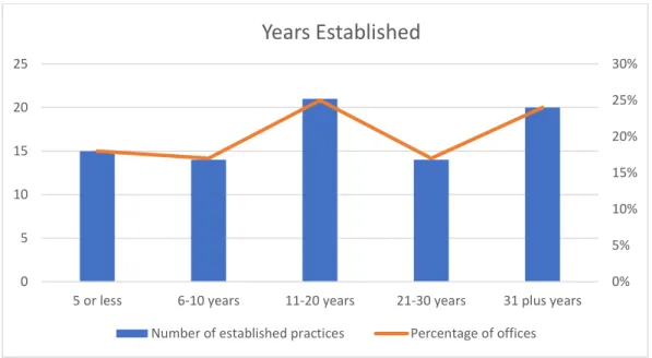
A total 104 respondents agreed to participate. Eighty-four participants (81%) answered Survey Question 2: “How many years has your office been at the current location?” Fifteen (18%) practices reported being established for less than five years. Fourteen (17%) practices reported being established in the six to ten years range. Twenty-one (25%) practices reported being established in the 11 to 20 years range. Fourteen (17%) practices reported being
established in the 21 to 30 years range. Twenty (24%) practices reported being established 31 plus years (Figure 2).
I AGREE I DO NOT AGREE
0.00% 20.00% 40.00% 60.00% 80.00% 100.00%
By selecting “I Agree”, you are consenting that you
have read the above information, volunteering to
participate, and at least18 years old
.42
Figure 2. Number of Years Current Practice Established
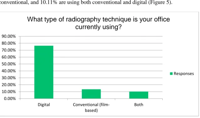
Eighty-nine of the 104 respondents answered Survey Question 3: “Where is your practice located?” Three choices of location resulted in 44.94% in rural areas, 32.58% in suburban areas, and 22.47% in urban areas (Figure 3).
Figure 3. Location of Respondents’ Practices
0% 5% 10% 15% 20% 25% 30% 0 5 10 15 20 25
5 or less 6-10 years 11-20 years 21-30 years 31 plus years
Years Established
Number of established practices Percentage of offices
0.00% 5.00% 10.00% 15.00% 20.00% 25.00% 30.00% 35.00% 40.00% 45.00% 50.00%
Urban Suburban Rural
43
Analysis of Data
The data was analyzed using Microsoft Excel. Descriptive statistics were computed to answer the research questions.
Research Question 1: Are private dental practices in Mississippi currently using electronic dental records?
Eighty-nine of the 104 respondents answered Survey Question 4: “What type of dental records is your office using?” which provided data related to Research Question 1. Electronic, electronic and paper, and paper were the available answers. Of the respondents, 46.07% are using electronic dental records. 42.70% are using electronic and paper dental records, and 11.24% are using paper dental records (Figure 4).
Figure 4. Type of Dental Records
Electronic Electronic and Paper Paper
0.00% 5.00% 10.00% 15.00% 20.00% 25.00% 30.00% 35.00% 40.00% 45.00% 50.00%
What type of dental records is your office using?
44
Research Question 2: What factors influence the selection of paper dental records or electronic dental records by private dental practices in Mississippi?
Eighty-four of the 104 respondents answered Survey Question 5: “What influenced the selection of your current type of dental records?” The respondents answered the question according to the type of dental record being used by the offices. Common factors among users of EDRs and EDRs with paper dental records were: keeping up with technology (10 offices), efficient (10 offices), ease of use (10 offices), time saver (4 offices), storage (8 offices), insurance processing (5 offices), and EHR mandate per government (4 offices). Common Factors among users of EDRs with paper records and paper records only were: cost of conversion (9 offices), computer issues (5 offices using EDRs with paper and 4 offices using paper only), and “old habits” or too old to change (15 offices).
Other factors provided by the respondents using electronic dental records were: convenient, standard of care, electronic health records mandated by government, and dental electronic software pre-existing in practice. Two mobile dental units use electronic dental records for easy access to dental records between the two mobile units when operating at different locations. One dental practice accepted grant funding from Medicaid that required the practice to convert to electronic dental records.
Several practices using EDRs with paper records were using EDRs only to send electronic insurance claims and to store digital radiographs. They were still using paper dental records to record patient notes and as a backup if computers failed. Additional factors for using paper dental records along with EDRs were time-consuming to scan old documents into the computer and paper dental records pre-existed in the practice.