RESEARCH
Circular RNA expression profile in blood
according to ischemic stroke etiology
Aiora Ostolaza
1†, Idoia Blanco‑Luquin
2*†, Amaya Urdánoz‑Casado
2, Idoya Rubio
1, Alberto Labarga
4,
Beatriz Zandio
1,3, Miren Roldán
2, Judith Martínez‑Cascales
2, Sergio Mayor
1,3, María Herrera
1,3, Nuria Aymerich
1,3,
Jaime Gallego
1,3, Roberto Muñoz
1,3†and Maite Mendioroz
1,2†Abstract
Background: The discovery of novel biomarkers of stroke etiology would be most helpful in management of acute ischemic stroke patients. Recently, circular RNAs (circRNAs) have been proposed as candidate biomarkers of neuro‑ logical conditions due to its high stability. circRNAs function as sponges, sequestering miRNAs and are involved in most relevant biological functions. Our aim was to identify differentially expressed circRNAs in acute ischemic stroke patients according to stroke etiology.
Methods: A comprehensive expression profile of blood circRNAs was conducted by Arraystar Human circRNA arrays (13,617 probes) on a discovery cohort of 30 stroke patients with different stroke etiologies by TOAST classification. Real‑time quantitative PCR (RT‑qPCR) was used to validate array results in a cohort of 50 stroke patients. Functional in silico analysis was performed to identify potential interactions with microRNAs (miRNAs) and pathways underlying deregulated circRNAs.
Results: A set of 60 circRNAs were found to be upregulated in atherotrombotic versus cardioembolic strokes (fold‑ change > = 1.5 and p‑value ≤ 0.05). Differential expression of hsa_circRNA_102488, originated from UBA52 gene, was replicated in the validation cohort. RNA‑binding proteins (RBPs) sites of hsa_circRNA_102488 clustered around AGO2 and FUS proteins. Further functional analysis revealed interactions between deregulated circRNAs and a set of miRNAs involved in stroke‑related pathways, such as fatty acid biogenesis or lysine degradation.
Conclusion: Different stroke subtypes show specific profiles of circRNAs expression. circRNAs may serve as a new source of biomarkers of stroke etiology in acute ischemic stroke patients.
Keywords: Stroke, Etiology, circRNA, miRNA, Biomarker, UBA52
© The Author(s) 2020. This article is licensed under a Creative Commons Attribution 4.0 International License, which permits use, sharing, adaptation, distribution and reproduction in any medium or format, as long as you give appropriate credit to the original author(s) and the source, provide a link to the Creative Commons licence, and indicate if changes were made. The images or other third party material in this article are included in the article’s Creative Commons licence, unless indicated otherwise in a credit line to the material. If material is not included in the article’s Creative Commons licence and your intended use is not permitted by statutory regulation or exceeds the permitted use, you will need to obtain permission directly from the copyright holder. To view a copy of this licence, visit http://creat iveco mmons .org/licen ses/by/4.0/. The Creative Commons Public Domain Dedication waiver (http://creat iveco mmons .org/publi cdoma in/ zero/1.0/) applies to the data made available in this article, unless otherwise stated in a credit line to the data.
Introduction
Stroke is a common vascular disease that causes death and disability and is therefore a major challenge to cur-rent healthcare systems [1]. Nowadays, stroke diagnosis
is still based on clinical criteria and imaging data, so cli-nicians are able to identify the etiology in about 75–80% of stroke cases by following the TOAST classification system [2]. In the remaining 20–25% of cases, the exact cause is unknown. It has been estimated that in about 25–30% of these cause-undetermined events, the under-lying source of stroke could be paroxysmal atrial fibril-lation (PAF) [1]. However, demonstrating this or other causes behind undetermined stroke events still remains a challenge in the clinical setting. Therefore, there is an increasing need for biomarkers capable of identify-ing stroke etiology in clinical practice. In recent years,
Open Access
*Correspondence: idoia.blanco.luquin@gmail.com
†Aiora Ostolaza, Idoia Blanco‑Luquin, Roberto Muñoz and Maite
Mendioroz contribute equally to this work
2 Neuroepigenetics Laboratory‑Navarrabiomed‑IdiSNA, Complejo
Hospitalario de Navarra, Universidad Pública de Navarra (UPNA), IdiSNA (Navarra Institute for Health Research), C/Irunlarrea, 3, 31008 Pamplona, Navarra, Spain
numerous studies have investigated a large amount of new blood biomarkers in relation with stroke etiology [3–7]. However, low sensitivity and specificity of the target biomarkers difficult their translation into clinical practice. Thus, the discovery of new molecules to aid the diagnosis of stroke etiology would be most helpful in this scenario.
By far, proteins represent the most widely studied class of molecules in the identification of biomarkers for cere-brovascular diseases. However, RNA molecules were also suggested as candidate stroke biomarkers about a decade ago [8, 9]. In particular, brain and blood profiling in rat models revealed differential expression of certain micro-RNAs (mimicro-RNAs) after transient focal ischemia [8] and blood miRNAs were proposed as diagnostic and prog-nostic biomarkers in acute stroke patients [9].
Since then, a wide range of RNA species have been discovered, including circular RNAs (circRNAs). cir-cRNAs are endogenous single-stranded RNA mol-ecules which form a circle through a covalent binding [10]. These molecules are evolutionary conserved and very abundant in the human transcriptome [11]. cir-cRNAs display a wide range of regulatory functions in RNA biology and gene expression. For instance, some circRNAs function as sponges that sequester miRNAs or RNA-binding proteins, regulating the expression of target genes [12]. In addition, given that circRNAs do not have free ends, they are resistant to endonucle-ase activity and therefore more stable than other lin-ear RNA species such as messenger RNA (mRNA) or even microRNAs (miRNAs) [13]. This features make circRNAs promising biomarkers in different medical
conditions. Indeed, circRNAs have recently been pro-posed as potential clinical biomarkers for neurologi-cal disorders [14]. However, comprehensive circRNAs expression has not been examined in stroke patients so far.
In this study, we have comprehensively profiled circR-NAs in the peripheral blood of ischemic stroke patients during the acute stage. To this end, we used Arraystar Human circRNA Array (8 × 15 K, Arraystar), which surveys up to 13,617 probes to identify human circR-NAs, in 30 acute stroke patients with different etiolo-gies by TOAST classification [2]. As a result, we show that distinct circRNAs are differentially expressed in atherotrombotic versus cardioembolic stroke patients, arising as promising candidate biomarkers of stroke etiology.
Methods
Study population
Patients with acute ischemic stroke admitted to the emergency department of the Hospital of Nav-arra within the first 4.5 h after symptoms onset were included in the study. Among a cohort of 700 consecu-tive patients recruited from January 2015 to December 2016, 30 patients were included in the discovery cohort (Table 1) and 50 patients were included in the valida-tion cohort (Addivalida-tional file 2: Table S1). All participants completely fulfilled the etiology testing protocol (see methods below). Informed consent was obtained from all participants and the study was approved by the local Ethics Committee.
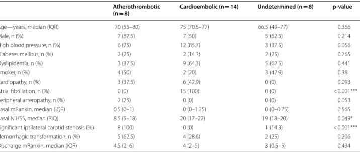
Table 1 Demographic and clinical characteristics of patients
IRQ interquartile range
Atherothrombotic
(n = 8) Cardioembolic (n = 14) Undetermined (n = 8) p-value
Age—years, median (IQR) 70 (55–80) 75 (70.5–77) 66.5 (49–77) 0.366
Male, n (%) 7 (87.5) 7 (50) 5 (62.5) 0.214
High blood pressure, n (%) 6 (75) 12 (85.7) 3 (37.5) 0.056
Diabetes mellitus, n (%) 2 (25) 2 (14.3) 2 (25) 0.765 Dyslipidemia, n (%) 3 (37.5) 9 (64.3) 5 (62.5) 0.441 Smoker, n (%) 4 (50) 2 (20) 3 (42.9) 0.38 Cardiopathy, n (%) 3 (37.5) 6 (42.9) 0 (0) 0.093 Atrial fibrillation, n (%) 0 (0) 15 (100) 0 (0) < 0.001*** Peripheral arteropathy, n (%) 2 (25) 0 (0) 0 (0) 0.053
Basal mRankin, median (IQR) 0.5 (0–1) 0 (0–1.25) 0 (0–0.75) 0.565
Basal NIHSS, median (RIQ) 8.5 (5–18) 20 (17–22) 19 (18–20) 0.049*
Significant ipsilateral carotid stenosis (%) 8 (100) 0 (0) 1 (14.3) < 0.001***
Hemorrhagic transformation, n (%) 5 (62.5) 4 (28.6) 2 (25) 0.206
Clinical, vascular and brain imaging protocol
A detailed history of vascular risk factors including hypertension, atrial fibrillation, diabetes mellitus, dys-lipidemia, tobacco, cardiovascular disease and periph-eral atherosclerosis was recorded for each patient. In order to determine stroke etiology, a set of tests was performed including electrocardiogram (EKG), chest radiography, complete blood count, blood biochemistry analysis, carotid ultrasonography, transcranial doppler (TCD) examination, non-contrast cranial tomography (CT) at baseline, echocardiogram and 24 h holter moni-toring. Patients were classified into etiological subgroups according to Trial of Org 10172 in Acute Stroke Treat-ment (TOAST) criteria [2].
Microarray expression of circRNAs
Venous blood samples were drawn from acute stroke patients within 24 h after admission. Total RNA was isolated from blood samples using miRNeasy Mini kit (QIAGEN, Redwood City, CA, USA) which enables purification of total RNA, including RNA from approxi-mately 18 nucleotides upwards, following manufacturer’s instructions. Genomic DNA was removed with a recom-binant DNase (TURBO DNA-free™ Kit, Ambion, Inc.,
Austin, TX, USA). RNA integrity was assessed by elec-trophoresis on a denaturing agarose gel. Concentration of RNA was determined by OD260 using a NanoDrop ND-1000 spectrophotometer. Array star Human cir-cRNA Array V2 analysis (Arraystar, Inc., Rockville, MD, USA) of the 30 selected stroke samples was performed as follows. Sample labeling and array hybridization were completed according to the manufacturer’s proto-col (Arraystar Inc.). Briefly, 2000 ng of total RNAs were digested with RNase R (Epicentre, Inc.) to remove lin-ear RNAs and enrich circular RNAs. Then, the enriched circular RNAs were amplified and transcribed into fluorescent cRNA utilizing a random priming method (Arraystar Super RNA Labeling Kit; Arraystar). The labeled cRNAs were purified by RNeasy Mini Kit (Qia-gen) and hybridized onto the Arraystar Human circRNA Array V2 (8 × 15 K, Arraystar). After having washed the
slides, the arrays were scanned by the Agilent Scanner G2505C (Agilent Technologies, Inc., Santa Clara, CA, USA).
circRNAs microarray data
Scanned images were imported into Agilent Feature Extraction software (version 11.0.1.1) for raw data extrac-tion. Quantile normalization of raw data and subsequent data processing were performed using the R software (R Project for Statistical Computing, Vienna, Austria) limma package. Normalized intensity values are shown in
Additional file 1: Fig. S1. After quantile normalization of the raw data, low intensity filtering was performed, and those circRNAs in which at least 8 out of 30 samples had flags in “P” or “M” (“All Targets Value”) were retained for further analyses. Three distinct groups by stroke etiol-ogy (cardioembolic, atherotrombotic and undetermined) were analyzed by pairs, so three different comparisons were performed. When comparing two groups of profile differences (such as cardioembolic versus atherotrom-botic), the fold change (FC) (i.e. the ratio of the group averages) between the groups for each circRNA was com-puted. Differentially expressed circRNAs between two groups were identified through FC filtering and statisti-cal significance of the difference between two groups was estimated by Student’s t-test. CircRNAs having FC ≥ 1.5 and p-values ≤ 0.01 were selected as significantly dif-ferentially expressed. Difdif-ferentially expressed circRNAs with statistical significance between two groups were shown by Scatter plots and Volcano plots.
Validation of array results by qPCR
Blood RNA isolated by miRNeasy Mini kit (QIAGEN, Redwood City, CA, USA) from the validation cohort (n = 50) was used to validate array results.
Complemen-tary DNA (cDNA) was reverse transcribed from 500 ng total RNA per sample with SuperScript® III First-Strand
Synthesis Reverse Transcriptase (Invitrogen, Carlsbad, CA, USA) and primed with random primers. Real time-qPCR (RT-time-qPCR) reactions were performed in triplicate for each sample using Power SYBR® Green PCR Master
Mix (Invitrogen, Carlsbad, CA, USA) in a QuantStudio 12 K Flex Real-Time PCR System (Applied Biosystems, Foster City, CA, USA). Divergent primer pairs were designed to overlap the backspliced junction by using Primer3 website (http://prime r3.ut.ee/). Furthermore, GAPDH housekeeping gene and convergent primer pairs to amplify the host mRNA were designed by Real Time PCR tool (IDT, Coralville, IA, USA). Primer sequences are listed in Additional file 2: Table S2. At the designing stage, verification of primers specificity was carried out by PCR tool at the UCSC Genome Browser [15]. After amplification, we also checked that RT-qPCR reaction had generated a single-peak in the melting curve and a single amplicon of the correct size by performing aga-rose gel electrophoresis. The thermal cycling conditions consisted of an initial denaturation step at 95 °C for 10 min followed by 40 cycles of 15 s at 95 °C and 1 min at 60 °C. Expression levels of each corresponding linear transcript, GAPDH or convergent amplicon of the host gene, was used to normalize circRNA levels [16]. Relative expression level of circRNA in a particular sample was calculated by the delta delta-CT method, as previously described [17]. Non-template reactions were included as
negative controls in each run. Finally, qPCR amplicons were subjected to Sanger sequencing [18] and checked for the presence of the predicted backspliced junctions in order to test their circularity.
Functional in silico analysis
Since certain circRNAs may function as sponges seques-tering miRNAs and, therefore, may be involved in the regulation of gene expression, it was interesting to explore the potential interactions between differen-tially expressed circRNAs in stroke and miRNAs. The circRNA/microRNA interaction was predicted with Arraystar’s home-made miRNA target prediction soft-ware based on TargetScan & miRanda [19], and the dif-ferentially expressed circRNAs were annotated in detail with the circRNA/miRNA interaction information. Then, overrepresented miRNAs (those linked to at least four differentially expressed circRNAs) were analyzed by DIANA-mirPath v.3 software [20] to predict the underly-ing pathways.
CircInteractome tool provided a list of miRNAs poten-tially targeted by the circRNA of interest and mapped binding sites for RNA-binding proteins (RBPs) on it [21]. Next, DIANA-mirPath v.3 software [20] analyzed the pathways in which the outcome miRNAs were involved based on TarBase v7.0, microT-CDS v5.0 and TargetScan. Kyoto Encyclopedia of Genes and Genomes (KEGG) and Gene Ontology (GO) analyses were used to predict cell signaling pathways and functions related with the set of outcome miRNAs.
Moreover, in order to assess the interactions betweem the RBPs predicted to bind a particular differentially expressed circRNA, protein list was uploaded to the Search Tool for the Retrieval of Interacting Genes (STRING) tool (Version 11.0) [22] and filtered for inter-actions of high confidence (score > 0.7) and PANTHER (Protein ANalysis THrough Evolutionary Relationships) Classification System (Version 14.1) [23]. Only those terms with a FDR-corrected p-value < 0.05 were reported.
Statistical Analysis
Statistical analysis was performed with SPSS 21.0 (IBM, Inc., USA). First, adjustment to normal distribution was tested for all continuous variables as per one-sample Kol-gomorov-Smirnov test and the normal quantil-quantil (QQ) plots. Data represents the mean ± standard devia-tion (SD) or the median (interquartile range). In the dis-covery cohort, univariate analysis of demographic and clinical characteristics was performed by ANOVA or Kruskall-Wallis test along with Chi square test. In the validation cohort, univariate analysis of demographic and clinical characteristics was performed by Student t-test or U Mann–Whitney test along with Chi square
test. circRNA expression differences between two groups was estimated by Mann–Whitney U test. For all the com-parisons, significance level was set at p-value < 0.05. SPSS 21.0 (IBM, Inc., USA) was used to draw graphs.
Results
Differential expression of circRNAs in etiologic stroke subtypes
To begin to ask whether circRNAs were differentially expressed in ischemic stroke depending on subtype eti-ology, we performed Arraystar Human circRNA Array V2 analysis, which included 13,617 distinct probes for human circRNAs, on a target group of 30 patients suffer-ing from acute ischemic stroke with different etiologies according to TOAST classification [2], i.e. 14 cardioem-bolic, 8 atherotrombotic and 8 undetermined stroke. Demographic and clinical characteristics of patients included in the discovery cohort are shown in Table 1. Normalized intensity values showed similar distribu-tions of the intensities (expression values) for all samples (Additional file 1: Fig. S1).
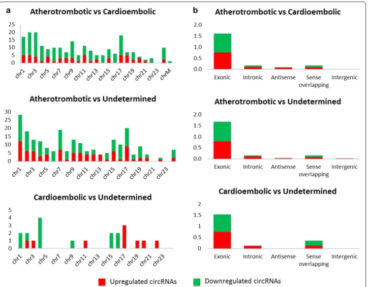
The main aim of this study was to identify differences in circRNA expression between atherotrombotic and cardioembolic stroke patients. We found 219 differen-tially expressed circRNAs (FC ≥ 1.5 and p-value ≤ 0.05)
in atherotrombotic versus cardioembolic stroke patients (Additional file 2: Tables S3 and S4). circRNA expression variation between the two compared groups is repre-sented as volcano plots and scatter plots (Fig. 1a, Addi-tional file 1: Fig. S2A). Despite we observed a higher number of downregulated (159) than upregulated (60) circRNAs in atherotrombotic compared to cardioem-bolic samples, the best findings in terms of statistical significance and magnitude of change were found among the upregulated circRNAs. Indeed, 11.7% (7) of upregu-lated circRNAs showed more than fourfold change in expression (Additional file 2: Table S3).
When comparing atherotrombotic versus undeter-mined strokes, 226 circRNAs were found to be differen-tially expressed (FC ≥ 1.5 and p-value ≤ 0.05), being 87
circRNAs upregulated and 139 circRNAs downregulated in atherotrombotic compared to undetermined strokes (Additional file 2: Tables S5 and S6, Fig. 1b and Addi-tional file 1: Fig. S2B). In this set, differences were not as strong as in the previous comparison of atherotrombotic versus cardioembolic strokes, and only one circRNA showed expression higher than fourfold change. Finally, a very few changes were found when comparing cardioem-bolic and undetermined strokes, as only 8 circRNAs were found to be upregulated and 9 circRNAs were down-regulated in this comparison (Additional file 2: Table S7, Fig. 1c and Additional file 1: Fig. S2C).
As shown in Fig. 2A, differentially expressed circR-NAs seem to cluster in chromosomes 1, 2, 3 and 17 for the comparison atherotrombotic versus cardioembolic stroke (> 15 hits); 1, 2, 7 and 17 for atherotrombotic ver-sus undetermined (> 15 hits) and 4 and 17 for cardioem-bolic versus undetermined (> 2.5 hits). With these results in mind, circRNAs from chromosome 17, and even 1 and 2, appear to be important for stroke etiology. In terms of distribution by chromosome regions, the number of dif-ferentially expressed circRNAs that are transcribed from protein-coding exons is the most abundant and compa-rable across downregulated and upregulated circRNAs (Fig. 2b). There were only a few intronic, antisense, sense overlapping and intergenic type circRNAs.
Validation of differentially expressed circRNAs by RT-qPCR
To validate the differences in circRNA expression, we focused on the atherotrombotic versus cardioembolic comparison since that was the main goal of our study. An expanded cohort which included samples previously analyzed in the microarray (n = 50; 25 atherotrombotic
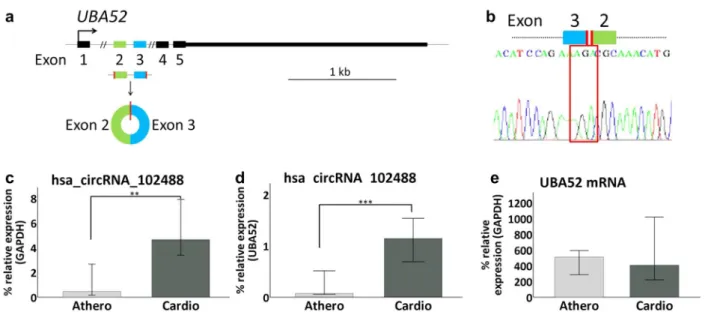
and 25 cardioembolic samples) was used to this end. Next, 3 upregulated and 2 downregulated circRNAs in atherotrombotic versus cardioembolic stroke patients were selected based on highest fold change and/or lowest p-value prioritization (Table 2). Then, we performed RT-qPCR to amplify and quantify the chosen circRNAs, their host mRNAs and GAPDH mRNA levels. Compared to the linear transcript, only hsa_circRNA_102488 showed a statistically significant change in expression between
etiology subtypes. hsa_circRNA_102488 is originated from the ubiquitin a-52 residue ribosomal protein fusion product 1 (UBA52) gene which is located at chromosome 19 (Fig. 3a). Sanger sequencing confirmed the presence of the backsplicing junction between UBA52´ exons 3 and 2 (Fig. 3b). Mann–Whitney U test revealed that expression levels of hsa_circRNA_102488 (alias hsa_circ_0005568) were lower for atherotrombotic compared to cardioem-bolic samples when normalizing to GAPDH mRNA (p-value < 0.01) (Fig. 3c), as well as to the corresponding UBA52 mRNA (p-value < 0.001) (Fig. 3d). Interestingly, we did not found significant differences in UBA52 mRNA expression levels between atherotrombotic and cardi-oembolic stroke samples (Fig. 3e).
Functional in silico analysis of differentially expressed circRNAs
In order to explore potential interactions between dif-ferentially expressed circRNAs in stroke subtypes and target miRNAs, a bioinformatics analysis was per-formed (see “Methods”). First, interactions between circRNAs and their target miRNAs were predicted by Arraystar’s home-made software. Then, we selected the overrepresented miRNAs in the three compari-sons (those miRNAs linked to at least four differen-tially expressed circRNAs), and characterized their related pathways by DIANA-mirPath (p-value thresh-old ≤ 0.05). Overrepresented miRNAs were
associ-ated with fatty acid biosynthesis and metabolism, lysine degradation, arrhythmogenic right ventricular
Fig. 1 Volcano‑plots of differential expression of circRNAs for stroke etiology comparisons. Graphs visualize the relationship between fold‑change (magnitude of change) in the X axes and statistical significance (which takes both magnitude of change and variability into consideration) in the Y axes. A high number of circRNAs are shown as differentially expressed between atherotrombotic versus cardioembolic strokes (a), and atherotrombotic versus undetermined strokes (b) whereas cardioembolic versus undetermined strokes show less differences suggesting both groups are similar in terms of circRNA expression. The vertical green lines correspond to the threshold of 1.5‑fold up and down, respectively, and the horizontal green line represents a p‑value = 0.05. Red points in the plot represent the differentially expressed circRNAs with statistical significance which crossed the fold‑change threshold
cardiomyopathy (ARVC), adrenergic signaling in car-diomyocytes or hypertrophic cardiomyopathy (HCM) as shown by KEGG analysis (p-value threshold < 0.05) (Additional file 1: Fig. S3A). Moreover, these miRNAs were linked to cellular nitrogen compound metabolic
process, homophilic cell adhesion via plasma mem-brane adhesion molecules, cell adhesion, blood coagulation or neurotrophin tyrosine-kinase (TRK) receptor signaling pathway in GO analysis (p-value threshold < 0.05) (Fig. 4a).
Fig. 2 Distribution by chromosomes (a) and chromosome’s region (b). Differentially expressed circRNA distribution in chromosomes and chromosome´s region (exonic, intronic, antisense, sense overlapping and intergenic) for each comparison
Table 2 Deregulated circRNA chosen for qPCR validation
CircRNA deregulated circRNA with greater intensity values in atherotrombotic stroke patients compared with cardioembolic; circRNA_type: the circRNAs are classified into 5 types: “exonic”, “intronic”, “antisense”, “sense overlapping” and “intergenic”; FC absolute ratio (no log scale) of normalized intensities between two conditions; p-value p-value calculated from unpaired t-test; Annotations, include chromosome position and Gene Symbol
Regulation CircRNA CircRNA type FC (abs) P-value Chromosome position Gene symbol
Up hsa_circRNA_001359 intronic 7.0479384 0.002611197 chr1 169663839 169664181 SELL hsa_circRNA_103559 exonic 4.8769307 0.035206182 chr3 196118683 196120490 UBXN7 hsa_circRNA_104220 exonic 4.8572966 0.026033314 chr6 150092297 150094305 PCMT1 Down hsa_circRNA_102488 exonic 2.7229035 8.17917E−05 chr19 18684102 18684558 UBA52 hsa_circRNA_104748 exonic 2.3292681 0.001448543 chr9 20907148 20929595 FOCAD
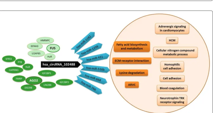
Regarding the validated circRNA, hsa_cir-cRNA_102488, CircInteractome tool [21] showed one 7mer-m8 or 7mer-1a type target site shared by the fol-lowing miRNAs: miR-1182, miR-1299, hsa-miR-431, hsa-miR-516b, hsa-miR-668 and hsa-miR-766. DIANA-mirPath analysis revealed that this set of miR-NAs converged on the pathways of fatty acid biosynthe-sis and metabolism, extracellular matrix (ECM)-receptor interaction, lysine degradation or arrhythmogenic right ventricular cardiomyopathy (ARVC) as shown by KEGG analysis (p-value threshold < 0.001 (Additional file 1: Fig. S3B, C). They seemed to have a common function as RNA-, ion- and enzyme-binding molecules, a protein and nucleic acid binding transcription factor activity and to be involved in cellular nitrogen compound meta-bolic, biosynthetic, cellular protein modification and gene expression processes (GO analysis, p-value thresh-old < 0.05) (Fig. 4b, c).
In addition, up to 10 RBPs sites matching to hsa_cir-cRNA_102488 were identified by CircInteractome: 3 for SFRS1, 2 for AGO2, HuR, IGF2BP2 and PTB and 1 for CAPRIN1, DGCR8, FMRP, IGF2BP1 and LIN28B. STRING clustered these RBPs around AGO2 (protein– protein interaction, PPI, enrichment p-value < 1.0e−16) (Additional file 1: Fig. S4A–C) and PANTHER related them to two categories of molecular functions: RNA binding and catalytic activity. Moreover, other RBPs
sites that matched to the hsa_circRNA_102488 flank-ing regions were identify for EIF4A3 (9 Tags), HNRNPC, HuR, U2AF65 and FUS genes. After functional analysis, we observed that these RBPs clustered around FUS (PPI enrichment p-value < 2.11e−10) and related to catalytic activity, IGF2BP1 and IGF2BP3 to binding molecular function and ZC3H7B separately but also to catalytic activity function (STRING and PANTHER tools) (Addi-tional file 1: Fig. S4D–F).
A diagram illustrating impact pathways mentioned above is shown in Fig. 5.
Discussion
In this study, we identified circRNAs that were differ-entially expressed in human blood according to stroke etiology. The strongest differences were found for the comparison between atherotrombotic and cardioem-bolic stroke. Differentially expressed circRNAs were pre-dicted to predominantly interact with a set of miRNAs involved in stroke-related pathways, including fatty acid biosynthesis, lysine degradation, arrhythmogenic right ventricular cardiomyopathy (ARVC) or hypertrophic car-diomyopathy (HCM), among others.
circRNAs are a new class of non-coding RNA mol-ecules with circular morphology due to the formation of a covalent junction between the 3′ and 5′ ends. These molecules are generated from a parental (also known as
Fig. 3 RT‑qPCR validation of hsa_circRNA_102488. a Schematic representation of the UBA52 gene, origin of hsa_circRNA_102488. The graph shows the map of hsa_circRNA_102488 which is originated from exons 2 and 3 of its host mRNA (NCBI RefSeq track shown in the UCSC Genome Browser). b hsa_circRNA_102488 backsplicing. The Sanger‑Sequencing electropherogram specifies the sequence of the backspliced junction that results in the circRNA formation. c–e hsa_circRNA_102488 and UBA52 mRNA expression levels between atherotrombotic vs cardioembolic samples. Percentage of hsa_circRNA_102488 expression relative to GAPDH mRNA (c) or UBA52 mRNA (d) is decreased in atherotrombotic with respect to cardioembolic stroke patients. However, we found similar expression levels of UBA52 mRNA in atherotrombotic and cardioembolic samples groups (e). Error bars: 95% CI. **p‑value < 0.01; ***p‑value < 0.001
Fig. 4 GO analysis by mirPath v.3. The image shows the heatmaps corresponding to the analysis performed with the overrepresented miRNAs in the three comparisons (atherotrombotic‑cardioembolic‑undetermined) by TargetScan (a) and the analysis involving the target miRNAs for hsa_ circRNA_102488 in accordance with molecular function (b, by Tarbase) or biological process (c, by microT‑CDS)
Fig. 5 hsa_circRNA_102488 impact pathways. The cartoon shows miRNAs (in blue) sharing a target site with hsa_circRNA_102488 (in black) and the pathways in which they converge (in dark orange), within those associated with the overrepresented miRNAs in the three comparisons (in light orange). RBPs sites matching to hsa_circRNA_102488 clustered around AGO2 (in dark green) and RBPs sites that matched to its flanking regions clustered around FUS (in light green). ARVC: Arrhythmogenic Right Ventricular Cardiomyopathy; HCM: Hypertrophic Cardiomyopathy
host) pre-messenger RNA through a particular alterna-tive splicing process known as backsplicing [24, 25]. The covalent bond confers resistance to ribonucleases pre-venting degradation and making circRNAs very stable molecules. Furthermore, circRNAs are expressed in most mammalian tissues, particularly in the brain and have been also detected in the human blood [26–28]. All these characteristics make circRNAs potential biomarkers of human diseases and, especially, of neurological diseases.
Indeed, previous reports suggesting the potential role of circRNAs as biomarkers or therapeutic targets in neu-rological conditions, such as multiple sclerosis or epi-lepsy, have been already published [29–31]. In the stroke field, a number of changes in circRNAs expression levels have been recently described. In 2017, it was observed for the first time that circRNAs are altered in the mouse brain after transient focal ischemia [32, 33]. Despite these initial observations, as far as we know, a comprehensive profiling of circRNAs in human stroke or its relationship to stroke etiology has not previously been reported.
In our study, a set of 219 differentially expressed circR-NAs between atherotrombotic and cardioembolic stroke was found in the blood of acute stroke patients. The most robust statistical differences were observed among the 60 circRNAs up-regulated in atherotrombotic compared to cardioembolic samples. Most of them tended to cluster in chromosome 17 and were of exonic origin, which is not surprising since the majority of known circRNA mol-ecules are exonic and therefore, there is an overrepresen-tation of exonic circRNAs in the Arraystar microarray.
Even though a large number of circRNAs were shown to be differentially expressed in atherotrombotic ver-sus undetermined stroke samples, there were hardly any differences between cardioembolic and undetermined strokes. This result suggests that the undetermined stroke group may harbor a great proportion of non-recognized cardioembolic strokes, which is in line with current knowledge in the clinical setting. In fact, it has been described that some patients with stroke of unde-termined etiology may have atrial structural or functional changes thus increasing the risk of cardioembolism [34].
In recent years, functions of circRNAs are beginning to be understood. It has been observed that circRNAs can act at different levels of gene regulation, including transcription, mRNA splicing or translation. However, the best known function so far is the regulation of gene expression by acting as miRNA sponges: circRNAs have binding sites for specific miRNAs and act by “sequester-ing” them to prevent miRNAs from exerting translational repression. Thus far, a few stroke-related circRNAs act-ing as miRNA sponges have been observed. circRNA DLGAP4 functions as a sponge for miRNA-143 and therefore, inhibits miRNA-143 activity. Interestingly,
overexpression of circRNA DLGAP4 ameliorates neu-rological deficit and infarct volume after transient focal ischemia in a stroke mouse model. circRNA DLGAP4 seems to significantly attenuate blood–brain barrier damage by regulating tight junction protein expression and endothelial-mesenchymal transition in endothelial cells [35]. Other example of deregulated circRNAs in stroke is circRNA Hectd1, which was found to be upreg-ulated in the brain of mice after transient middle cerebral artery occlusion (tMCAO). circRNA HECTD1 partici-pates in the inhibition of astrocyte activation via macro-autophagy/autophagy by acting as a sponge for MIR-142 [36]. Interestingly, circRNA HECTD1 was increased in peripheral blood mononuclear cells from acute ischemic stroke patients compared to controls and higher levels of circRNA HECTD1 were related with stroke recurrence [37]. The circRNA TLK1, which acts as an endogenous miR-335-3p sponge, has been proved to be detrimental in ischemic brain injury. It was increased in the mouse brain after tMCAO and knocking down circRNA TLK1 improve neurological deficits and infarct volume in this stroke mouse model. Remarkably, circRNA TLK1 was observed to be also elevated in human patients after ischemic stroke [38].
In the present study, differentially expressed circR-NAs were predicted to interact with numerous miR-NAs. Functional study showed that those miRNAs were involved in pathways associated with stroke etiology or pathogenesis, such as fatty acids biogenesis, lysine deg-radation or development of cardiopathy, including ARVC and HCM. Elevated levels of free fatty acids have been associated with cardioembolic etiology of stroke irre-spective of the presence of atrial fibrillation [39, 40] and predict stroke recurrence in patients with atrial fibrilla-tion [41]. Although the underlying mechanisms remain unclear, fatty acids are major components of epicardial fat and it is known that thickness of epicardial fat is related with the presence of atrial fibrillation [42]. According to that, serum fatty acid binding proteins are proposed as biomarkers for atrial fibrillation [43]. However, fatty acids seem independent predictors of stroke events so they may be directly related to thrombogenesis [39, 40].
Interestingly, lysine degradation pathway showed a strong association with overrepresented miRNAs in our study. Lysine is an essential aminoacid with pleiotropic functions in humans. In addition to proteinogenesis, lysine is involved in the crosslinking of collagen peptides, the uptake of calcium and iron and is the precursor of carnitine, which in turn is essential in the metabolism of fatty acids [44]. Lysine degradation differs in brain from that of in extracerebral tissues and it is known to be regulated by thyroid hormones [45]. For now, its role in stroke is not yet well understood. A recent study among
hypertensive subjects revealed altered metabolic path-ways, including lysine degradation, related to incident ischemic stroke [46]. Moreover, a metabolomic study showed that levels of serum lysine catabolites were low in patients at high-risk of suffering ischemic stroke [47].
Despite ARVC is not among the classical embolic sources of stroke events, ARVC pathways have been recently associated with ischemic stroke in a compre-hensive long non-coding RNA transcriptomic study [48] and this is consistent with our results. HCM is a common hereditary cardiomyopathy frequently associated with sudden death. It has been described that atrial fibrillation with risk for progressive heart failure and embolic stroke occurs in 20% of patients with HCM [49].
In this study, we were able to validate hsa_cir-cRNA_102488 in an expanded cohort of atherotrom-botic versus cardioembolic strokes. This circRNA is formed from the pre-mRNA of UBA52 gene and has a 456 bp genomic length. hsa_circRNA_102488 was found to be highly expressed in brain tissues [27]. No previ-ous records of the association between UBA52 gene and stroke have been reported to our knowledge.
Functional analysis revealed that hsa_circRNA_102488 harbors a binding site for 6 different miRNAs. Intrigu-ingly, this set of 6 miRNAs is enriched in pathways similar to those found for the overrepresented miRNAs, namely fatty acids biogenesis, lysine degradation and ARVC. This result suggests that hsa_circRNA_102488 may be representative of the altered circRNA network and its related pathways underlying stroke etiology and makes hsa_circRNA_102488 an interesting candidate worth to be explored in futures studies.
Among known functions, circRNAs interact with RNA binding proteins (RBPs) to influence gene transcription and translation [50]. In hsa_circRNA_102488, the analy-sis revealed RBPs binding sites that cluster around two RBPs: argonaute 2 (Ago2) and RNA-binding protein FUS. Argonaute 2 is a critical component of the RNA-induced silencing complex (RISC) and, as a consequence, a master regulator of miRNAs-dependent gene silencing pathway. Interestingly, it has been observed that Ago2 accumu-lates and undergoes hydroxylation following hypoxia, thus Ago2 emerges as a major regulator of the pathologi-cal responses to hypoxia [51]. In fact, stroke substantially modifies Ago2-associated miRNA profiles in the brain in a stroke rat model [52]. On the other hand, FUS partici-pates in DNA repair mechanisms [53] and is frequently mutated in amyotrophic lateral sclerosis [54]. However, no previous works on the role of FUS in stroke has been previously reported.
The determination of circRNA as biomarkers in the eti-ology of stroke would be a great advance in the diagnostic process of cerebral vascular diseases. It should be noted
that circRNAs are especially interesting as biomarkers because they represent a gene regulatory mechanism for several genes at once and, despite being RNA molecules, they are extremely stable. A large part of the study car-ried out on these patients is aimed to find the cause of the stroke, and in up to 25% of cases a clear etiology can-not be established. The risk factors that predispose to presenting an atherothrombotic stroke are widely known and current guidelines make general recommendations to prevent them. However, patients who remain without a specific etiological diagnosis would not benefit from these guidelines. Moreover, knowing the cause of stroke in a rapid and non-invasive way would help to improve secondary prevention and avoid unnecessary studies.
As a discovery study, where typically a large number of molecules are measured in a reduced number of sub-jects, an obvious limitation of this study is the limited sample size. Therefore, we should be cautious with our conclusions. For the sake of external validity, these find-ings should be replicated in blood samples of an inde-pendent cohort of ischemic stroke patients with different etiologies.
Conclusions
The present study showed a set of differentially expressed circRNAs in peripheral blood from different stroke etio-logic subtypes. These circRNAs revealed interesting pathways underlying stroke etiology, such as fatty acid biogenesis or lysine degradation. Due to their molecular features, circRNAs may be useful as candidate biomark-ers for stroke etiology. Large and independent cohort studies will be needed to further investigate the role of circRNAs as stroke biomarkers.
Supplementary information
Supplementary information accompanies this paper at https ://doi. org/10.1186/s1357 8‑020‑00394 ‑3.
Additional file 1. Additional figures.
Additional file 2. Additional tables.
Acknowledgements
We want to kindly thank technical staff from Neuroradiology Angiography Room and the Stroke Unit Team and of the Complejo Hospitalario de Navarra, Spain, for the support to collect blood samples. We also want to thank person‑ nel of the Navarrabiomed Biobank, including Valle Coca, Leticia San José, Ana Purroy and Isabel Gil MD, PhD, for their support. We are very grateful to David Otaegui, PhD from Biodonostia Institute, and his PhD student Leire Iparragu‑ irre, for their technical and scientific support. Finally, we are very grateful to the patients that generously donor samples for this study.
Authors’ contributions
AO and IBL contributed to running experiments, acquisition, analysis and interpretation of data, and drafting/revising the manuscript for content. AU contributed to analysis of data, figure drawing and drafting/revising the manuscript for content. IR contributed to revising subject diagnosis, clas‑ sification of patients and drafting/revising the manuscript for content. AL
contributed to bioinformatics analysis and drafting the manuscript. BZ con‑ tributed to analyze results and drafting/revising the manuscript for content. MR and JMC contributed to running experiments and data collection. SM, MH, NA and JG contributed to analyze results and drafting/revising the manuscript for content. RM and MM contributed to study concept and design, statistical analysis, analysis and interpretation of data, drafting/revising the manuscript for content, study supervision and obtaining funding. All authors read and approved the final manuscript.
Funding
This work was supported by Navarre Government Funding (Indus‑ try and Health department) through ADITECH and by RED INVICTUS (RD16/0019/0024) from the Institute of Health Carlos III, jointly funded by European Regional Development Fund (ERDF), European Union. AUC received a grant “Doctorados industriales 2018–2020” founded by Government of Navarra and MM received a grant “Programa de intensificación” founded by “LaCaixa Foundation” and Fundación Caja‑Navarra.
Availability of data and materials
The datasets used and/or analysed during the current study are available from the corresponding author on reasonable request.
Ethics approval and consent to participate
All procedures performed in studies involving human participants were in accordance with the ethical standards of the institutional and/or national research committee (clinical research ethics committee of Navarre; Pyto 2015/21) and with the 1964 Helsinki declaration and its later amendments or comparable ethical standards.
Informed consent
Informed consent was obtained from all individual participants included in the study.
Competing interest
The authors declare that they have no competing interests.
Author details
1 Department of Neurology, Complejo Hospitalario de Navarra‑IdiSNA (Navarra
Institute for Health Research), 31008 Pamplona, Navarra, Spain. 2 Neuroepige‑
netics Laboratory‑Navarrabiomed‑IdiSNA, Complejo Hospitalario de Navarra, Universidad Pública de Navarra (UPNA), IdiSNA (Navarra Institute for Health Research), C/Irunlarrea, 3, 31008 Pamplona, Navarra, Spain. 3 Stroke Unit,
Department of Neurology, Complejo Hospitalario de Navarra‑ IdiSNA (Navarra Institute for Health Research), 31008 Pamplona, Navarra, Spain. 4 Bioinformat‑
ics Unit, Navarrabiomed, Public University of Navarre (UPNA), IdiSNA (Navarra Institute for Health Research), C/Irunlarrea, 3, 31008 Pamplona, Navarra, Spain. Received: 8 January 2020 Accepted: 24 February 2020
References
1. Szegedi I, Szapáry L, Csécsei P, Csanádi Z, Csiba L. Potential biological markers of atrial fibrillation: a chance to prevent cryptogenic stroke. Biomed Res Int. 2017;2017:8153024. https ://doi.org/10.1155/2017/81530 24.
2. Adams HP, Bendixen BH, Kappelle LJ, Biller J, Love BB, Gordon DL, et al. Classification of subtype of acute ischemic stroke. Definitions for use in a multicenter clinical trial. TOAST. Trial of Org 10172 in acute stroke treat‑ ment. Stroke. 1993;24(1):35–41.
3. Yin R, Ma A, Pan X, Yang S. Biomarkers of cerebral microembolic signals. Clin Chim Acta. 2017;475:164–8. https ://doi.org/10.1016/j. cca.2017.10.028.
4. Sun H, Zhao J, Zhong D, Li G. Potential serum biomarkers and meta‑ bonomic profiling of serum in ischemic stroke patients using UPLC/Q‑ TOF MS/MS. PLoS ONE. 2017;12(12):e0189009. https ://doi.org/10.1371/ journ al.pone.01890 09.
5. Dagonnier M, Cooke IR, Faou P, Sidon TK, Dewey HM, Donnan GA, et al. Discovery and longitudinal evaluation of candidate biomarkers for ischaemic stroke by mass spectrometry‑based proteomics. Biomark
Insights. 2017;12:1177271917749216. https ://doi.org/10.1177/11772 71917 74921 6.
6. Llombart V, Garcia‑Berrocoso T, Bustamante A, Fernandez‑Cadenas I, Montaner J. Cardioembolic stroke diagnosis using blood biomarkers. Curr Cardiol Rev. 2013;9(4):340–52.
7. Jauch EC, Barreto AD, Broderick JP, Char DM, Cucchiara BL, Devlin TG, et al. Biomarkers of acute stroke etiology (BASE) study methodology. Transl Stroke Res. 2017. https ://doi.org/10.1007/s1297 5‑017‑0537‑3. 8. Jeyaseelan K, Lim KY, Armugam A. MicroRNA expression in the blood
and brain of rats subjected to transient focal ischemia by middle cerebral artery occlusion. Stroke. 2008;39(3):959–66. https ://doi.org/10.1161/strok eaha.107.50073 6.
9. Tan KS, Armugam A, Sepramaniam S, Lim KY, Setyowati KD, Wang CW, et al. Expression profile of MicroRNAs in young stroke patients. PLoS ONE. 2009;4(11):e7689. https ://doi.org/10.1371/journ al.pone.00076 89. 10. Chen LL, Yang L. Regulation of circRNA biogenesis. RNA Biol.
2015;12(4):381–8. https ://doi.org/10.1080/15476 286.2015.10202 71. 11. Salzman J, Gawad C, Wang PL, Lacayo N, Brown PO. Circular RNAs are
the predominant transcript isoform from hundreds of human genes in diverse cell types. PLoS ONE. 2012;7(2):e30733. https ://doi.org/10.1371/ journ al.pone.00307 33.
12. Chen L, Huang C, Wang X, Shan G. Circular RNAs in eukaryotic cells. Curr Genomics. 2015;16(5):312–8. https ://doi.org/10.2174/13892 02916 66615 07071 61554 .
13. Bolha L, Ravnik‑Glavač M, Glavač D. Circular RNAs: biogenesis, func‑ tion, and a role as possible cancer biomarkers. Int J Genomics. 2017;2017:6218353. https ://doi.org/10.1155/2017/62183 53.
14. Lu D, Xu AD. Mini review: circular RNAs as potential clinical biomarkers for disorders in the central nervous system. Front Genet. 2016;7:53. https :// doi.org/10.3389/fgene .2016.00053 .
15. Kent WJ, Sugnet CW, Furey TS, Roskin KM, Pringle TH, Zahler AM, et al. The human genome browser at UCSC. Genome Res. 2002;12(6):996–1006. https ://doi.org/10.1101/gr.22910 2.
16. Vandesompele J, De Preter K, Pattyn F, Poppe B, Van Roy N, De Paepe A, et al. Accurate normalization of real‑time quantitative RT‑PCR data by geometric averaging of multiple internal control genes. Genome Biol. 2002;3(7):34.
17. Livak KJ, Schmittgen TD. Analysis of relative gene expression data using real‑time quantitative PCR and the 2(‑Delta Delta C(T)) Method. Methods (San Diego, Calif ). 2001;25(4):402–8. https ://doi.org/10.1006/ meth.2001.1262.
18. Sanger F, Nicklen S, Coulson AR. DNA sequencing with chain‑terminating inhibitors. Proc Natl Acad Sci USA. 1977;74(12):5463–7.
19. Pasquinelli AE. MicroRNAs and their targets: recognition, regulation and an emerging reciprocal relationship. Nat Rev Genet. 2012;13(4):271–82. https ://doi.org/10.1038/nrg31 62.
20. Vlachos IS, Zagganas K, Paraskevopoulou MD, Georgakilas G, Karagkouni D, Vergoulis T, et al. DIANA‑miRPath v3.0: deciphering microRNA function with experimental support. Nucleic Acids Res. 2015;43(W1):W460–6. https ://doi.org/10.1093/nar/gkv40 3.
21. Dudekula DB, Panda AC, Grammatikakis I, De S, Abdelmohsen K, Gorospe M. CircInteractome: a web tool for exploring circular RNAs and their interacting proteins and microRNAs. RNA Biol. 2016;13(1):34–42. https :// doi.org/10.1080/15476 286.2015.11280 65.
22. Salwinski L, Miller CS, Smith AJ, Pettit FK, Bowie JU, Eisenberg D. The database of interacting proteins: 2004 update. Nucleic Acids Res. 2004;32(Database issue):D449–51. https ://doi.org/10.1093/nar/gkh08 6. 23. Thomas PD, Campbell MJ, Kejariwal A, Mi H, Karlak B, Daverman R, et al. PANTHER: a library of protein families and subfamilies indexed by func‑ tion. Genome Res. 2003;13(9):2129–41. https ://doi.org/10.1101/gr.77240 3.
24. Ashwal‑Fluss R, Meyer M, Pamudurti NR, Ivanov A, Bartok O, Hanan M, et al. circRNA biogenesis competes with pre‑mRNA splicing. Mol Cell. 2014;56(1):55–66. https ://doi.org/10.1016/j.molce l.2014.08.019. 25. Ragan C, Goodall GJ, Shirokikh NE, Preiss T. Insights into the biogenesis
and potential functions of exonic circular RNA. Sci Rep. 2019;9(1):2048. https ://doi.org/10.1038/s4159 8‑018‑37037 ‑0.
26. Xie L, Mao M, Xiong K, Jiang B. Circular RNAs: a novel player in develop‑ ment and disease of the central nervous system. Front Cell Neurosci. 2017;11:354. https ://doi.org/10.3389/fncel .2017.00354 .
•fast, convenient online submission •
thorough peer review by experienced researchers in your field • rapid publication on acceptance
• support for research data, including large and complex data types •
gold Open Access which fosters wider collaboration and increased citations maximum visibility for your research: over 100M website views per year •
At BMC, research is always in progress. Learn more biomedcentral.com/submissions
Ready to submit your research? Choose BMC and benefit from:
27. Rybak‑Wolf A, Stottmeister C, Glažar P, Jens M, Pino N, Giusti S, et al. Circular RNAs in the mammalian brain are highly abundant, conserved, and dynamically expressed. Mol Cell. 2015;58(5):870–85. https ://doi. org/10.1016/j.molce l.2015.03.027.
28. Hanan M, Soreq H, Kadener S. CircRNAs in the brain. RNA Biol. 2017;14(8):1028–34. https ://doi.org/10.1080/15476 286.2016.12553 98. 29. Iparraguirre L, Muñoz‑Culla M, Prada‑Luengo I, Castillo‑Triviño T, Olas‑
coaga J, Otaegui D. Circular RNA profiling reveals that circular RNAs from ANXA2 can be used as new biomarkers for multiple sclerosis. Hum Mol Genet. 2017;26(18):3564–72. https ://doi.org/10.1093/hmg/ddx24 3. 30. Gong GH, An FM, Wang Y, Bian M, Wang D, Wei CX. Comprehensive
circular RNA profiling reveals the regulatory role of the CircRNA‑0067835/ miR‑155 pathway in temporal lobe epilepsy. Cell Physiol Biochem. 2018;51(3):1399–409. https ://doi.org/10.1159/00049 5589. 31. Ojha R, Nandani R, Chatterjee N, Prajapati VK. Emerging role of
circular RNAs as potential biomarkers for the diagnosis of human diseases. Adv Exp Med Biol. 2018;1087:141–57. https ://doi. org/10.1007/978‑981‑13‑1426‑1_12.
32. Mehta SL, Pandi G, Vemuganti R. Circular RNA expression profiles alter significantly in mouse brain after transient focal ischemia. Stroke. 2017;48(9):2541–8. https ://doi.org/10.1161/strok eaha.117.01746 9. 33. Liu C, Zhang C, Yang J, Geng X, Du H, Ji X, et al. Screening circular RNA
expression patterns following focal cerebral ischemia in mice. Onco‑ target. 2017;8(49):86535–47. https ://doi.org/10.18632 /oncot arget .21238 . 34. Fonseca AC, Alves P, Inácio N, Marto JP, Viana‑Baptista M, Pinho‑E‑Melo
T, et al. Patients with undetermined stroke have increased atrial fibrosis: a cardiac magnetic resonance imaging study. Stroke. 2018;49(3):734–7. https ://doi.org/10.1161/strok eaha.117.01964 1.
35. Bai Y, Zhang Y, Han B, Yang L, Chen X, Huang R, et al. Circular RNA DLGAP4 ameliorates ischemic stroke outcomes by targeting miR‑143 to regulate endothelial‑mesenchymal transition associated with blood–brain barrier integrity. J Neurosci. 2018;38(1):32–50. https ://doi.org/10.1523/jneur osci.1348‑17.2017.
36. Han B, Zhang Y, Bai Y, Chen X, Huang R, Wu F, et al. Novel insight into circular RNA HECTD1 in astrocyte activation via autophagy by targeting MIR142‑TIPARP: implications for cerebral ischemic stroke. Autophagy. 2018;14(7):1164–84. https ://doi.org/10.1080/15548 627.2018.14581 73. 37. Peng X, Jing P, Chen J, Xu L. The role of circular RNA HECTD1 expression
in disease risk, disease severity, inflammation, and recurrence of acute ischemic stroke. J Clin Lab Anal. 2019. https ://doi.org/10.1002/jcla.22954 . 38. Wu F, Han B, Wu S, Yang L, Leng S, Li M, et al. Circular RNA TLK1 aggra‑
vates neuronal injury and neurological deficits after ischemic stroke via miR‑335‑3p/TIPARP. J Neurosci. 2019. https ://doi.org/10.1523/jneur osci.0299‑19.2019.
39. Seo WK, Jung JM, Kim JH, Koh SB, Bang OY, Oh K. Free fatty acid is associ‑ ated with thrombogenicity in cardioembolic stroke. Cerebrovasc Dis. 2017;44(3–4):160–8. https ://doi.org/10.1159/00047 8895.
40. Seo WK, Kim J, Kim YH, Kim JH, Oh K, Koh SB, et al. Elevated free fatty acid is associated with cardioembolic stroke subtype. Can J Neurol Sci. 2011;38(6):874–9.
41. Choi JY, Jung JM, Kwon DY, Park MH, Kim JH, Oh K, et al. Free fatty acid as an outcome predictor of atrial fibrillation‑associated stroke. Ann Neurol. 2016;79(2):317–25. https ://doi.org/10.1002/ana.24568 .
42. Cho KI, Kim BJ, Cho SH, Lee JH, Kim MK, Yoo BG. Epicardial fat thickness and free fatty acid level are predictors of acute ischemic stroke with atrial fibrillation. J Cardiovasc Imag. 2018;26(2):65–74. https ://doi.org/10.4250/ jcvi.2018.26.e1.
43. Golaszewska K, Harasim‑Symbor E, Polak‑Iwaniuk A, Chabowski A. Serum fatty acid binding proteins as a potential biomarker in atrial fibrillation. J Physiol Pharmacol. 2019. https ://doi.org/10.26402 /jpp.2019.1.11. 44. Hoppel C. The role of carnitine in normal and altered fatty acid metabo‑
lism. Am J Kidney Dis. 2003;41(4 Suppl 4):S4–12.
45. Hallen A, Jamie JF, Cooper AJ. Lysine metabolism in mammalian brain: an update on the importance of recent discoveries. Amino Acids. 2013;45(6):1249–72. https ://doi.org/10.1007/s0072 6‑013‑1590‑1. 46. Guo X, Li Z, Zhou Y, Yu S, Yang H, Zheng L, et al. Metabolic profile for pre‑
diction of ischemic stroke in chinese hypertensive population. J Stroke Cerebrovasc Dis. 2019;28(4):1062–9. https ://doi.org/10.1016/j.jstro kecer ebrov asdis .2018.12.035.
47. Lee Y, Khan A, Hong S, Jee SH, Park YH. A metabolomic study on high‑risk stroke patients determines low levels of serum lysine metabolites: a retrospective cohort study. Mol BioSyst. 2017;13(6):1109–20. https ://doi. org/10.1039/c6mb0 0732e .
48. He W, Wei D, Cai D, Chen S, Li S, Chen W. Altered long non‑coding rna transcriptomic profiles in ischemic stroke. Hum Gene Ther. 2018;29(6):719–32. https ://doi.org/10.1089/hum.2017.064. 49. Maron BJ, Maron MS. Hypertrophic cardiomyopathy. Lancet.
2013;381(9862):242–55. https ://doi.org/10.1016/s0140 ‑6736(12)60397 ‑3. 50. Zang J, Lu D, Xu A. The interaction of circRNAs and RNA binding proteins:
an important part of circRNA maintenance and function. J Neurosci Res. 2018. https ://doi.org/10.1002/jnr.24356 .
51. Wu C, So J, Davis‑Dusenbery BN, Qi HH, Bloch DB, Shi Y, et al. Hypoxia potentiates microRNA‑mediated gene silencing through posttransla‑ tional modification of Argonaute2. Mol Cell Biol. 2011;31(23):4760–74. https ://doi.org/10.1128/mcb.05776 ‑11.
52. Liu XS, Fan BY, Pan WL, Li C, Levin AM, Wang X, et al. Identification of miRNomes associated with adult neurogenesis after stroke using Argo‑ naute 2‑based RNA sequencing. RNA Biol. 2017;14(5):488–99. https ://doi. org/10.1080/15476 286.2016.11963 20.
53. Baechtold H, Kuroda M, Sok J, Ron D, Lopez BS, Akhmedov AT. Human 75‑kDa DNA‑pairing protein is identical to the pro‑oncoprotein TLS/FUS and is able to promote D‑loop formation. J Biol Chem. 1999;274(48):34337–42. https ://doi.org/10.1074/jbc.274.48.34337 . 54. Zhou Y, Liu S, Liu G, Oztürk A, Hicks GG. ALS‑associated FUS mutations
result in compromised FUS alternative splicing and autoregulation. PLoS Genet. 2013;9(10):e1003895. https ://doi.org/10.1371/journ al.pgen.10038 95.
Publisher’s Note
Springer Nature remains neutral with regard to jurisdictional claims in pub‑ lished maps and institutional affiliations.