A Practical Manual on Visual
Screening for Cervical Neoplasia
R. Sankaranarayanan, MD Ramani S. Wesley, MD
World Health Organization - International Agency for Research on Cancer (IARC) World Health Organization Regional Office for Africa (AFRO)
International Network for Cancer Treatment and Research (INCTR)
A
Practical Manual on
V
isual Screening for Cervical Neoplasia
IARC
Screening for Cervical
Neoplasia
The International Agency for Research on Cancer (IARC) was established in 1965 by the World Health Assembly, as an independently financed organization within the framework of the World Health Organization. The headquarters of the Agency are at Lyon, France.
The Agency conducts a programme of research concentrating particularly on the epidemiology of cancer and the study of potential carcinogens in the human environment. Its field studies are supplemented by biological and chemical research carried out in the Agency’s laboratories in Lyon and, through collaborative research agreements, in national research institutions in many countries. The Agency also conducts a programme for the education and training of personnel for cancer research.
The publications of the Agency are intended to contribute to the dissemination of authoritative information on different aspects of cancer research. Information about IARC publications and how to order them is available via the Internet at: http://www.iarc.fr/
WORLD HEALTH ORGANIZATION
A Practical Manual on Visual
Screening for Cervical
Neoplasia
R. Sankaranarayanan, MD
International Agency for Research on Cancer
Lyon, France
Ramani S. Wesley, MD
Regional Cancer Centre,
Thiruvananthapuram, India
IARC Technical Publication No. 41
IARCPress
Lyon, 2003
© International Agency for Research on Cancer, 2003
Distributed by IARCPress (fax: +33 04 72 73 83 02; E-mail: press@iarc.fr)
and by the World Health Organization Distribution and Sales, CH-1211 Geneva 27 (fax: +41 22 791 4857). Publications of the World Health Organization enjoy copyright protection in accordance with the provisions of Protocol 2 of the Universal Copyright Convention. For rights of reproduction or translation
of WHO publications, in part or in toto, application should be made to the Office of Publications, World Health Organization, Geneva, Switzerland. The World Health Organization welcomes such
applications.
The designations employed and the presentation of the material in this publication do not imply the expression of any opinion whatsoever on the part of the Secretariat of the World Health Organization concerning the legal status of any country, territory, city, or area or of its authorities, or concerning the
delimitation of its frontiers or boundaries.
The mention of specific companies or of certain manufacturers' products does not imply that they are endorsed or recommended by the World Health Organization in preference to others of a similar nature
that are not mentioned. Errors and omissions excepted, the names of proprietary products are distinguished by initial capital letters.
The authors alone are responsible for the views expressed in this publication.
IARC Library Cataloguing in Publication Data
Sankaranarayanan, R. (Rengaswamy)
A practical manual on visual screening for cervical neoplasia / R. Sankaranarayanan, Ramani S. Wesley (IARC technical publication ; no. 41)
1. Cervical Intraepithelial Neoplasia - diagnosis 2. Cervical Intraepithelial Neoplasia - prevention and control 3. Manuals I. Wesley, Ramani S, II.Title III. Series
ISBN 92 832 2423 X (NLM Classification WP 480)
Design and layout by: M J Webb Associates • Newmarket • England Printed in France
Foreword
. . . .
vii
Acknowledgements
. . . .
ix
Chapter 1
Anatomical and pathological basis of visual
. . . .
1
inspection with acetic acid (VIA) and with
Lugol’s iodine (VILI)
Chapter 2
Testing and reporting the results of visual
. . . .
15
inspection with 5% acetic acid (VIA)
Chapter 3
Testing and reporting the results of visual
. . . .
27
inspection with Lugol's iodine (VILI)
Appendix 1
FIGO staging of cervical carcinomas
. . . .
37
Appendix 2
Informed consent
. . . .
39
Appendix 3
Format for reporting results of VIA and VILI
. . . .
40
Appendix 4
Cleaning of instruments and materials used for
. . . .
43
early detection and treatment of cervical neoplasia
Appendix 5
Preparation of 5% acetic acid, Lugol’s iodine solution,
. . . . .
45
and Monsel’s paste
Suggestions for further reading
. . . .
48
Cervical cancer is the second most common cancer among women worldwide, accounting for 452 000 new cases per year. In many developing countries, where an estimated three-fourths of the world burden occurs, it is the most common cancer among women and the most common cause of death among middle-aged women. Despite its public health importance, there are no effective prevention programmes in most developing countries and hence the risk of disease and death from cervical cancer remains largely uncontrolled. Invasive cervical cancers are preceded by a long phase of precancerous lesions that can be detected by screening, and treated effectively by simple treatments which prevent invasive cancer. Cytology-based screening is effective, but beyond the capacity of the health services in many of these countries. Hence, other methods of early detection of cervical neoplasia, particularly those based on visual inspection, are being investigated.
Two simple, low-technology screening tests, namely visual inspection with acetic acid (VIA) and with Lugol's iodine (VILI), which are based on the ability of the trained health-care personnel to detect acetowhite areas or yellow non-iodine uptake areas in the cervical transformation zone, are currently being evaluated in experimental settings as alternatives to cervical cytology. Published results show that VIA has similar sensitivity, but somewhat lower specificity, when compared to quality cytology. For VILI, initial results from several studies in progress indicate that it is another promising screening test.
This manual is intended to help in the training of a range of health-care personnel such as health workers, nurses and physicians to perform VIA and VILI. Draft versions of this manual have been used over the last three years to train health-care personnel in 22 training courses in the context of specific evaluation studies of cervical cancer prevention, supported by the Bill & Melinda Gates Foundation through the Alliance for Cervical Cancer Prevention (ACCP) in Angola, Burkina Faso, Congo, Guinea, India, Mali, Mauritania, Nepal, Laos, Senegal and Tanzania. Feedback from the participants in these courses and test providers has been particularly useful in revising draft versions of the manual, which is expected to fulfil the long-felt need for a simple learning resource to provide proper and adequate training of health-care personnel and to ensure skilled practice of VIA and VILI in experimental and clinical detection settings.
P. Kleihues MD Director, IARC
The authors gratefully acknowledge the following colleagues who reviewed draft versions of this manual and provided useful suggestions and advice for revision:
Dr Geethanjali Amin, Tata Memorial Hospital, Mumbai, India
Dr Parthasarathi Basu, Chittaranjan National Cancer Institute, Kolkata, India Dr Martha Jacob, EngenderHealth, New York, NY, USA
Dr José Jeronimo Guibovich, Ginecologia Oncologia, Patalogia Mamaria, Colposcopia, Instituto de Enfermedades Neoplasicas, Peru
Dr B.M. Nene, Nargis Dutt Memorial Cancer Hospital, Barshi, India
Dr R. Rajkumar, Christian Fellowship Community Health Centre, Ambillikai, India Dr John Sellors, Program for Appropriate Technology in Health, Seattle, WA, USA Dr Sudha S. Sundar, John Radcliffe Hospital, Oxford, UK
The authors are much indebted to the following colleagues for their valuable and tireless contribution in the preparation of this manual:
Dr John Cheney, IARC, Lyon, France, who undertook the task of editing this manual; Mrs Evelyn Bayle, IARC, Lyon, France, who carried out the preliminary editing and typing of the several draft versions of this manual;
Ms Krittika Pitaksaringkarn, IARC, Lyon, France, who helped in the preparation of the diagrams;
Mrs Lakshmi Sankaranarayanan, Lyon, France, who helped with the line drawings.
The authors are grateful to all the students at the training courses where this manual was used, for their helpful suggestions.
Anatomical and pathological basis of visual
inspection with acetic acid (VIA) and with
Lugol’s iodine (VILI)
Introduction
Naked-eye visual inspection of the uterine cervix, after application of 5% acetic acid (VIA) and/or of Lugol’s iodine (VILI), provides simple tests for the early detection of cervical precancerous lesions and early invasive cancer. VILI is similar to the Schiller’s iodine test, which was used for early detection of cervical neoplasia in the third and fourth decades of the 20thcentury, but discontinued after
the advent of cervical cytology testing. The potential difficulties in implementing cervical cytology-based screening in low-resource settings have prompted the investigation of the accuracy of alternative low-technology tests such as VIA and VILI in the early detection of cervical neoplasia.
The results of VIA and VILI are immediately available and do not require any laboratory support. The categorization of the results of VIA or VILI depends upon the colour changes observed on the cervix. A clear understanding of the anatomy, physiology and pathology of the cervix is absolutely essential to understand the basis and to interpret the outcome of screening using VIA and VILI. The objective of this manual is to help a range of health-care providers such as doctors, nurses, midwives and health workers to acquire
the skills and competence in administering and reporting the results of these tests by describing their basis and practice.
Gross anatomy of the uterine
cervix
The cervix, constituting the lower portion of the uterus, is cylindrical or conical in shape, and measures 3-4 cm in length and 2.5-3.5 cm in diameter. It varies in size and shape depending on the age, parity and hormonal status of the woman. The lower half of the cervix, called portio vaginalis, protrudes into the vagina through its anterior wall, and the upper half, called the supravaginal portion, remains above the vagina (Figure 1.1). The cervix opens into the vagina through the external os. The supravaginal portion meets the body of the uterus at the internal os. In parous women, the cervix is bulky and the external os appears as a wide, gaping, transverse slit. In nulliparous women, the external os resembles a small circular (pinhole) opening.
The portion of the cervix that is exterior to the external os is called the ectocervix, which is readily visible during speculum examination. The portion above the external os is called the endocervix. The endocervical canal, which traverses the endocervix, connects
the uterine cavity with the vagina and extends from the internal to the external os. The portion of the upper vaginal cavity that surrounds the portio vaginalis is called the fornix.
The stroma of the cervix is composed of dense, fibro-muscular tissue traversed by the vascular, lymphatic and nerve
supplies to the cervix. The arteries of the cervix, derived from internal iliac arteries through the cervical and vaginal branches of the uterine arteries, descend in the lateral aspects of the cervix at 3 and 9 o’clock positions. The veins run parallel to the arteries and drain into the hypogastric venous plexus. The lymphatic
Endocervical canal Portio vaginalis Cervix Bladder Anterior fornix Pubic bone Urethra
FIGURE 1.1: Gross anatomy of the uterine cervix
Fundus Fallopian tube Body of uterus Supravaginal cervix Internal os Endocervix Lateral fornix External os Ectocervix Vagina Uterus Posterior fornix Rectum Sacrum Vagina
vessels from the cervix drain into the common, internal and external iliac nodes, obturator and the parametrial nodes. The nerve supply is derived from the hypogastric plexus. The endocervix has extensive sensory nerve endings, while there are very few in the ectocervix. Hence, procedures such as biopsy and cryotherapy are well tolerated in most women, without local anaesthesia. Since sympathetic and parasympathetic fibres are also abundant in the endocervix, manipulation of the endocervix may stimulate these nerve endings, occasionally leading to giddiness
or fainting attacks.
Microscopic anatomy
Squamous epithelium
The cervix is covered by two types of epithelium, stratified squamous epithelium and columnar epithelium, which meet at the squamocolumnar junction. A large area of ectocervix is covered by the stratified, non-keratinizing, glycogen-containing squamous epithelium. It is opaque, has multiple (15-20) layers of cells (Figure 1.2) and appears pale pink in colour on visual examination. It consists of a single layer of round basal cells with a large dark-staining
FIGURE 1.2: Stratified squamous epithelium (x20) Superficial cell layer
Parabasal layer
Basal cell layer Intermediate cell layer
Stromal papilla
Stroma Basement
membrane
FIGURE 1.3: Columnar epithelium (x40)
Columnar cells
Stroma Basement membrane
nucleus and little cytoplasm at the basement membrane. The basement membrane separates the epithelium from the underlying stroma. The basal cells
divide and differentiate to form the parabasal, intermediate and superficial layers. From the basal to the superficial layer, the cells undergo an increase in
FIGURE 1.4: Location of squamocolumnar junction (SCJ)
(a) Original squamocolumnar junction (SCJ) in a young woman in the early reproductive age group. The SCJ is located far away from the external os. Note the presence of everted columnar epithelium occupying a large portion of the ectocervix producing ectropion.
(b) The new SCJ has moved much closer to the external os in a woman in her 30s. The SCJ is visible as a distinct white line after the application of 5% acetic acid due to the presence of immature squamous metaplastic epithelium adjacent to the new SCJ.
(c) The new SCJ is at the external os in a perimenopausal woman.
(d) The new SCJ is not visible and has receded into the endocervix in a postmenopausal woman. Mature metaplastic squamous epithelium occupies most of the ectocervix.
Ectropion External os Columnar epithelium Original SCJ Original squamous epithelium Metaplastic squamous epithelium New SCJ Columnar epithelium External os Mature metaplastic
squamous epithelium External os New SCJ External os Mature metaplastic squamousepithelium
a b
cytoplasm and a reduction of nuclear size. The intermediate and superficial layer cells contain abundant glycogen in their cytoplasm. Since iodine readily stains with glycogen, application of Lugol’s iodine on the squamous epithelium will result in mahogany brown or black coloration. In postmenopausal women, the cells in the squamous epithelium do not mature beyond the parabasal layer, and do not accumulate as multiple layers of intermediate and superficial cells. Consequently, the squamous epithelium becomes thin and atrophic. Thus, it appears pale and brittle, with sub-epithelial petechiae, as it is easily prone to trauma.
Columnar epithelium
The endocervical canal is lined by the columnar epithelium (sometimes referred to as glandular epithelium), composed of a single layer of tall cells with dark-staining nuclei (Figure 1.3). On visual examination, it appears as a grainy, strikingly reddish area because the thin single cell layer allows the coloration of the underlying stroma to be seen more easily. It forms several invaginations into the substance of the cervical stroma, resulting in the formation of endocervical crypts (sometimes referred to as endocervical glands). The columnar cells secrete the mucus that lubricates the cervix and vagina. At its upper limit, it merges with the endometrial epithelium in the body of uterus, and at its lower limit, it meets with the squamous epithelium at the squamocolumnar junction. A localized proliferation of the columnar epithelium in the form of a polyp may occasionally be visible as a reddish mass protruding from the external os (Figure 1.5). As columnar epithelium does not produce glycogen, it
does not change colour after the application of Lugol’s iodine, or remains slightly discoloured with a thin film of iodine solution.
Squamocolumnar junction
The squamocolumnar junction (Figure 1.4) appears as a sharp line. The location of the squamocolumnar junction in relation to the external os varies, depending upon factors such as age, hormonal status, birth trauma and certain physiological conditions such as pregnancy (Figure 1.4). During childhood and perimenarche, it is located at, or very close to, the external os. After puberty and during the reproductive period, the female genital organs grow under the influence of estrogen. Thus, the cervix enlarges and the endocervical canal elongates. This leads to the eversion of the columnar epithelium onto the ectocervix, particularly on the anterior and posterior lips, resulting in ectropion or ectopy. Thus, the squamocolumnar junction is located in the ectocervix, far away from the external os during the reproductive years and
pregnancy (Figure 1.4a). On visual
inspection, ectropion is seen as a strikingly reddish ectocervix (Figure 1.4a).
The buffer action of the mucus covering the columnar cells is interfered with when the everted columnar epithelium is exposed to the acidic vaginal environment. This leads to the destruction and eventual replacement of the columnar epithelium by the newly formed metaplastic squamous epithelium. Metaplasia refers to the change or replacement of one type of epithelium by another. As a woman passes through her reproductive life to the perimenopusal age group, the location of the squamocolumnar junction progressively starts moving on the ectocervix towards the external os (Figures 1.4band c). Thus, it is located at variable
distances from the external os, as a result of the progressive formation of the new metaplastic squamous epithelium in the exposed areas of the columnar epithelium in the ectocervix. From the perimenopausal period and after the onset of menopause, the cervix shrinks, due to the lack of estrogen, and, consequently, the movement
of the squamocolumnar junction towards the external os and into the endocervical canal is further accelerated (Figure 1.4c). In postmenopausal women, the squamo-columnar junction is located in the endocervical canal and, hence, often cannot be seen on visual examination (Figure 1.4d).
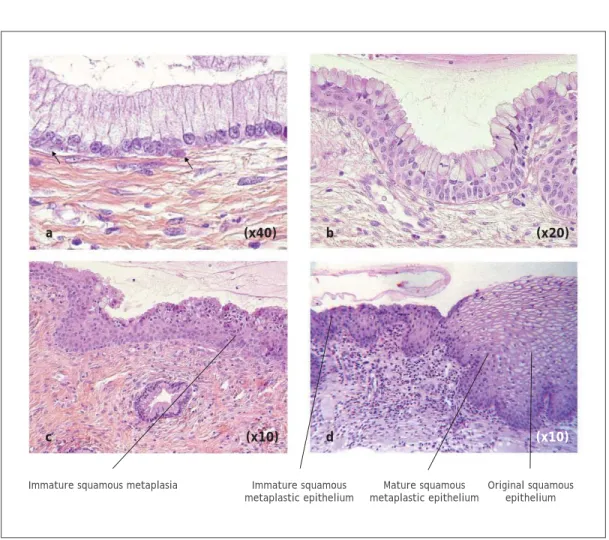
FIGURE 1.5: Development of squamous metaplastic epithelium
(a) The arrows indicate the appearance of the subcolumnar reserve cells.
(b) The reserve cells proliferate to form two layers of reserve cell hyperplasia beneath the overlying layer of columnar epithelium.
(c) The reserve cells further proliferate and differentiate to form immature squamous metaplastic epithelium. There is no evidence of glycogen production.
(d) Mature squamous metaplastic epithelium is indistinguishable from the original squamous epithelium for all practical purposes.
Immature squamous metaplasia Immature squamous metaplastic epithelium Original squamous epithelium a b c d (x40) (x20) (x10) (x10) Mature squamous metaplastic epithelium
Squamous metaplasia
The earliest event in squamous metaplasia is the appearance of small, round, sub-columnar cells in the exposed areas of the columnar epithelium, called reserve cells (Figure 1.5a). These reserve cells proliferate (Figure 1.5b) and differentiate to form a thin, non-stratified, multicellular epithelium called immature squamous epithelium (Figure 1.5c). The cells in the immature squamous metaplastic epithelium do not produce glycogen and, hence, do not stain brown or black with Lugol’s iodine solution. Numerous foci of immature squamous metaplasia may arise at the same time.
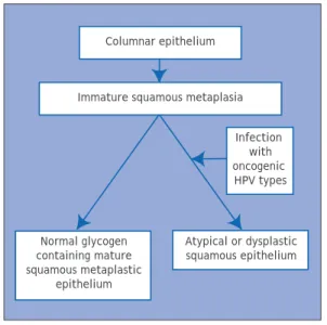
Further development of the newly formed immature metaplastic epithelium may take either one of two directions (Figure 1.6). In the vast majority of women, it develops into a mature, stratified, glycogen-producing, squamous metaplastic epithelium, which is similar to the squamous epithelium found on the ectocervix, for all practical purposes (Figure 1.5d). Thus, it stains brown or black after the application of Lugol’s iodine. Several cysts, called nabothian cysts, may be observed in the mature metaplastic squamous epithelium (Figure 2.3). These are retention cysts that develop as a result of the occlusion of the crypt openings in the trapped columnar epithelium by the overlying metaplastic squamous epithelium. The buried columnar epithelium in the cysts may continue to secrete mucus, which eventually distends the cysts. The entrapped mucus gives an ivory-white hue to the cyst on visual examination.
In a very small minority of women, the immature squamous metaplasia may turn into a dysplastic epithelium (an altered
epithelium showing precancerous cellular changes), due to infection with certain human papillomavirus (HPV) types (Figure 1.6).
Transformation zone
The transformation zone is the area of the cervix where the columnar epithelium has been replaced and/or is being replaced by the metaplastic squamous epithelium. With the naked eye, one can identify the inner border of the transformation zone by tracing the squamocolumnar junction and the outer border by locating the distal most nabothian cysts (if present) or crypt openings (usually visible under magnification). In premenopausal women, the transformation zone is primarily located on the ectocervix. After menopause, and through old age, the cervix shrinks with the decreasing levels of oestrogens. Consequently, the transformation zone may move partially,
Columnar epithelium
Immature squamous metaplasia Infection with oncogenic HPV types Atypical or dysplastic squamous epithelium Normal glycogen containing mature squamous metaplastic epithelium
FIGURE 1.6: A schematic diagram of further maturation of immature squamous metaplasia.
and later fully, into the endocervical canal. Almost all cervical neoplasia occurs in this zone, close to the squamocolumnar junction.
Inflammation of the uterine cervix
(Figure 1.7)
The most common pathological condition affecting a woman’s cervix is inflammation. This is caused mostly by infection (usually polymicrobial) and, less commonly, by foreign bodies (retained tampon, etc.), trauma and chemical irritants such as gels and creams. The infectious agents causing inflammation in the cervix include:
Trichomonas vaginalis; Candida albicans;
overgrowth of anaerobic bacteria such as
Gardnerella vaginalis, G. mobilluncus and
peptostreptococcus; other bacterial infections such as Haemophilus ducreyi,
Neisseria gonorrhoeae, Chlamydia
trachomatis, Escherichia coli, streptococci,
and staphylococci; and viral infections such as Herpes simplex.
Columnar epithelium is more prone to infection than squamous epithelium. We use the term cervicitis in this manual to denote all cervicovaginal inflammatory conditions. Clinically, cervicitis may be associated with symptoms such as excessive discharge, itching of the vulva and vagina, pain and a burning sensation during sexual intercourse and lower abdominal pain. Clinical signs include excessive, coloured (greyish, greyish-white, curdy-white (in the case of candidial infection), yellow or greenish-yellow), malodorous or odorous, frothy or non-frothy secretions, tender, reddish cervix with or without vesicles, ulcerations and/or fibrosis; the columnar epithelium may look flattened; and there may be
FIGURE 1.7:
(a) An inflamed cervix, with ulceration, bleeding, necrosis, greenish-yellow discharge and inflammatory exudate.
(b) A reddish angry-looking, inflamed cervix with loss of the villi in the columnar epithelium and covered with inflammatory exudate.
excoriation marks on the vulva, vulval erythema and oedema, vagina and inner thigh and perineum. Microscopically, cervicitis is characterized by cellular debris and excessive secretions covering the epithelium, swollen and inflamed cells, desquamation of the glycogen-containing superficial and intermediate cells, epithelial denudation, superficial or deep ulceration and congestion of the underlying cervical stroma. Chronic inflammation results in recurrent ulceration and may lead to healing by fibrosis.
A diagnosis of cervicitis can be made based on the clinical features. On visual examination, cervicitis due to non-candidial infection may be characterized by vulval erythema and oedema, excoriation marks in the vulva and vagina and a reddish, tender cervix with malodorous, greenish yellow or greyish-white mucopurulent discharge, with or without ulceration. In the case of gonococcal cervicitis, painful urethral discharge is also observed. Candidial cervicitis is characterized by vulval oedema and erythema, excoriation, and thick, curdy-white, non-odorous discharge. Herpes infection is associated with the presence of vesicles and ulcers in the external genitalia, vagina and the cervix,
as well as cervical tenderness. Women with non-candidial cervicitis may be treated with a combination of metronidazole 400 mg plus doxycycline 100 mg orally, two times a day for seven days. Those with candidial cervicitis may be treated with clotrimazole or micanazole 200 mg intravaginally, daily for three days.
Cervical neoplasia
Invasive cervical cancers are usually preceded by a long phase of preinvasive disease, characterized microscopically as a spectrum of precursor lesions progressing from cellular atypia to various grades of cervical intraepithelial neoplasia (CIN) before progression to invasive carcinoma. Epidemiological studies have identified a number of risk factors that contribute to the development of CIN and cervical cancer. These include infection with certain types of human papillomavirus (HPV), sexual intercourse at an early age, multiple sexual partners, multiparity, long-term oral contraceptive use, tobacco smoking, low socioeconomic status, infection with Chlamydia trachomatis, micronutrient deficiency and a diet deficient in vegetables and fruits. HPV types 16, 18, 31, 33, 35, 39, 45, 51, 52, 56, 58, 59 and 68 are strongly associated with
CIN 1
Mild dysplasia
Low-grade squamous intraepithelial lesion (LSIL)
CIN 2
Moderate dysplasia
High-grade squamous intraepithelial lesion (HSIL)
CIN 3
Severe dysplasia Carcinoma in situ
High-grade squamous intraepithelial lesion (HSIL)
CIN and invasive cancer. Persistent infection with one or more of the above HPV types is considered to be a necessary cause for cervical neoplasia.
Infection with one or more of the oncogenic HPV types may result in the integration of the viral genome into the host cellular genome resulting in the formation of cervical neoplastic cells, the proliferation of which leads to various grades of CIN (synonyms: dysplasia or squamous intraepithelial lesions (SIL)), which may progress to invasive cervical cancer. The correlation between the CIN terminology, used in this manual, and other terminologies is given in Table 1.
Cervical intraepithelial neoplasia
There are no specific symptoms or visible signs associated with CIN. However, the presence of CIN may be suspected by the naked-eye detection of well defined, acetowhite areas in the transformation zone, close to or abutting the squamocolumnar junction, after the application of 3-5% acetic acid or of well defined mustard or saffron yellow iodine non-uptake areas in the transformation zone
after application of Lugol’s iodine solution. The final diagnosis of CIN is established
FIGURE 1.9:
Histology of CIN 2: Atypical cells are found mostly in the lower two-thirds of the epithelium x10.
FIGURE 1.10:
Histology of CIN 3: Dysplastic cells are distributed in the full thickness of the epithelium with loss of polarity of cells x20. FIGURE 1.8:
Histology of CIN 1: The dyplastic cells are confined to the lower third of the epithelium x20.
by histopathological examination of tissue specimens from the cervix. The undifferen-tiated cells in CIN are characterized by enlarged nuclei, increased intensity of nuclear staining, nuclear polymorphism and variation in nuclear size, and a decreased amount of cytoplasm, resulting in a higher nuclear cytoplasmic ratio. The proportion of the thickness of the epithelium showing undifferentiated cells is used for grading CIN. In CIN 1 the undif-ferentiated cells are confined to the deeper layers (lower third) of the epithelium (Figure 1.8). Mitotic figures are present, but not very numerous. CIN 2 is characterized by dysplastic cellular changes mostly restricted to the lower half or the lower two-thirds of the epithelium, with more marked nuclear abnormalities than in CIN 1 (Figure 1.9). Mitotic figures may be seen throughout the lower half of the epithelium. In CIN 3, differentiation and stratification may be totally absent or present only in the superficial quarter of the epithelium with numerous mitotic
figures (Figure 1.10). Nuclear
abnormalities extend throughout the thickness of the epithelium. Many mitotic figures have abnormal forms.
It is well established that most CIN 1 lesions are transient; most of them regress to normal, within relatively short periods, or do not progress to higher grades. High-grade CIN (CIN 2-3), on the other hand, carries a much higher probability of progressing to invasive cancer, although a large proportion of such lesions also regress or persist. It is assumed that the mean interval for progression of cervical precursors to invasive cancer may be as long as 10 to 20 years.
Women with CIN are treated with cryotherapy, loop electrosurgical excision procedure (LEEP) or cold-knife conization.
Women with CIN 1 may be advised to undergo immediate treatment (e.g., in situations where follow-up of women cannot be assured) or treated later if two follow-up visits at six or nine months apart reveal persistent or progressive disease.
The precursor lesion that arises from the columnar epithelium is referred to as adenocarcinoma in situ (AIS). In AIS, normal columnar epithelium is replaced by abnormal epithelium showing abnormal, irregularly arranged cells with increased size of cells and nuclei, nuclear hyperchromasia, mitotic activity, reduction of cytoplasmic mucin expression and cellular stratification.
Invasive cancer
In very early phases of invasion, cervical cancer may not be associated with obvious symptoms and signs, and, therefore, is known as preclinical invasive cancer. Women with moderately advanced or advanced invasive cervical cancer often present with one or more of the following symptoms: intermenstrual bleeding, postcoital bleeding, excessive seropurulent discharge, recurrent cystitis, backache, lower abdominal pain, oedema of the lower extremities, obstructive uropathy, bowel obstruction, breathlessness due to severe anaemia and cachexia.
As the stromal invasion progresses, the disease becomes clinically obvious, showing several growth patterns, which are visible on speculum examination. Early lesions may present as a rough, reddish, granular area that bleeds on
touch (Figure 1.11) More advanced
cancers may present as a proliferating, bulging, mushroom- or cauliflower-like growth with bleeding and foul-smelling discharge (Figure 1.12). Occasionally they may present without much surface
growth, resulting in a grossly enlarged, irregular cervix with a rough, granular surface.
As the invasion continues further, it may involve the vagina, parametrium, pelvic sidewall, bladder and rectum. Compression of the ureter, due to advanced local disease, causes ureteral obstruction, which results in hydronephrosis and, ultimately, renal failure. Regional lymph node metastasis occurs along with local invasion. Metastatic cancer in para-aortic nodes may extend through the node capsule and directly invade the vertebrae and nerve roots causing back pain. Direct invasion of the branches of the sciatic nerve roots causes low back pain and leg aches, and encroachment of the pelvic wall veins and lymphatics causes oedema of the lower limbs. Distant metastases occur late in the disease, usually involving para-aortic nodes, lungs, liver, bone and other structures.
Histologically, approximately 90-95% of invasive cervical cancers in developing
countries are squamous cell cancers (Figure 1.13) and 2-8% are adenocarcinomas (Figure 1.14). It is obligatory that all invasive cancers be clinically staged. The most widely used staging system for cervical cancer was developed by the International Federation of Gynecology and Obstetrics (FIGO) (see Appendix 1). This is primarily a clinical staging system based on tumour size and extension of the disease in the pelvis. The extent of growth of cancer is assessed clinically, as well as by various investigations, to categorize the disease stages I through IV. Stage I represents growth localized on the cervix, while stage IV represents the growth phase in which the cancer has spread to distant organs by metastasis.
Women with early invasive cancers (stages I and II A) may be treated with radical surgery and/or radiotherapy. Those with stage IIB and III cancers may be treated with radiotherapy with or without cisplatinum-based chemotherapy. Women
FIGURE 1.11:
Early invasive cervical cancer: note the irregular, granular, nodular surface with bleeding on touch.
FIGURE 1.12:
Advanced invasive cervical cancer: note the bulging, cauliflower-like, ulceroproliferative growth with bleeding and necrosis.
with stage IV cancers are usually treated with palliative radiotherapy and/or chemotherapy and with symptomatic measures.
Other conditions
Leukoplakia (hyperkeratosis) is a well
demarcated white area on the cervix (before the application of acetic acid), due to keratosis, visible to the naked eye. Usually leukoplakia is idiopathic, but it may also be caused by chronic foreign body irritation, HPV infection, or squamous neoplasia. Condylomata or genital warts are often multiple, exophytic lesions that are usually found on the cervix, and occasionally in the vagina and on the vulva, caused by infection with certain HPV types such as 6 and 11. They may also present as a diffuse, greyish-white lesion involving areas of the cervix and vagina. Condylomata may be obvious to the naked eye (before the application of acetic acid).
Pathophysiological basis of VIA
Application of 5% acetic acid is believed to cause a reversible coagulation, or
precipitation of the cellular proteins. It also causes swelling of the epithelial tissue, columnar and any abnormal squamous epithelial areas in particular, dehydration of the cells, and it helps in coagulating and clearing the mucous secretions on the cervix. The normal squamous epithelium appears pink and the columnar epithelium red, due to the reflection of light from the underlying stroma, which is rich in blood vessels. If the epithelium contains a lot of cellular proteins, acetic acid coagulates these proteins, which may obliterate the colour of the stroma. The resulting acetowhitening is seen distinctly as compared with the normal pinkish colour of the surrounding normal squamous epithelium of the cervix, an effect that is commonly visible to the naked eye. Thus, the effect of acetic acid depends upon the amount of cellular proteins present in the epithelium. Areas of increased nuclear activity and DNA content exhibit the most dramatic white colour change.
When acetic acid is applied to normal squamous epithelium, little coagulation
FIGURE 1.13:
Histology – Keratinizing well differentiated invasive squamous cell carcinoma. Note the stroma is infiltrated by sheets of malignant cells x10.
FIGURE 1.14:
Histology – Well differentiated invasive adenocarcinoma. Note the malignant cells lining the cervical crypts x20.
↓
↓
↓
↓
occurs in the superficial cell layer, as this is sparsely nucleated. Although the deeper cells contain more nuclear protein, the acetic acid may not penetrate sufficiently and, hence, the resulting precipitation is not sufficient to obliterate the colour of the underlying stroma. Areas of CIN and invasive cancer undergo maximal coagulation due to their higher content of nuclear protein (in view of the large number of undifferentiated cells contained in the epithelium) and prevent light from passing through the epithelium. As a result, the sub-epithelial vessel pattern is obliterated and the epithelium appears densely white. In CIN, acetowhitening is restricted to the transformation zone close to the squamocolumnar junction, while in cancer it often involves the entire cervix.
The acetowhite appearance is not unique to CIN and early cancer. It is also seen in other conditions when increased nuclear protein is present, as in immature squamous metaplasia, in healing and regenerating epithelium (associated with inflammation), leukoplakia (hyperkeratosis) and condyloma. While the acetowhite epithelium associated with CIN and early invasive cancer is more dense, thick and opaque with well demarcated margins from the surrounding normal epithelium, the acetowhitening associated with immature squamous metaplasia, inflammation and regenerating epithelium is less pale, thin, often translucent, and patchy with ill-defined margins. Acetowhitening due to inflammation and healing is usually distributed widely in the cervix, not restricted to the transformation zone and may quickly disappear (within a minute). Leukoplakia and condylomata appear intensely greyish-white after the application of acetic acid.
The acetic acid effect reverses much more slowly in CIN lesions and in early preclinical invasive cancer than in immature squamous metaplasia and inflammation. It appears rapidly and may last for 3-5 minutes in the case of CIN 2-3 and invasive cancer.
Pathophysiological basis of VILI
Squamous metaplastic epithelium is glycogenated, whereas CIN and invasive cancer cells contain little or no glycogen. Columnar epithelium does not contain glycogen. Immature squamous metaplastic epithelium usually lacks glycogen or, occasionally, may be partially glycogenated. Iodine is glycophilic and hence the application of iodine solution results in uptake of iodine in glycogen-containing epithelium. Therefore, the normal glycogen-containing squamous epithelium stains mahogany brown or black after application of iodine. Columnar epithelium does not take up iodine and remains unstained, but may look slightly discoloured due to a thin film of iodine solution; areas of immature squamous metaplastic epithelium may remain unstained with iodine or may be only partially stained. If there is shedding (or erosion) of superficial and intermediate cell layers associated with inflammatory conditions of the squamous epithelium, these areas do not stain with iodine and remain distinctly colourless in a surrounding black or brown background. Areas of CIN and invasive cancer do not take up iodine (as they lack glycogen) and appear as thick mustard-yellow or saffron coloured areas. Areas with leukoplakia (hyperkeratosis) do not stain with iodine either, and condylomata may not, or occasionally may only partially, stain with iodine.
Testing and reporting the results of visual
inspection with 5% acetic acid (VIA)
Test provider skills
The test provider must have a good knowledge of the anatomy, physiology and pathology of the cervix in relation to its visual examination. He/she should know the clinical features of benign conditions, inflammation, precancerous lesions and invasive cancer of the cervix.
Procedure
Women coming for testing should have the screening procedure explained to them in detail. Written informed consent should be obtained before screening. An example of a written informed consent form is given in Appendix 2. Relevant obstetric and gynaecological history should be obtained and recorded with the help of a form for this purpose (Appendix 3). The woman should be reassured that the procedure is painless, and every effort should be made to ensure that she is fully relaxed and remains at ease during testing.
The woman is invited to lie down in a modified lithotomy position on a couch with leg rests or knee crutches or stirrups. After proper positioning of the woman, observe if there is any vaginal discharge. Observe the external genitalia and perineal region for any signs of excoriations, oedema, vesicles, papules, sores, ulceration and warts.
Instruments and materials required: • Examination table with knee crutches or
leg rests or stirrups;
• Good light source (preferably a bright halogen lamp that can be easily directed at the cervix or a bright halogen torch light);
• Sterile bivalved speculum: Cusco’s, Grave’s or Collin’s;
• Pair of gloves;
• Cotton swabs, cotton-tipped buds, gauze;
• Ring forceps, pickup forceps;
• 5% freshly prepared acetic acid or vinegar (check the strength of acetic acid in vinegar);
• A steel/plastic container with 0.5% chlorine solution in which to immerse the gloves;
• A plastic bucket or container with 0.5% chlorine solution to decontaminate instruments;
• A plastic bucket with a polythene bag to dispose of contaminated swabs and other waste items.
Preparation of 5% dilute acetic acid
5% acetic acid is prepared by adding 5 ml of glacial acetic acid into 95 ml of distilled water.
If vinegar bought from a store is used, check the strength to ensure that it is 5%.
Look for any swelling in the inguinal/femoral region.
Afterwards, gently introduce a sterile vaginal speculum which has been immersed in warm water and open the blades of the speculum to view the cervix. Adjust the light source so that there is adequate light in the vagina and on the cervix. As the speculum is gently opened and the lips are fixed, the cervix comes into view. Observe the size and shape of the cervix.
Identify the external os, columnar epithelium (red in colour), squamous epithelium (pink) and the squamocolumnar junction. Proceed to identify the transformation zone, the upper limit of which is formed by the squamocolumnar junction. Remember that cervical neoplasias occur in the transformation zone nearest to the squamocolumnar junction.
Look for ectropion, cervical polyp, nabothian cysts, healed laceration of the cervical lips, leukoplakia, condylomata and signs of cervicitis. You may note that in postmenopausal women, the cervix appears pale and brittle, due to thinning and atrophy of the squamous epithelium. Assess the characteristics of discharge in terms of quantity, colour, odour and thickness. Thread-like, thin mucinous discharge from the external os indicates ovulation. If heavy blood flow through the external os is observed in women during menstruation, they may be subjected to VIA after 5-15 days.
In ectropion, the cervix has a large area of red appearance around the external os and the squamocolumnar junction far away from the os. Nabothian cysts appear as bulging blue-white or yellow-white nodules, having a smooth delicate lining with branching blood vessels. In some
women, nabothian cysts can become large and distort the shape of the cervix. A cervical polyp appears as a smooth mass protruding from the cervical canal beyond the external os, which may appear dark red or pink-white. Sometimes a necrotic polyp resembles a cervical cancer. Healed lacerations appear as tears on the lips of the cervix, with the external os appearing irregular. Leukoplakia appears as a smooth-surfaced, white area on the cervix that cannot be removed or scraped off. Cervical condylomata appear as raised, grey-white areas within or outside the transformation zone in the squamous epithelium and may be accompanied by similar lesions in the vagina and vulva.
Look for small blisters containing fluid or multiple, small ulcers on the cervix. Extensive erosive red areas may be present on the cervix, extending to the vagina in instances of severe cervical infection and inflammation. Observe whether there is any bleeding from the cervix, especially on touch, or ulceroproliferative growth. A very early invasive cancer may present as a rough, reddish, granular area that may bleed on touch. More advanced invasive cancers may present as a large exophytic growth with an ulceroproliferative, bulging mass with polypoid or papillary excrescences, arising from the cervix or as a predominantly ulcerating growth replacing most of the cervix. In both of these types, bleeding on touch and necrosis are predominant clinical features. Foul-smelling discharge is also common due to superadded infection. Occasionally, invasive cancer can present as an infiltrating lesion with a grossly enlarged irregular cervix.
Now, gently, but firmly, apply 5% acetic acid using a cotton swab soaked in acetic acid. The secretions should be gently wiped
off. The swabs after use should be disposed of in the waste bucket. The curdy-white discharge associated with candidiasis is particularly sticky, and if particular care is not taken to remove it properly, it may mimic an acetowhite lesion, thus leading to a false-positive result. After removing the swab, carefully look at the cervix to see whether any white lesions appear, particularly in the transformation zone close to the squamocolumnar junction, or dense, non-removable acetowhite areas in the columnar epithelium. The results one minute after application of acetic acid should be reported. Note how rapidly the acetowhite lesion appears and then disappears.
Carefully observe:
• The intensity of the white colour of
the acetowhite lesion: if it is shiny-white, cloudy-shiny-white, pale-white or dull-white.
• The borders and demarcations of the
white lesion: distinctly clear and sharp or indistinct diffuse margins; raised or flat margins; regular or irregular margins.
• Whether the lesions are uniformly
white in colour, or the colour intensity varies across the lesion, or if there are areas of erosion within the lesion.
• Location of the lesion: is it in, near or far away from the transformation zone? Is it abutting (touching) the squamocolumnar junction? Does it extend into the endocervical canal? Does it occupy the entire, or part of, the transformation zone? Does it involve the entire cervix (which usually indicates early preclinical invasive cancer)?
• Size (extent or dimensions) and
number of the lesions.
If in doubt, it is safe to repeat the test a few times, taking care not to induce bleeding. Women with suspected invasive cancers should be referred for further investigations and treatment.
Conclusion of the examination
Contaminated swabs, gauze and other waste material should be disposed of in the plastic bag in the plastic bucket.
Withdraw the speculum gently, and inspect the vaginal walls for condyloma and acetowhite lesions. Before removing the soiled gloves, immerse the hands briefly in a container filled with 0.5% chlorine solution. Decontaminate the used gloves by soaking in the 0.5% chlorine in a plastic bucket for 10 minutes. Preparation of 0.5% chlorine solution is described in Appendix 4.
The speculum and other instruments used for VIA should be immersed in 0.5% chlorine solution for 10 minutes’ deconta-mination, before cleaning with detergent and water. The cleaned instruments may be reused after high-level disinfection by immersing them in boiling water for 20 minutes or by sterilizing the instruments using an autoclave.
Documentation of findings and
advising the woman
Carefully document the outcome of testing in the reporting form (Appendix 3). Explain the outcome of the test to the woman, as well as any further course of follow-up actions. If the test is negative, the woman is reassured and she may be advised to repeat testing after five years. If the test is positive, she should be referred for further investigations such as colposcopy and biopsy as well as treatment for any confirmed lesions. If invasive cancer is
suspected, she should be referred to a cancer diagnosis and treatment facility.
Reporting the outcome of VIA
VIA negative (-)
VIA screening is reported as negative in the case of any of the following observations:
• No acetowhite lesions are observed on
the cervix (Figure 2.1).
• Polyps protrude from the cervix with
bluish-white acetowhite areas (Figure 2.2).
• Nabothian cysts appear as button-like
areas, as whitish acne or pimples (Figure 2.3).
• Dot-like areas are present in the
endocervix, which are due to grape-like columnar epithelium staining with acetic acid (Figure 2.4).
• There are shiny, pinkish-white, cloudy-white, bluish-cloudy-white, faint patchy or doubtful lesions with ill-defined, indefinite margins, blending with the rest of the cervix (Figures 2.5-2.7).
• Angular, irregular, digitating
acetowhite lesions, resembling geographical regions, distant (detached) from the squamocolumnar junction (satellite lesions) (Figure 2.8).
• Faint line-like or ill-defined
acetowhitening is seen at the
squamocolumnar junction (Figures
2.8-2.10).
• Streak-like acetowhitening is visible in the columnar epithelium (Figure 2.8).
• There are ill-defined, patchy, pale,
discontinuous, scattered acetowhite areas (Figures 2.10-2.11).
FIGURE 2.1:
VIA negative: No acetowhite area seen. Note the advancing edges of squamous metaplasia in the anterior and posterior lips (arrows).
FIGURE 2.2:
VIA negative. There are no acetowhite areas on the polyp and the cervix after the application of acetic acid.
FIGURE 2.6:
VIA negative: There is an ill-defined pinkish-white hue with indefinite margins blending with the rest of the epithelium. The squamocolumnar junction is fully visible. FIGURE 2.3:
VIA negative. The nabothian cysts appear as pimple- or button-like areas after the application of acetic acid.
FIGURE 2.4:
VIA negative: There is dot-like acetowhitening in the columnar epithelium in the anterior lip. The squamocolumnar junction is fully visible.
FIGURE 2.5:
VIA negative: There are ill-defined pinkish-white and cloudy-pinkish-white areas with indefinite margins blending with the rest of the epithelium. The squamocolumnar junction is fully visible.
↓
↓
↓
FIGURE 2.7:
VIA negative: There is an ill-defined pinkish-white hue, with indefinite margins, blending with the rest of the epithelium. The squamocolumnar junction is fully visible.
FIGURE 2.8:
VIA negative: There are pale white, satellite, geographical lesions with angular margins (narrow arrow) far away from the squamocolumnar junction (dense arrow). Note the streak-like acetowhitening in the columnar epithelium (within the oval area).
FIGURE 2.9:
VIA negative: There is dense, thick, mucus on the cervix before the application of acetic acid. After the application of acetic acid, the mucus is cleared and the squamocolumnar junction becomes prominent.
VIA positive (+)
The VIA test outcome is reported as positive in any of the following situations:
• There are distinct, well-defined,
dense (opaque, dull- or oyster-white) acetowhite areas with regular or irregular margins, close to or abutting the squamocolumnar junction in the transformation zone or close to the external os if the squamocolumnar junction is not visible (Figures 2.12-2.20).
• Strikingly dense acetowhite areas are
seen in the columnar epithelium (Figures 2.21-2.22).
• The entire cervix becomes densely
white after the application of acetic acid (Figure 2.23).
• Condyloma and leukoplakia occur
close to the squamocolumnar junction, turning intensely white after application of acetic acid.
FIGURE 2.10:
VIA negative: The squamocolumnar junction is prominent after the application of acetic acid. Note the ectropion.
FIGURE 2.11:
VIA negative: The cervix is unhealthy, inflamed with ulceration, necrosis, bleeding and inflammatory exudate.There is ill-defined, diffuse, pinkish-white acetowhitening with indefinite margins blending with the rest of epithelium (arrows).
FIGURE 2.12:
VIA positive: There is a well-defined, opaque acetowhite area, with irregular digitating margins, in the anterior and posterior lips abutting the squamocolumnar junction and extending into the cervical canal.
↓
↓
FIGURE 2.13:
VIA positive: There is a well-defined, opaque acetowhite area, with bleeding on touch, in the anterior lip, abutting the squamocolumnar junction, which is fully visible.
FIGURE 2.14:
VIA positive: There is a well-defined, opaque acetowhite area, with regular margins, in the anterior lip, abutting the squamocolumnar junction, which is fully visible.
FIGURE 2.15:
VIA positive: There is a well-defined, opaque acetowhite area, with regular margins, in the lower lip, abutting the squamocolumnar junction, which is fully visible.
FIGURE 2.16:
VIA positive: There is a well-defined, opaque acetowhite area, with regular margins, in the anterior lip, abutting the squamocolumnar junction, which is fully visible. Note the satellite lesions in the lower lip.
FIGURE 2.17:
VIA positive: There is a well-defined, opaque acetowhite area, with regular margins, in the anterior lip, abutting the squamocolumnar junction, which is fully visible. Note the somewhat ill-defined white area in the lower lip. The lesion is extending into the cervical canal.
FIGURE 2.18:
VIA positive: There is a well-defined, dull, dense, opaque acetowhite area in the anterior lip abutting the squamocolumnar junction which is fully visible.
FIGURE 2.19:
VIA positive: There is a well-defined, dull, dense, opaque acetowhite area, with raised and rolled-out margins in the anterior lip abutting the squamocolumnar junction which is fully visible. The lesion is extending into the cervical canal.
FIGURE 2.20:
VIA positive: There is a well-defined, dull, dense, opaque acetowhite area in the posterior lip extending into the endocervical canal.
VIA positive, invasive cancer
The test outcome is scored as invasive cancer when:
• There is a clinically visible
ulcero-proliferative growth on the cervix that turns densely white after application of acetic acid and bleeds on touch (Figures 2.24-2.27).
Self-evaluation of the test
providers
The test providers are encouraged to correlate the results of their VIA testing with those of colposcopy and histology. They are strongly advised to participate in the colposcopy sessions with doctors and review the findings. Such measures improve the skills of the test providers. A benchmark to assess one’s own skill is to estimate what proportion of women examined are scored as aceto-positive and what proportion of aceto-positive
FIGURE 2.21:
VIA positive: There is an acetowhite area in the columnar epithelium in the anterior and posterior lips.
FIGURE 2.22:
VIA positive: There are dense acetowhite areas in the columnar epithelium in the anterior lip.
FIGURE 2.23:
VIA positive: There is a dense acetowhite area all over the cervix involving all the four quadrants and extending into the cervical canal.
FIGURE 2.25:
VIA positive, invasive cancer: There is a proliferative growth with dense acetowhitening and bleeding.
FIGURE 2.26:
VIA positive, invasive cancer: There is a dense acetowhite area with irregular surface contour.
FIGURE 2.24:
VIA positive, invasive cancer: There is a dull, opaque, dense acetowhite area, with raised and rolled-out margins, irregular surface and bleeding on touch in the posterior lip. The lesion is extending into the cervical canal. The bleeding obliterates acetowhitening.
FIGURE 2.27:
VIA positive, invasive cancer: There is an ulceroproliferative growth with acetowhitening and bleeding.
women are ultimately diagnosed with CIN. A sufficiently skilled examiner will categorize 8-15% of women examined as aceto-positive, and 20-30% of the
acetowhite lesions identified on VIA by the test provider harbour CIN of any grade.
Testing and reporting the results of visual
inspection with Lugol's iodine (VILI)
Test provider skills
The test provider must have a good knowledge on the anatomy, physiology and pathology of the cervix in relation to its visual examination. He/she should know the clinical features of benign conditions, inflammation, precancerous lesions and invasive cancer of the cervix.
Procedure
Women coming for testing should have the screening procedure explained to them in detail. Written informed consent should be obtained before screening. An example of a written informed consent form is given in Appendix 2. Relevant obstetric and gynaecological history should be obtained and recorded with the help of a form for this purpose (Appendix 3). The woman should be reassured that the procedure is painless, and every effort should be made to ensure that she is fully relaxed and remains at ease during testing.
The woman is invited to lie down in a modified lithotomy position on a couch with leg rests or knee crutches. After proper positioning of the woman, observe if there is any vaginal discharge. Observe the external genitalia, and perineal region for any signs of excoriations, oedema, vesicles, papules, sores,
Instruments and materials required: • Examination table with knee crutches or
leg rests or stirrups;
• Good light source (preferably a bright halogen lamp that can be easily directed at the cervix);
• Sterile speculum: Cusco’s, Grave’s or Collin’s;
• Pair of gloves;
• Cotton swabs; cotton-tipped buds, gauze;
• Ring forceps, pickup forceps;
• 5% Lugol’s iodine solution;
• An aluminium, steel or plastic container with 0.5% chlorine solution in which to immerse the gloves;
• Plastic bucket or container with 0.5% chlorine solution for decontamination of instruments;
• A plastic bucket with a plastic bag to dispose of contaminated swabs and other waste items.
Preparation of Lugol's iodine
Dissolve 10 g of potassium iodide in 100 ml distilled water. Add 5 g of iodine after the potassium iodide is fully dissolved. Stir well until all the iodine flakes have fully
dissolved. The solution should be stored in a sealed container to prevent evaporation of iodine and loss of staining activity.
ulceration and warts. Look for any swelling in the inguinal/femoral region. Then proceed to wipe the vaginal introitus and the surrounding perineum with a cotton swab or gauze soaked with an antiseptic solution.
Afterwards, gently introduce a sterile vaginal speculum, which has been immersed in warm water, and open the blades of the speculum to view the cervix. Adjust the light source so that there is adequate light in the vagina and on the cervix. As the speculum is gently opened and the lips are fixed, the cervix comes into view. Observe the size and shape of the cervix.
Identify the external os, columnar epithelium (red in colour), squamous epithelium (pink) and the squamocolumnar junction. Proceed to identify the transformation zone, the upper limit of which is formed by the squamocolumnar junction. Look for ectropion, cervical polyp, nabothian cysts, healed laceration of the cervical lips, leukoplakia, condylomata and signs of cervicitis. You may note that in postmenopausal women, the cervix appears pale and brittle, due to thinning and atrophy of the squamous epithelium. Assess the characteristics of discharge in terms of quantity, colour, odour and thickness. Thread-like, thin mucinous discharge from the external os indicates ovulation. Blood flow through the external os is observed in women during menstruation, and they may be subjected to VILI after 10-15 days.
In ectropion, the cervix has a large area of gross red appearance around the external os and the squamocolumnar junction far away from the os. Nabothian cysts appear as bulging blue-white or yellow-white nodules, having a smooth delicate lining with branching blood
vessels. In some women, nabothian cysts can become large and distort the shape of the cervix; however, they seldom ulcerate and become necrotic. A cervical polyp appears as a smooth mass protruding from the cervical canal beyond the external os, which may appear dark red or pink-white. Sometimes a necrotic polyp resembles a cervical cancer. Healed lacerations appear as tears on the lips of the cervix, with the external os appearing irregular. Leukoplakia appears as a smooth-surfaced, white area on the cervix that cannot be removed or scraped off. Cervical condylomata appear as raised, grey-white areas within, or outside, the transformation zone in the squamous epithelium and may be accompanied by similar lesions in the vagina and vulva.
Look for the presence of blisters with fluid or small ulcers on the cervix. Extensive erosive red areas may be present in the cervix, extending to the vagina in instances of severe cervical infection and inflammation. Observe whether there is any bleeding from the cervix, especially on touch, or ulceroproliferative growth. A very early invasive cancer may present as a rough, reddish, granular area, that may bleed on touch. More advanced invasive cancers may present as a large exophytic growth with an ulceroproliferative, bulging mass with polypoid or papillary excrescences, arising from the cervix or as a predominantly ulcerating growth replacing most of the cervix. In both of these types, bleeding on touch and necrosis are predominant clinical features. Foul-smelling discharge is also common due to superadded infection. Occasionally, invasive cancer can present as an infiltrating lesion with a grossly enlarged irregular cervix.
After carefully recording the visual findings, liberally and gently apply Lugol's iodine with a cotton swab on the cervix. Take care not to stain either the woman's or your own clothes with iodine! After removing the swab, carefully look at the cervix for any iodine non-uptake (non-staining) areas in the form of pale or yellowish-white areas, particularly in the transformation zone, close to the squamocolumnar junction. Once the examination is completed, the excess iodine in the vaginal fornices should be mopped up with dry cotton.
Conclusion of the examination
Contaminated swabs, gauze and other waste material should be disposed of in the plastic bag in the plastic bucket.
Withdraw the speculum gently, and inspect the vaginal walls for condyloma and iodo-negative lesions. Before removing the soiled gloves, immerse the hands briefly in a container filled with 0.5% chlorine solution. Decontaminate the used gloves by soaking in the 0.5% chlorine in a plastic bucket for 10 minutes. Preparation of 0.5% chlorine solution is described in Appendix 4.
The speculum and other instruments used during VILI should be immersed in 0.5% chlorine solution for 10 minutes’ decontamination, before cleaning with water. The cleaned instruments may be reused after high-level disinfection by immersing them in boiling water for 20 minutes or by sterilizing the instruments using an autoclave.
Documentation of findings and
advising the woman
Carefully document the outcome of testing in the reporting form (Appendix 3). Explain the outcome of the test to the
woman, as well as any further course of follow-up actions. If the test is negative, the woman is reassured and she may be advised to repeat testing after five years. If the test is positive, she should be referred for further investigations such as colposcopy and biopsy as well as treatment for any confirmed lesions. If invasive cancer is suspected, she should be referred to a cancer diagnosis and treatment facility.
Reporting the outcome of VILI
The patterns associated with the outcomes after VILI are shown in Figures 3.1-3.21.
VILI negative (-):
VILI screening is reported as negative in the case of any of the following observations after iodine application:
• A normal cervix; the squamous
epithelium turns mahogany brown or black and the columnar epithelium does not change colour (Figure 3.2).
• Patchy, indistinct, ill-defined,
colourless or partially brown areas are seen (Figures 3.3-3.6).
• Pale areas of no or partial iodine
uptake are present on polyps (Figure 3.7).
• A leopard-skin appearance (Figure 3.8) is associated with T. vaginalis infection.
• Pepper-like non-iodine uptake areas
are seen in the squamous epithelium, far away from the squamocolumnar junction (Figure 3.9)
• Satellite, thin, yellow, non-iodine
uptake areas with angular, or digitating margins, resembling geographical areas, are seen far away from the squamocolumnar junction (Figure 3.10).
FIGURE 3.1:
Visual inspection with Lugol’s iodine (VILI). VILI Negative
PINHOLE OS
VILI Positive
NORMAL ECTROPION POLYP
NABOTHIAN CYST CERVICITIS CERVICITIS NON-SPECIFIC INFECTIONS
SQUAMOUS
FIGURE 3.2:
VILI negative: The squamous epithelium is black and the columnar epithelium does not change colour after the application of iodine.
FIGURE 3.3:
VILI negative: There are patchy, ill-defined, scattered non-iodine uptake areas in the cervix; these are not restricted to the transformation zone. This appearance is due to inflammation of the cervix.
FIGURE 3.4:
VILI negative: There are patchy, ill-defined areas of iodine non-uptake (narrow arrows) and partial iodine uptake (dense arrows).
FIGURE 3.5:
VILI negative: Squamous epithelium remains brown. There are patchy areas of no or partial uptake of iodine in the transformation zone corresponding to areas of immature squamous metaplasia and inflammation.
Squamous epithelium
Columnar epithelium has not taken up iodine
↓
↓
↓↓
FIGURE 3.9:
VILI negative: There are pepper-like areas of iodine non-uptake in the squamous epithelium, due to cervical ulceration following inflammation.
FIGURE 3.6:
VILI negative: There are patchy, ill-defined areas of iodine non-uptake (narrow arrow) and partial iodine uptake (dense arrows).
FIGURE 3.7:
VILI negative: There are areas of iodine non-uptake (narrow arrow) and partial non-uptake (dense arrow) in the polyp. Squamous epithelium is black.
FIGURE 3.8:
VILI negative: There are patchy areas of iodine non-uptake, scattered all over the cervix, not restricted to the transformation zone. This is characteristic of chronic cervicitis.
↓↓
↓↓
↓
↓
↓↓
↓
↓
VILI positive (+):
The outcome is scored as positive if dense, thick, bright, mustard-yellow or saffron-yellow iodine non-uptake areas are seen in the transformation zone, close to or abutting the squamocolumnar junction or close to the os if the squamocolumnar junction is not seen (Figures 3.11-3.15) or when the entire cervix turns densely yellow (Figure 3.16).
VILI positive, invasive cancer:
Invasive cancer is reported when a frank, nodular, irregular, ulceroproliferative growth is visible on the cervix which turns densely yellow on application of iodine (Figures 3.17-3.19).
Self-evaluation of the test
providers
The test providers are encouraged to correlate the results of their VILI testing with histological findings (if done). A sufficiently skilled examiner will categorize 10-15% of women as VILI +, and 20-30% of the women VILI+ harbour CIN of any grade.
FIGURE 3.10:
VILI negative: Iodine-negative, irregular yellow areas are detached from the squamocolumnar junction and constitute ‘satellite’ lesions.
FIGURE 3.11:
VILI positive: There is a saffron-yellow area of iodine non-uptake in the anterior lip abutting the squamocolumnar junction.
FIGURE 3.12:
VILI positive: There is a mustard-yellow lesion with no iodine uptake in the anterior lip touching the squamocolumnar junction.
FIGURE 3.13:
VILI positive: There is a mustard-yellow area of iodine non-uptake in the anterior lip abutting the squamocolumnar junction.
FIGURE 3.14:
VILI positive: There is a dense, mustard-yellow area of iodine non-uptake abutting the squamocolumnar junction in the anterior lip of the cervix with irregular, angular margins.
FIGURE 3.15:
VILI positive: There are large, thick, mustard-yellow areas of iodine non-uptake in the upper and lower lip of the cervix extending into the cervical canal.
FIGURE 3.16:
VILI positive: There is a large, dense, saffron-yellow area of iodine non-uptake with irregular surface, involving all the four quadrants and extending into the cervical canal.
FIGURE 3.18:
VILI positive, invasive cancer: There is a large, thick, irregular, nodular mustard-yellow lesion suggestive of invasive cancer.
FIGURE 3.19:
VILI positive, invasive cancer: There is a large, thick mustard-yellow area in the cervix with irregular, nodular surface contour.
FIGURE 3.17:
VILI positive, invasive cancer: There is a dense, thick, extensive mustard-yellow area, with irregular and nodular surface, involving all the quadrants of the cervix. The external os is obliterated.