Perspectives
Genesis of non-coding RNA genes- a sequence
connection with protein genes separated by evolutionary
time
Nicholas Delihas
Department of Microbiology and Immunology, Renaissance School of Medicine, Stony Brook University, Stony Brook, New York, 11794-5222 USA
Email:[email protected]
Abstract: A small phylogenetically conserved sequence of 11,231 bp termed FAM247 is repeated in human chromosome 22 by segmental duplications. This sequence forms part of diverse genes that span evolutionary time, the protein genes being the earliest as they are present in zebrafish and/or mice genomes, the long non-coding RNA genes and pseudogenes the most recent as they appear to be present only in the human genome. We propose that the conserved sequence provides a nucleation site for new gene development at evolutionary conserved chromosomal loci where the FAM247 sequences reside. The FAM247 sequence also carries information in its open reading frames that provides protein exon amino acid sequences; one exon plays an integral role in immune system regulation, specifically, the function of ubiquitin specific protease (USP18) in the regulation of interferon. An analysis of this multifaceted sequence and the genesis of genes that contain it are presented.
Keywords: gene evolution; gene formation; long non-coding RNA genes; pseudogenes; USP18; GGT5
1. Introduction
The genesis of genes has been a major topic of interest for several decades [1, 2]. One mechanism of
gene origins is the formation of genes from the duplication of existing genes [1, 3]. This is considered one
of the major processes for new gene formation, but it has also been shown that there is a prevalence of
gene birth from non-coding DNA via de novo gene formation [4-7]; this pathway also significantly
contributes to new gene formation [4, 7].
In this treatise we present and analyze gene development by an evolutionary conserved ancestral
sequence. This is a repeat element, previously termed clincRNA [8] and now termed FAM247. The long
intergenic non-coding RNA (lincRNA) FAM247A gene sequence has been used as a guide to find
homologous sequences and heretofore FAM247 is used in place of FAM247A. We propose that FAM247
carries information to form nucleation sites for gene development and this is exemplified with the
maturation of pseudogenes by addition of chromosomal sequences at specific sites on FAM247. The
FAM247 sequence also carries information in terms of its open reading frames. These open reading
frames are found to form exon sequences of proteins.
FAM247 forms part of non-coding RNA genes that appear to be human specific. The sequence is also
found in protein genes, USP18 (ubiquitin specific protease) and GGT5 (gamma glytamyltransferase). Both these genes date back in evolutionary time, USP18 over 350 million years ago (MYA) and GGT5 over 90
MYA. Thus, the FAM247 sequence has formed a part of genes through much of vertebrate evolution.
2. Background on conserved linked sequences
FAM247 is present in different segmental duplications or low copy repeats (LCR22) in human
chromosome 22 (chr22) as part of phylogenetically conserved linked gene sequences [8]. Figure 1 is a
representation of these linked sequences and shows conserved nearest neighbor sequence signatures
found in humans. The linked gene sequences are repeated in chr 22 and generate gene families. The
signatures are also representative of ancestral primate linked-sequences, e.g., the sequence arrangement
in Figure 1b is present in the Rhesus Old World monkey ( Macaca mulatta) where FAM247 and spacer
sequences are linked to GGT1 on chr10. The spacer sequence (3953 bp) depicted in Figure 1 is also
evolutionarily conserved. It is present in Pan troglodytes (chimpanzee), Papio Anubis (olive baboon), Pongo
abelii (Sumatran orangutan), and Macaca mulatta (Rhesus monkey) genomes but it does not encode genes or form part of genes [8]. That it is evolutionarily conserved indicates it may have a function. FAM247 is
the common denominator in Figure 1a and 1b. In Figure 1c, the FAM247 sequence depicted is embedded
Figure 1. Schematic representation of evolutionarily conserved linked sequences with different colors
depicting different sequences, as described in [8]. In Figure 1c, the FAM247 sequence (green highlight) is
embedded the USP18 gene.
Table 1 contains a list of human gene families that are found in repeat units shown in Figure 1 and
indicates the sequence or chromosomal locus of origin. For example, GGT represents the locus of origin
of GGT1, the gamma-glutamyltransferase and gamma-glutamyltransferase light chain genes and their
respective pseudogenes; FAM247 is the sequence/locus of origin of GGT5.
Table 1. Human genes and gene families found in linked sequences: FAM247-GGT,
FAM230-USP.
Gene/gene family Type Locus origin
*FAM230A-J lincRNA FAM230
FAM247A-D lincRNA FAM247
POM121L9P, POM121L10P pseudogene FAM247
BCRP3 pesudogene FAM247
GGT1, GGT2 protein GGT
GGTLC2 protein GGT
GGTLC3 protein GGT
GGT3P pseudogene GGT
GGT4P pseudogene GGT
GGTLC4P pseudogene GGT
GGTLC5P pseudogene GGT
GGT5 protein FAM247
USP18 protein FAM247
*Some FAM230 family members such as FAM230J do not have the linked sequence signatures.
A description of genes is as follows. POM121L9P and POM121L10P are member of the POM121 transmembrane nucleoporin like 1 pseudogene family. BCRP3 is a member of the BCR pseudogene
family. BCR, a large gene of 137,529 bp is the activator of RhoGEF and GTPase and formerly termed breakpoint cluster region protein. The BCR gene is important clinically as it is associated with the production the Philadelphia chromosome in chronic myelogenousleukaemia [9, 10]. POM121L9P and
BCRP3 stem from the FAM247 sequence at chromosomal loci where the GGT sequence is found, as represented in Figure 1b. USP18 is the ubiquitin specific peptidase gene, a member of
the deubiquitinating protease family; the protein product plays a major role in interferon regulation [11] and has multiple functions [12].
3. lincRNA gene families
The FAM230 lincRNA and FAM247 lincRNA gene families (https://www.genenames.org/) exemplify
how segmental duplications or low copy repeats in chromosome 22 are a driving element in the genesis and proliferation of lincRNA gene families. Ten FAM230 and five FAM247 genes are present in chr22 low
copy repeats (LCR22) that originate from sequence duplications [8]. FAM230 family genes differ from one another in sequence, transcript sequence and exon number, and in RNA expression in various fetal
developing tissues [8, 13, 14]. Their functions are not known. FAM230 sequences are also present in primates, but these sequences are annotated as predicted protein genes or pseudogenes, not as lincRNA genes, e.g., more than eleven genes that contain the FAM230 sequence in chimpanzee and gorilla are
annotated as an uncharacterized pseudogene and resides in a chr locus that is homologous to that of human FAM230D linked to USP18 in humans; the Rhesus LOC106992440 has 58% identity with the
human lincRNA FAM230D. In this and in other cases, the detection of an experimental transcript that verifies the computational prediction of a protein gene or pseudogene is essential. Evolutionarily, the FAM230 sequence may have originated in the Rhesus monkey or Old World monkeys as the FAM230
sequence is present in the Rhesus genome but is not found in the genome of the Prosimian primate ancestor, Philippine tarsier.
The FAM247 lincRNA gene family may have newly formed in humans as there are few or no differences in gene sequence or in RNA transcript expression in somatic and fetal developing tissues [8,
13]. An homologous sequence to FAM247 is present in chimpanzee and is linked to GGT2. It has the full
length FAM247 sequence [8], but the chimpanzee sequence has not been annotated as encoding a gene.
Segments of sequences homologous to FAM247 are found in other primate genomes (gorilla, orangutan,
Rhesus monkey, Philippine tarsier), and these FAM247 sequences may date back evolutionarily to the
house mouse and zebrafish (discussed below).
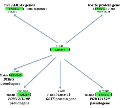
4. The FAM247 sequence is present in diverse genes
A significant property of the FAM247 sequence is that it forms part of diverse genes. Sequences
homologous to FAM247 form genes that include lincRNA genes, pseudogenes, and protein genes (Figure
2). These genes stem from phylogentically conserved nearest neighbor gene loci where the FAM247
sequence is linked to adjacent genes that form signatures containing gene families e.g.,
FAM230E-FAM247C-GGT3P present in segmental duplication LCR22A and FAM230B-FAM247A-GGT2 in LCR22D. Other than the FAM247 lincRNA family genes, which contain the entire 11,231 bp, only segments of
FAM247 are found to be part of other genes. The ends of these segments represent sequence breaks, i.e.
bp positions ~5958 and ~8000-8200 (Figure 2, see numbers above green highlighted FAM247). These are
regions that contain Alu sequences at their ends (Figure 2, caption), and they may provide sites for
Figure 2. Protein genes, lincRNA genes and pseudogenes that stem from FAM247 sequence and contain
different sections of the FAM247 sequence (shown in bp position numbers above FAM247 highlighted in green). Analysis of FAM247 by the RepeatMasker program:
RepeatMasker http://www.repeatmasker.org/cgi-bin/WEBRepeatMasker shows the presence of Alu
sequences in the FAM247 sequence at bp positions 6007-6285 and at 8063-8374, regions close to breaks.
4.1. USP18
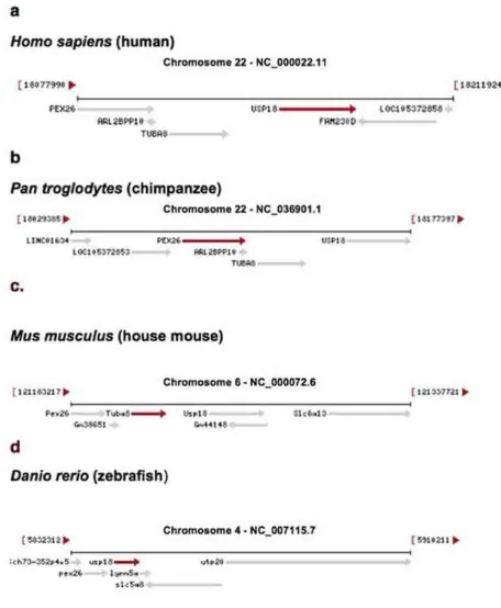
A comparison of USP18 chromosomal coordinates at loci in different species shows that the position is
evolutionarily conserved relative to adjacent genes (Figure 3). This provides nearest neighbor signatures where evolutionary history and origins of USP18 can be assessed. Two neighbor genes, PEX26
(peroxisomal biogenesis factor 26) and TUBA8 (tubulin alpha 8) are in homologous loci that show a
conserved orientation with respect to each other in the chromosomes of mice [Mus musculus (house mouse)] and primates (Figure 3a-c). In zebrafish (Danio rerio, a member the Cyprinidae family of
freshwaterfish), the tubulin gene (termed tuba8l4 tubulin, alpha 8 like 4) appears to have moved to a different chromosome and developed into two genes, TUBAa and TUBAb; this results in PEX28 and
Figure 3. Genes that are adjacent to USP18 are found in different species and are shown in the gene maps. Drawings of gene arrangement are taken directly from the NCBI website:
https://www.ncbi.nlm.nih.gov/gene [15}.
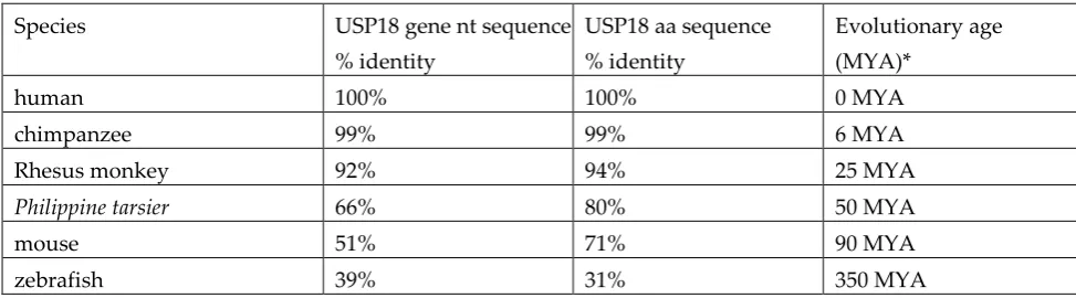
To analyze the phylogenetic relatedness of USP18 gene nt and aa sequences, sequences were aligned
from zebrafish, the house mouse, three primate species and humans. The resultant percent sequence
identities mimic evolutionary distances between species (Table 2) with a linear change in nt and aa
of gene nt and protein aa sequence change with evolutionary time and is consistent with a common
lineage of the USP18 gene that dates to an ancestor of zebafish, more than about 350 million years ago
(MYA). This parallels the nearest neighbor gene history of USP18.
Table 2. USP18 gene and protein sequence identities and evolutionary time between species
Species USP18 gene nt sequence
% identity
USP18 aa sequence % identity
Evolutionary age (MYA)*
human 100% 100% 0 MYA
chimpanzee 99% 99% 6 MYA
Rhesus monkey 92% 94% 25 MYA
Philippine tarsier 66% 80% 50 MYA
mouse 51% 71% 90 MYA
zebrafish 39% 31% 350 MYA
*approximate age in million years ago (MYA) [16]
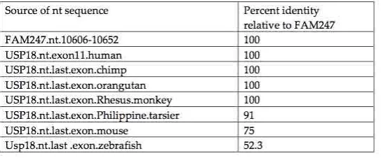
4.2. USP18 exon 11
Both human exon 11, which encodes the last 14 aa (the carboxy terminal end) of the USP18 peptidase, and the 3’ UTR of the USP18 mRNA sequence are provided by the FAM247 sequence [8]. The identity
between the FAM247 nt sequence and the human/primate exon11 nt sequences is 100%, with the exception of that of Philippine tarsier (Figure 4). The sequence of the carboxy terminal exon is more stable
than that of the sequence of entire gene (compare with Table 2). The identities of the USP18 3’UTR sequences from various species compared to FAM247 (Table 3) shows the 3’ UTR sequence is also conserved in primates, but to a lesser extent than that of that of exon 11 and is more similar to the USP18
gene.
Figure 4. Alignment of the USP18 terminal exon nt sequences from seven species compared with the
FAM247 sequence. Data obtained using the EBI Clustal Omega sequence alignment and phylogeny programs. The EMBL-EBI Clustal Omega Multiple Sequence Alignment program [17] at website:
http://www.ebi.ac.uk/Tools/msa/clustalo/ was used for nt sequence alignment. A. Phylogenetic tree of USP18 terminal exon sequences from seven species and the FAM247 sequence. B. The percent identities created using Clustal 2.1. C. Alignment of nt sequences.
The sequence similarity of 52% between FAM247 and zebrafish exon 11 (Figure 4B), the presence of a
number of invariant nt positions (Figure 4C) and the similarity with the 3’UTR sequence (53%) (Table 3) suggests that this part of the FAM247 sequence was present in the USP18 sequence of zebrafish. The
invariant nt residues of exon 11, e.g., positions nt 5-9 (Figure 4C) may relate to the functional importance of the USP18 carboxy terminal end in its role in the regulation of the immune system by USP18 [11, 12].
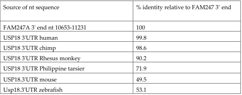
Table 3. Nucleotide sequence identities of 3’UTR of USP18 from different species
Source of nt sequence % identity relative to FAM247 3’ end
FAM247A 3' end nt 10653-11231 100
USP18 3'UTR human 99.8
USP18 3'UTR chimp 98.6
USP18 3'UTR Rhesus monkey 90.2
USP18 3'UTR Philippine tarsier 71.9
USP18.3'UTR mouse 49.5
Usp18.3'UTR zebrafish 53.1
Figure 5 shows USP18 aa sequence percent identity, sequence alignment and a phylogenetic tree
produced from an alignment of USP18 terminal exon aa sequences from different species with the
translated aa sequence of FAM247. Eight of the 14 amino acid residues that form the terminal exon are
totally conserved from primates to zebrafish, together with FAM247 translated aa sequence. (Figure 5c).
The USP18 carboxy terminal peptide sequence interacts with the INFAR2 interferon receptor and is an
important regulator of IFN signaling [11]; in addition, the carboxyl end sequence functions in
delSGlyation [18, 19, 12]. A mutation in L365 in the exon 11 sequence 359QETAYLL365VYMKMEC372
abolishes deISGylation and INAFR2 binding [19]; L365 is one of the evolutionary conserved amino acids of
exon 11 (Figure 5). The mutation may alter the protein conformation necessary for function. On the other
hand, the high number aa residues conserved relative to the FAM247 translated aa sequence adds to the
Figure 5. Alignment of the USP18 terminal exon amino acid sequences from seven species compared with the FAM247 translated amino acid sequence. Data obtained using the EBI Clustal Omega sequence
alignment and phylogeny programs. The EMBL-EBI Clustal Omega Multiple Sequence Alignment program [17] at website: http://www.ebi.ac.uk/Tools/msa/clustalo/ was used for aa sequence alignment.
Top. Phylogenetic tree of USP18 terminal exon aa sequences from seven species and the FAM247 aa sequence. Middle. The percent identities created using Clustal 2.1. Bottom. Alignment of aa sequences.
4.3. GGT5
The human GGT5 protein gene resides in chromosomal segmental duplication LCR22G and is linked to
pseudogene POM121L9P with a spacer sequence and the pseudogene GGTLC4P situated between GGT5
and POM121L9P (Figure 6) [8]. The GGT5 nearest gene/sequence arrangement is more complex than that
of the signatures shown in Figure 1b. GGT5 is an anomaly as its sequence does not stem from a GGT
locus, as other GGT family members do, but from the chromosomal site containing the FAM247 sequence
[8]. GGT5 carries a sequence homologous to the 5’ half of the FAM247A sequence, bp positions 1-5958
(Figures 2 and 6) and POM121L9P contains part of the 3’ half of FAM247 (bp 5949-8219). The GGTLC4P
Figure 6. The genes linked to GGT5 in LCR22G with nearest neighbor arrangements (top schematic) and
the source of sequences found in human linked genes GGT5-GGTLC4P-POM121L9P.
GGT5 and POM121L9P appear to have formed at very different evolutionary times. FAM247 is part of
GGT5 genes in non-human primates, including Philippine tarsier. In addition, FAM247 provides the sequence for exon 1 of GGT5. There is a significant similarity between the FAM247 nt sequence and that
of the mouse GGT5 exon 1 (Figure 7A). There is not enough evidence to suggest that the mouse GGT5
contains the entire 5’ half of the FAM247 sequence but alignment of the mouse exon 1 nt sequence with
FAM247 shows that a significant number of nucleotides are invariant (Figure 7B). Although there is
invariance in 50 out of 173 nt between the FAM247 sequence and zebrafish GGT5 exon 1, the zebrafish
exon sequence shows significant differences, which makes it difficult to further assess a sequence
similarity. The exon 1 data are consistent with the formation of the GGT5 gene with the FAM247 sequence
that occurred before the evolutionary appearance of primates and appearing in mice or an ancestor to
Figure 7. A. The percent identity of FAM247 sequence with that of GGT5 exon 1 sequences from four
species. Data were obtained using Clustal 2.1. B. Alignment of the GGT5 exon nucleotide sequences from
the four species, compared with the FAM247 sequence, positions 192-567. Data obtained using the EBI
Clustal Omega sequence alignment and phylogeny programs. The EMBL-EBI Clustal Omega Multiple
Sequence Alignment program [17] at website: http://www.ebi.ac.uk/Tools/msa/clustalo/
4.4. POM121L9P
POM121L9P has a very different sequence from the other POM1211LP family pseudogenes and it is a unique gene. A schematic of the compositional make-up of the POM121L9P gene shows that the pseudogene contains most of the sequence homologous to the putative parent gene, protein gene
POM121L1 (LOC101929738 putative POM121-like protein 1, 2379 bp) on its 5’ side, and the BCRP1 pseudogene sequence (that is homologous to the 3’ section of the BCR gene that includes BCR terminal exons 19-23) on its 3’ side (Figure 8). FAM247 may have formed a nucleation site for addition of these
motifs, which are copies of sequences from different regions of the genome. The sequence motifs are found attached onto 5’ and 3’ ends of FAM247 at bp positions where there are Alu sequences (FAM247 bp
position 5949 and 8219). Also, the complete POM121L-1 sequence has an Alu sequence at bp positions 2309 -2379 (the end of POM121L-1 is position 2304, the attachment site with FAM247). Alu sequences may
have facilitated the addition of POM121L-1 to FAM247. The BCR sequence addition to POM121L9P is more complex as there is an undefined sequence between the two (bp position 4479-5779 on
tissues [13, 14]. Its functions are not known but should be of interest in view of the strong RNA expression levels.
Figure 8. A schematic of the compositional make-up of the pseudogene POM121L9P. The numbers under
the motifs shown represent the bp positions on the POM121L9P sequence. The FAM247 sequence that forms part of POM121L9P consists of FAM247 positions 5949-8219, where there are Alu sequences at both
ends.
In an homologous nearest neighbor gene arrangement present in chimpanzee chr22 that are annotated as glutathione hydrolase light chain 2 gene (LOC749018) and putative POM121-like protein 1 gene,
LOC112206778; these are linked to GGT5 through the spacer sequence (Figure 9). Thus the human pseudogenes GGTLC4P and POM121L9P sequences are annotated as protein genes in the homologous
chromosomal loci of chimpanzee. This is another example of ncRNA genes in humans annotated as
protein genes in non-human primates and is of significance, but isolation of protein products from the
chimpanzee genes is essential to confirm this.
Of importance is that 69% of the POM121L9P sequence is present in the chimpanzee genome with a
98% identity, at the genomic region where the comparable chromosomal locus resides in chimpanzee.
There areno FAM247 or POM121L9P sequences that have been found linked to GGT5 in Rhesus. It
appears that the development of the POM121L9P sequence began in the chimpanzee, with but a partial sequence.
Figure 9. Nearest neighbor gene arrangements in human and chimpanzee chromosomal loci where the GGT5 gene resides.
Human BCRP3 and POM121L10P are linked to GGT1 in the gene/sequence arrangement, GGT1-spacer-BCRP3-POM121L10P, which is present in chr22 LCR22H. FAM247 forms part of the two pseudogenes: BCRP3, which has the FAM247 positions 33-5958 and POM121L10, positions 5957- 8219 (Figure 2). Thus parts of the 5’ and 3’ regions of FAM247 are found in genes found on these linked genes, which is similar
to the presence of FAM247 in genes GGT5 and POM121L9P.
BCRP3 is a member of the BCRP pseudogene family consisting of eight pseudogenes, all of which contain the homologous sequence of the 3’ end sequence of BCR protein gene. However, BCRP3
differs as it contains additional sequence motifs (Figure 10) and is the only BCRP family member that contains the FAM247 sequence. The BCRP3 gene appears to have a unique sequence. The compositional
make-up of BCRP3 shows that its 5’ side has the FAM247 sequence, which is followed by a 4255 bp segment of the immunoglobulin lambda locus (IGL) and the 3’ end of the BCR sequence (Figure 10). The
IGL sequence is homologous to the IGL locus V segments and three C segments, which are known to not encode immunoglobulin proteins. The IGL sequence has an Alu sequence at the junction with FAM247, which may relate to the process of attachment of IGL to FAM247. In terms of RNA expression, the
pseudogene shows broad expression of linear RNA in 27 normal somatic tissues and a broad expression of circular RNA in developing fetal tissues [13, 14].
Figure 10. Sequence motif of the BCRP3 gene. Positions 6226 -10480 of BCRP3 span the IGL insert. The
total length of BCRP3 is 20446 bp.
The POM121L10P sequence is linked to BCRP3 on chr 22. It also contains the FAM247 sequence (Figure
2). POM121L10P is compositionally made up of nearly the entire sequence of the related pseudogene POM121L1P, but has a 1062 bp sequence at its 3’ end that consists of a copy of the 3’ end of the BCR gene. POM121L10P also appears to be a unique gene construct. The POM121L10P linear RNA transcript is strongly expressed in testes; circular RNAs are broadly expressed fetal tissues. [13, 14]. Thus both this gene and BCRP3 show a robust RNA expression. It should be pointed out that there are additional
POM121LP pseudogene family members that carry the FAM247 sequence but are not addressed here.
In the rhesus genome, GGT1 is linked to the spacer sequence and followed by the FAM247 sequence,
which is similar to that of the human GGT1 gene/sequence arrangement (Figure 1B). Rhesus gene
LOC107000612, annotated as a "breakpoint cluster region protein-like protein” is situated close to GGT1. This is part of the homologous chromosomal region where the pseudogene BCRP3 resides in the human
genome; approximately 78% of the human BCRP3 sequence is present in the Rhesus genome at this locus.
The BCRP3 sequence has not been detected in the early primate Philippine tarsier. The earliest appearance
pseudogene in humans. There was a large chromosomal expansion of the rhesus monkey genome
between genes GGT1 and GGT5. The chromosomal length between genes GGT1 and GGT5 in Philippine
tarsier is 2872 bp; in the rhesus monkey it is 216,200 bp. Thus there is a 75-fold sequence expansion between GGT1 and GGT5 in rhesus. Segments of the BCRP3 gene may have formed with this
chromosomal expansion. This may account for the source of the BCRP3 sequence in Rhesus, but the
sequence is not found in Philippine tarsier.
5. Conclusions
Both the FAM247 lincRNA gene family and the various pseudogenes appear to have the repeat FAM247 sequence as a foundation for gene development, however, the mechanism of formation and the
compositional make-up between the lincRNA genes and pseudogenes greatly differs. The FAM247 family
(as well as the FAM230 lincRNA gene family) formed by gene duplication and family members display sequences that are “variations on a theme”. Although pseudogenes BCRP3, POM121L9P and POM121L10P
contain duplications of part of or entire portions of parent protein genes, they formed differently by a process of addition of large unrelated genomic sequences to the FAM247 sequence, with the resultant
formation of unique pseudogene sequences. Alu elements are present in FAM247 at sites of attachment; these may contribute to the process of sequence addition. As these pseudogenes are unique with large
sequences unrelated to the parent protein gene, the question is whether they should be called
pseudogenes. How USP18 and GGT5 protein genes developed is not know but a putative ancient FAM247
sequence was likely involved. A separate but important aspect of the FAM247 sequence in cellular and molecular functions is that it contributes the amino acid sequence for protein exons, the first exon of GGT5 and last exon of USP18. The functions of the carboxyl terminal aa sequence of USP18 are of major
significance because of the role in the regulation of the immune system.
Funding: This research received no external funding
Conflicts of Interest: The author declares no conflict of interest.
References
1. Ohno, S. Gene duplication and the uniqueness of vertebrate genomes circa 1970-1999. Semin Cell Dev
Biol. 1999, 10, 517-522.
2. Jacob, F. Evolution and tinkering. Science 1977, 196, 1161–1166
3. Wang, W.; Yu, H.; Long ,M. Duplication-degeneration as a mechanism of gene fission and the origin of
new genes in Drosophila species. Nat Genet. 2004, 36, 523–527.
4. Carvunis, A.R.; Rolland, T.; Wapinski, I.; Calderwood, M.A.; Yildirim, M.A.; Simonis, N.; Charloteaux,
B.; Hidalgo, C.A.; Barbette, J.; Santhanam, B.; et al. Proto-genes and de novo gene birth. Nature 2012, 487,
370-374.
5. McLysaght, A.; Guerzoni, D. New genes from non-coding sequence: the role of de novo protein-coding
genes in eukaryotic evolutionary innovation. Philos Trans R Soc Lond B Biol Sci. 2015, 370, 20140332.
6. Schlotterer, C. Genes from scratch—the evolutionary fate of de novo genes. Trends Genet. 2015, 31, :215–
7. Van Oss, S.B.; Carvunis, A.R.; De novo gene birth. PLoS Genet. 2019, 15(5):e1008160.
8. Delihas, N. Formation of human long intergenic non-coding RNA genes, pseudogenes, and protein
genes: Ancestral sequences are key players. PLoS One, 2020, 15(3):e0230236.
9. Nowell, P.; Hungerford, D.; A minute chromosome in human chronic granulocytic leukemia. Science,
1960, 132, 1497.
10. de Klein, A.; van Kessel, A.G.; Grosveld, G.; Bartram, C.R.; Hagemeijer. A.; Bootsma, D.; Spurr, N.K.;
Heisterkamp, N.; Groffen, J.; Stephenson, J.R. A cellular oncogene is translocated to the Philadelphia
chromosome in chronic myelocytic leukaemia. Nature, 1982, 300, 765–767.
11. Arimoto, KI.; Löchte, S.; Stoner ,S.A.; Burkart, C,.; Zhang, Y.; Miyauchi, S.; Wilmes, S.; Fan, J.B.;
Heinisch, J.J.; Li, Z. STAT2 is an essential adaptor in USP18-mediated suppression of type I interferon
signaling. Nat. Struct. Mol. Biol. 2017, 24, 279-289.
12. Honke, N.; Shaabani, N.; Zhang, D.E.; Hardt, C.; Lang, K.S. Multiple functions of USP18. Cell Death
Dis. 2016, 7(11):e2444.
13. Szabo, L.; Morey, R.; Palpant, N.J.; Wang, P.L.; Afari ,N.; Jiang, C.; Parast, M.M.; Murry, C.; Laurent ,
L.C.; Salzman, J. Statistically based splicing detection reveals neural enrichment and tissue-specific
induction of circular RNA during human fetal development. Genome Biol. 2015, 16(1):126.
14. Fagerberg, L.; Hallstro¨m, B.M.; Oksvold, P.; Kampf, C.; Djureinovic, D.; Odeberg, J.; Habuka, M.; Tahmasebpoor, S.; Danielsson, A,; Edlund, K.; et al. Analysis of the human tissue-specific expression by genome-wide integration of transcriptomics and antibody based proteomics. Mol. Cell Proteomics 2014, 13,
397–406.
15. O’Leary, N.A.; Wright, M.W.; Brister, J.R.; Ciufo, S.; Haddad, D.; McVeigh, R.; Rajput B,; Robbertse, B.;
Smith-White, B.; Ako-Adjei, D.; et al. Reference sequence (RefSeq) database at NCBI: current status,
taxonomic expansion, and functional annotation. Nucleic Acids Res. 2016, 44, D733–D745.
16. Siepel, A. Phylogenomics of primates and their ancestral populations. Genome Res. 2009, 19, 1929-1941.
17. Madeira, F.; Park, Y.M.; Lee, J.; Buso, N.; Gur, T.; Madhusoodanan, N.; Basutkar, P.; Tivey, ARN.;
Potter, S.C.; Finn, R.D. The EMBL-EBI Search and Sequence Analysis Tools APIs in 2019, Nucleic Acids
Res. 2019, 47(W1):W636- W641.
18. Malakhov, M.P.; Malakhova, O.A.; Kim, K.I.; Ritchie, KJ.; Zhang, D.E. Protein ISGylation Modulates
the JAK-STAT Signaling Pathway. J Biol Chem. 2002, 277, 9976-9981.
19. Dauphinee, S.M.; Richer, E.; Eva, M.M.; McIntosh, F.; Paquet ,M.; Dangoor, D.; Burkart, C.; Zhang,
D.E.; Gruenheid, S.; Gros, P. Contribution of increased ISG15, ISGylation and deregulated type I IFN
![Figure 1. Schematic representation of evolutionarily conserved linked sequences with different colors depicting different sequences, as described in [8]](https://thumb-us.123doks.com/thumbv2/123dok_us/1004245.1600268/3.595.78.355.71.298/schematic-representation-evolutionarily-conserved-sequences-different-depicting-different.webp)