RVC OPEN ACCESS REPOSITORY – COPYRIGHT NOTICE
This is an Accepted Manuscript of an article published by Taylor & Francis in Connective Tissue Research on 22 July 2016, available online:
http://dx.doi.org/10.1080/03008207.2016.1215442.
The full details of the published version of the article are as follows:
TITLE: Modulation of mesenchymal stem cell genotype and phenotype by extracellular matrix proteins
AUTHORS: Lucy E. Clements, Elaine R. Garvican, Jayesh Dudhia & Roger K. W. Smith JOURNAL TITLE: Connective Tissue Research
PUBLISHER: Taylor & Francis
Modulation of mesenchymal stem cell genotype and phenotype by extracellular matrix proteins
LE Clements, ER Garvican, J Dudhia*, RKW Smith
The Royal Veterinary College, Department Clinical Sciences and Services, Hawkshead Lane, North Mymms, Hatfield, Herts, AL9 7TA, UK.
Tel: +44 (0)1707 666333 Fax: +44 (0)1707 652090
*corresponding author: Jayesh Dudhia [email protected]
The Royal Veterinary College, Department Clinical Sciences and Services, Hawkshead Lane, North Mymms, Hatfield, Herts, AL9 7TA, UK.
Tel: +44 (0)1707 666333 Fax: +44 (0)1707 652090
Abstract
Aim: To investigate the effect of extracellular matrix proteins (ECM) on characteristics of mesenchymal stem cells (MSCs) and tendon-derived cells (TDCs).
Materials and Methods: MSCs and TDCs, cultured in monolayer (2D) or hydrogels (3D), with or without ECM protein supplementation, and on non-cellular native tendon matrix (NNTs) were assayed for adhesion, proliferation, gene expression and integrin expression.
Results: MSCs exhibited a fibroblastic, spindle-shaped morphology on 2D matrices except in the presence of fibronectin. In 3D matrices, MSCs displayed a rounded phenotype except when cultured on NNTs where cells aligned along the collagen fibrils but, unlike TDCs, did not form inter-cellular cytoplasmic processes. MSC proliferation was significantly (p<0.01) increased by collagen type I in 2D culture and fibronectin in 3D culture. TDC proliferation was unaffected by substrata. MSCs and TDCs differentially expressed α2 integrin. Adhesion to substrata was reduced by RGD-blocking peptide and β1 integrin antibody. The presence of collagen I or fibronectin upregulated MSC expression of collagen type I and collagen type III, COMP, decorin, osteopontin and fibronectin.
Introduction
Mesenchymal stem cells (MSCs) are now routinely used in clinics to treat overstrain injury of equine tendons, following establishment of a protocol (Smith 2003; Frisbie 2010) and
demonstration of treatment efficacy (Godwin 2012). To what extent their therapeutic effect is as a result of subsequent in situ differentiation and how much is due to paracrine activity is unclear. MSCs can adhere to equine tendon surfaces in vitro and to migrate into the tendon extracellular matrix (ECM) (Garvican 2014). Cell-ECM interactions have been shown to effect changes in cell behaviour including migration, proliferation, differentiation, survival and quiescence (Behonick 2003) but the impact of the tendon matrix on implanted MSCs is not known.
ECM proteins have been shown to regulate MSC behaviour by modulating both endogenous and exogenous growth factor stores (Bi 2005; Chen 2004). Cellular differentiation is highly dependent on external stimuli and the presence of extracellular matrix (ECM) proteins can influence the differentiation status, (Mauney 2004; Salasznyk 2004; Mauney 2005)
proliferative response (Kantlehner 2000; Hashimoto 2006) and morphology (Allen 2006) of cells cultured in vitro. In particular, the use of ECM proteins as a substrate on which to culture human MSCs has shown that specific lineage differentiation can be enhanced by the presence of certain proteins (Klees 2004).
While MSCs are cultured frequently in 2D culture, they are known to behave differently in 3D culture. Adoption of a 3D culture system, more analogous to that found in vivo, may enable maintenance or enhancement of desirable cell characteristics. Evidence suggests that although gross effects of ECM culture on cell characteristics are non-specific (Marinkovic 2015) the presence of components of the particular cellular “niche” may be critical for the initiation of more sophisticated cellular attributes.
Materials and Methods
Isolation of cells
The collection of equine tendons at post mortem or from a local abattoir was carried out under approval from the Ethics and Welfare Committee at the Royal Veterinary College (URN 2013 1230 R 2005). No horses were euthanised for the sole purpose of obtaining tissues for this study. Macroscopically normal superficial digital flexor tendons (SDFT) were aseptically harvested from skeletally mature horses (age 4 – 10 years) euthanised for reasons other than orthopaedic disease, finely diced and digested with pronase (1% w/v) for 1 h, then collagenase (0.5% w/v; Worthingtons, UK) for 18 h. Cells were recovered following
straining of the digest through a 70 µm filter and centrifugation, and seeded in culture flasks at 5,000 cells/cm2. Cells were expanded in culture in D10 medium (Dulbecco’s Modified Eagle Medium, supplemented with fetal bovine serum (FBS, 10% v/v), 100 U/mL penicillin, and 100 U/mL streptomycin (all from Invitrogen, Paisley, UK)). Cells between passage 0 and 2 (P0 to P2) (Goodman 2004) were re-suspended in cell freezing medium (90% FBS, 10% DMSO) and stored in liquid nitrogen until use. Cells of the same passage number were used in each paired 2D and 3D experiment. Equine bone marrow-derived mesenchymal stem cells (MSCs) used in this study were derived from surplus stocks used to treat clinical cases of overstrain SDFT injury examined at the Royal Veterinary College, for which MSC tri-lineage differentiation capacity was determined (Smith 2013). Cells chosen for use were obtained from horses aged 4 – 10 years of age and stored frozen, as before, until use.
2D culture systems
Glass coverslips (VWR International, Dorset, UK) were cleaned with 70% ethanol and sterilised by autoclaving prior to coating with matrix proteins or cells (Inoue 2005).
(collagen I, fibronectin (Roman 2004)) or poly-L-lysine) or left uncoated to act as controls. It should be noted that serum proteins within the cell culture medium (D10) will adsorb to glass and plastic surfaces, thus the uncoated coverslips cannot be considered totally devoid of protein (Brodkin 2004). Poly-L-lysine was selected over poly-D-lysine as the latter has been reported to cause a significant decrease in the proliferation rate of human MSCs when compared to poly-L-lysine and ECM proteins (Qian 2004). Cells (n=3 horses) were seeded onto coverslips at a concentration of 60,000 cells/cm2 and cultured for 24 or 72 h.
Photomicrographs were taken to evaluate differences in morphology (Olympus BX60F5 microscope and QICAM FAST 1394 digital camera, QImaging).
3D culture systems
Hydrogels: MSCs and TDCs (both n=3 horses) were suspended in 1 mL of 10% sucrose (in Tissue culture grade water) at a concentration of 1×10⁶ cells/mL. Aliquots of 20 µL cell suspension were then added to 40 µL of 1% Puramatrix (BD Biosciences, Bedford, UK) to give a final concentration of 2 x 10⁴ cells in each hydrogel (recommended by the
manufacturer). Hydrogels were prepared as detailed in Table 1; components were combined within a 1 mL syringe, left to set at room temperature for 1 h then transferred into 24-well plates. Collagen I (1% w/v, Sigma-Aldrich, Dorset, UK) was supplied dissolved in acetic acid which was brought to pH 7 by the addition of sodium hydroxide, then combined with 30% sucrose (in Tissue culture grade water) to give a final concentration of 0.1% collagen type I. Hydrogels were cultured in 0.5 mL of D10 for 24 or 72 h before mechanical disruption by aspiration with a pipette and re-suspension in 0.5 mL Trizol reagent (Sigma-Aldrich, Dorset, UK) for RNA extraction. Separate groups of hydrogels were prepared for each experiment.
(SDFT) that had undergone repeated freeze-thaw cycles and therefore contained no viable cells (Dudhia 2007). Longitudinal sections of 75 µm and 10 µm thickness were cut in a cryostat (Bright Instrument, Huntingdon, UK) and immediately placed into sterile PBS. These dimensions were previously determined to balance explant integrity and prevent tears during handling whilst also being thin enough to allow nutrient diffusion within the explant. Sections were then sterilised in 70% ethanol for 90 minutes before rehydrating for 2 h in PBS containing 1% Fungizone (Gibco, Paisley, UK) and 1% penicillin and streptomycin (PAA, Somerset, UK) and 10 – 12 NNTs placed in a 12-well suspension culture plate in 2.5 mL D10 containing 4 x 10⁶ MSCs or TDCs (per NNT). Cells were allowed to adhere to the tissue for 18 h (Garvican 2014). The D10 was then discarded and NNTs washed with D10 to remove loosely adherent cells, then placed in a new 12-well plate (1 explant per well with 2 mL D10 per well) and cultured for 7 or 14 days to allow migration of cells into the matrix (Garvican 2014). On conclusion of the culture period, explants were frozen at -80 ºC, homogenised with a Braun Mikrodismembranator (Sartorius, Epsom, UK) and stored in Trizol reagent (0.5 mL per NNT) at -80ºC.
Assessment of cell viability and proliferation
Hydrogels were incubated in PBS containing 5.6 mM glucose, 0.5 mM MgCl2, 0.9 mM CaCl, 4 µM ethidium homodimer and 2 µM Calcein AM (Molecular Probes, Oregon, USA) for 1 h protected from light. The hydrogels were then imaged using a confocal laser scanning microscope (Leica SP2 AOBS, Leica Microsystems, Milton Keynes) with dual-channel fluorescence (laser wavelengths 488nm and 543nm).
spectrophotometer (Spectramax 250, Molecular Devices Ltd, Berkshire, UK). Cell proliferation on NNTs has previously been reported (Garvican 2014).
Cell adhesion assay
Pre-coated coverslips were incubated with 0.5mL containing 120,000 cells (n=3 horses; density 60,000 cells/cm2) and incubated for 15, 30, 45 or 60 minutes before washing with D10 (two washes with 500 µL D10, flow rate 3 mL/minute). Resultant monolayers were fixed with 4% paraformaldehyde and stained with Haematoxylin for cell counting and the percentage of cells adhered was calculated using image analysis software (Image-Pro Plus 5, Media Cybernetics, Berkshire, UK.).
Gene expression
Total cellular RNA was isolated from hydrogel or NNT preparations (n=3 horses) using RNeasy minicolumns and reagents (Qiagen Ltd., Crawley, Surrey, UK). Residual DNA contamination was removed by performing an on-column DNAse digestion using an RNase-Free DNAse kit (Qiagen Ltd). The quality and quantity of RNA eluted from the column was assessed by spectrophotometry (260 nm). cDNA was synthesized using the RNA as template with the Superscript first-strand synthesis system for RT-PCR (Invitrogen, Paisley, UK). Aliquots of cDNA were amplified by polymerase chain reaction (Opticon II DNA engine thermocycler, MJ Research Inc, Massachusetts, USA), using gene specific primers (Table 2) in a 25 µL reaction volume with a SYBR® Green Core kit for detection (Eurogentec, Seraing, Belgium). Relative expression levels were normalized with GAPDH and calculated with the 2-ΔΔCT method (Livak 2001).
Immunocytochemistry
containing 0.1% Tween 20. Primary monoclonal antibody diluted in the blocking buffer (anti-vinculin (final dilution at 1:100; Sigma, Poole, UK) or anti-human integrin alpha 2 (final dilution 1:500; Chemicon International, CA, USA) or anti-human integrin alpha 5 ( final dilution 1:1000; Chemicon) was added and cells were incubated for 1 h at room temperature then washed three times for 5 minutes each. Secondary antibody (AlexaFluor 488 conjugated goat anti-mouse antibody, 15 µg/mL) was added and coverslips were incubated in the dark for 1 h, washed in PBS and counterstained with Hoechst (diluted 1:2000 in PBS) for 1 minute. Following a final wash, coverslips were mounted in Vectashield H-1000 mountant (Vector Laboratories, Peterborough, UK) and multiple images obtained using a fluorescent microscope (Leica SP2 AOBS, Leica Microsystems, Milton Keynes). Cells cultured on Thermanox plastic coverslips were not assessed using immunocytochemistry because these coverslips exhibit a background fluorescence that interferes with the cell analysis. Equine liver was used as a positive control for anti-vinculin antibody binding (Kawai 2003) and the secondary antibody alone used as a negative control (for non-specific binding).
Integrin-mediated cell binding assays
MSCs and TDCs (n=3 horses) were detached from the substrate using trypsin-EDTA and maintained in suspension at 2 ×10⁵ cells/mL of D10 at 37˚C (humidified 5% CO2 and air) for 2 h to allow for re-expression of integrins that may have been cleaved from the cell
adhesion was evaluated as above and total cell numbers assessed using methylene blue dye staining (Dent 1995).
Statistical analysis
Results
Morphology and viability of cells on different substrata
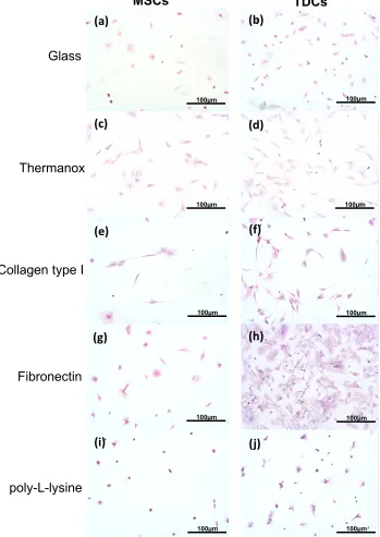
2D culture: Both MSCs and TDCs exhibited cell morphologies that were small and rounded or flattened on glass and on poly-L-lysine surfaces (Figure 1 a, b, i, j) compared to growth on Thermanox and collagen type I surfaces where the morphology was characteristically
elongated and spindle-shaped with long cell processes (Figure 1c, d, e, f). In contrast, while MSCs grown on fibronectin were small and rounded, TDCs were more elongated and formed denser cultures on this substrate (Figure 1 g, h).
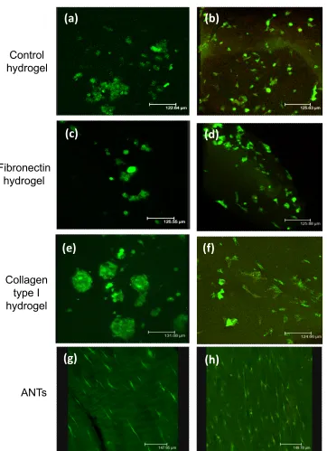
3D culture: MSCs retained a rounded morphology in all types of hydrogel (Figure 2a, c, e) and in collagen type I hydrogels cells were distributed in focal clusters. In contrast, TDCs displayed a spindle-shaped morphology with long cell processes in collagen type I hydrogels only (Figure 2f). When cultured on ANTs, MSCs also adopted a spindle-shaped morphology and aligned with resident collagen fibres (Figure 2g). TDCs also demonstrated alignment to the collagen fibres but in addition formed networks of elongated processes that were not observed in MSCs (Figure 2h). Viable cells were visible throughout the hydrogels. Cell adhesion
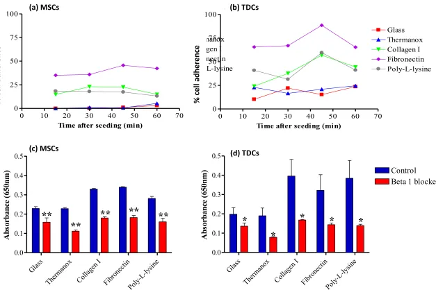
2D culture: Significant preferential adhesion was demonstrated on fibronectin over all other substrates for both cell types (p < 0.05; Figure 3a, b). In addition, MSCs demonstrated significantly greater adhesion to collagen I and poly-L-lysine for all time points compared to glass and Thermanox controls (p < 0.05). RGD peptide inhibited binding in MSCs but not in TDCs (data not shown). On all substrata, a significant reduction in cell adherence occurred after blocking with β1 integrin antibody for both MSCs (p < 0.001) and TDCs (p < 0.01) (Figure 3c, d).
Cell proliferation
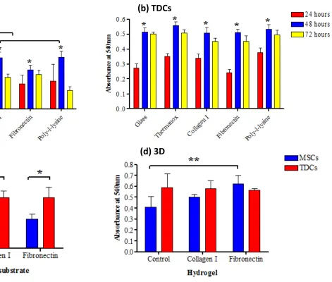
peaking by 48 h (Figure 4a,b). Reduced rates at 72 h may have been due to cell confluency resulting in contact inhibition of further proliferation. MSCs cultured on collagen type I and poly-L-lysine exhibited a significantly higher proliferation rate (p ≤ 0.01) than those on untreated coverslips after 48 h (Figure 4c). TDCs exhibited a significantly greater
proliferation rate compared to MSCs for all time points (p < 0.001), consistent with the faster growth rates observed on plastic surfaces, but substrata did not significantly affect TDC proliferation rate (Figure 4b).
3D culture: MSCs proliferated at a significantly greater rate in fibronectin hydrogels compared to control hydrogels (p < 0.02, Figure 4d) and at a significantly greater rate in hydrogels compared to 2D cultures (p < 0.05, data not shown). There were no significant differences in proliferation between TDCs in 2D and 3D culture (data not shown). Gene Expression
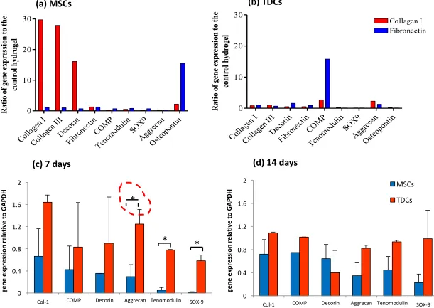
2D culture: MSCs cultured on all substrata expressed an up-regulation of Col I and III, COMP, decorin and fibronectin mRNA at 24 h. In contrast, only Col I expression was up-regulated in TDCs. After 72 h, expression of Col I by MSCs cultured on collagen type I increased 2-fold with no other notable changes in gene expression. Fibronectin expression by TDCs cultured for 72 h on collagen type I substrate increased 7-fold, while expression in those cultured on poly-L-lysine increased 2-fold. Culture of TDCs for 72 h on fibronectin substrate resulted in a 2-fold decrease of Col I mRNA expression compared to that expressed at 24 h (data not shown).
ECM-supplemented hydrogels and the control. COMP mRNA expression by TDCs was 16-fold greater in fibronectin hydrogels (Fig 5b).
3D culture:ANTs: After 7 days of culture on ANTs, there were significant differences between MSCs and TDCs in the expression levels of aggrecan (p<0.05), tenomodulin (p<0.001) and Sox-9 (p < 0.001, Figure 5c), all of which were greater in MSCs that TDCs, but by 14 days of culture, no significant differences remained (Figure 5d).
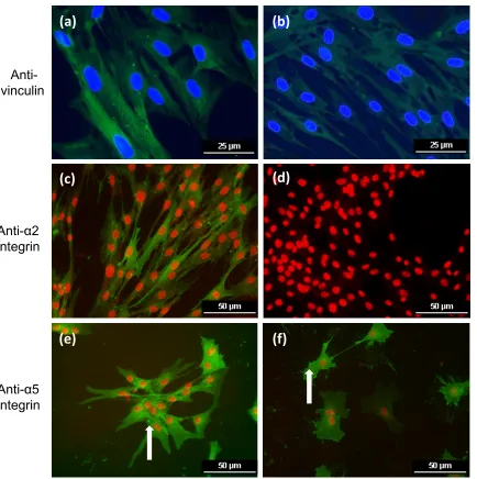
Immunocytochemistry (2D)
MSCs and TDCs cultured on all substrates stained positive for vinculin which was
concentrated in focal areas in the cytoplasm, although TDCs displayed only weak staining for vinculin on collagen type I (Figure 6a, b). Positive staining for the α2 integrin sub-unit was only present in MSCs cultured on collagen type I whereas TDCs were negative on all
substrata (Figure 6c, d). All cells, cultured on all substrata, stained positive for the α5 integrin subunit. Particularly intense staining (punctate signal) around the nuclei and the membrane of the cell was observed in both MSCs and TDCs cultured on the poly-L-lysine substrate
Discussion
This study demonstrated the interaction of both progenitor and differentiated cells with the extracellular matrix influences their phenotype in different ways. This may not only impact homeostatic mechanisms of resident cells but may also be of direct relevance for implantation of MSCs in cell therapy.
3D culture environments are believed to represent an environment of greater relevance to the in vivo situation. In this study, 3D environments resulted in a more rounded morphology while 2D environments resulted in the more spindle-shaped cell, characteristic of fibroblasts and MSCs. The rounded morphology of both cell types in poly-L-lysine may have arisen through decreased adherence or promotion of a chondrogenic phenotype (Malda 2003). Decreased adhesion is unlikely as greater number of cells were seen on this surface. In support of the latter explanation, a poly-L-lysine scaffold has previously demonstrated potential for supporting the initiation of chondrogenic differentiation of MSCs (Jung 2014).
by the presence of fibronectin in culture media serum. The α2β1 integrin is involved in cell adhesion to fibrillar collagens and its expression in MSCs cultured on collagen type I substrate is in agreement with previously published studies of human MSCs (Heckmann 2006; Popov 2011). The presence of these integrins in MSCs may be important for transplanted cells in binding to the tendon matrix after implantation. Given the loss of cells after implantation in vivo (Becerra 2013) and the loss of integrins from the cell surface after release of the cells from tissue culture passage, retention of implanted cells may be improved by allowing re-expression of these integrins on the cell surface.
The constitutive expression profile of the undifferentiated, unstimulated MSC in vitro has been shown to include osteogenic and chondrogenic genes (Tremain 2001; Liu 2014) which is confirmed here. Cartilage oligomeric matrix protein (COMP) is highly abundant in tendon and cartilage and appears to be important for the development of optimal tendon tissue with optimal mechanical properties (Smith 2002; Smith 2002). It is most abundantly expressed in growing collagen-rich matrices under load and hence may be a useful biomarker for differentiation towards these tissues (Barry 2001). However, MSCs cultured in monolayer expressed COMP in the absence of cytokine or growth factor stimulation. Culture in fibronectin greatly enhanced COMP expression in TDCs but this effect was not seen in MSCs suggesting fibronectin induced this effect only after differentiation has occurred.
(Kasashima Y, unpublished data). Thus, it is possible that the tendon matrix inhibits MSC proliferation.
MSC differentiation initiated by 3D culture systems has a well-established precedence in chondrogenesis (Bosnakovski 2006; Mathieu 2014) and MSC-seeded collagen scaffolds have shown encouraging initial results for future clinical use (Awad 2000; Kovacevic 2008). Although elevation of expression in some genes associated with a tenogenic phenotype occurred in MSCs cultured in collagen type I hydrogels, cell morphology remained rounded rather than fibroblastic, thus the presence of individual cell matrix interactions is insufficient to induce specific differentiation to a tenogenic phenotype and suggesting the need for the presence of multiple matrix component simultaneously. It is interesting that OPN expression increased in MSCs cultured in hydrogels containing collagen or fibronectin. Assessment of a more extensive gene expression profile, including more in-depth osteogenic gene expression, would be interesting but was outwith the scope of this study.
Conclusions
List of Abbreviations
MSC: Mesenchymal stem cell
ECM: Extracellular matrix
TDC: Tendon-derived cell
NNT: Non-cellular Native Tendon matrix
2D/ 3D: Two/three dimensional
Acknowledgments
LR was in receipt of an institutional stipend (RVC). This work was funded in part by the BBSRC (48-ERA16303) and the Technology Strategy Board (BY120B). Neither funding body had any involvement in study design; collection, analysis and interpretation of data; writing of this manuscript or in the decision to submit for publication.
Declaration of interest
Figure Legends
Figure 1: Cell morphology in 2D culture. Mesenchymal stem cells (MSCs) and tendon derived cells (TDCs) cultured in 2D (monolayer) on a range of substrata for 24 h. Cells were stained with haematoxylin and eosin.
Figure 2: Cell morphology in 3D culture. Mesenchymal stem cells (MSCs) and tendon derived cells (TDCs) cultured in a range of 3D systems for 24 h. Cells were stained with Calcein AM for fluorescent imaging.
Figure 3: Integrin-mediated cell adhesion. Effect of culture substrates on adhesion of a) MSC (n = 6) and b) TDC (n = 6) populations in short-term culture. Effect of β1 integrin subunit antibody blocking on adhesion to culture substrates of c) MSCs and d) TDCs (** denotes p<0.001; * denotes p = 0.01). Error bars represent standard error of the mean.
Figure 4: Cell proliferation in 2- and 3D culture. Proliferation rates of a) MSCs (n = 6) and b) TDCs (n = 6) cultured on various substrata, at 24, 48 and 72 h post-seeding; c) MSCs and TDCs (both n = 3) cultured in 2D and d) 3D with fibronectin or collagen type I
supplementation (** denotes p < 0.02; * denotes p < 0.05). Error bars represent standard error of the mean.
Figure 5: Effect of culture substrata on gene expression. mRNA expression was normalised to GAPDH and values shown are in comparison to control hydrogel cultures. a) MSCs (n = 3) and b) TDCs (n = 3) after 72 h cultured in hydrogels supplemented with collagen type I or fibronectin. c) MSCs (n = 3) and d) TDCs (n = 3) cultured for 7 and 14 days respectively, on non-cellular native tendon matrices (* denotes p<0.05). Error bars represent standard error of the mean.
References
Allen LT, Tosetto M, Miller IS, O'Connor DP, Penney SC, Lynch I, Keenan AK, Pennington SR, Dawson KA, Gallagher WM (2006) Surface-induced changes in protein adsorption and implications for cellular phenotypic responses to surface interaction. Biomaterials 27: 3096-3108.
Awad HA, Butler DL, Harris MT, Ibrahim RE, Wu Y, Young RG, Kadiyala S, Boivin GP (2000) In vitro characterization of mesenchymal stem cell-seeded collagen scaffolds for tendon repair: effects of initial seeding density on contraction kinetics. J Biomed Mater Res 51(2): 233-40.
Baker TK, Carfaqna MA, Gao H, Dow ER, Li Q, Searfoss GH, Ryan TP (2001) Temporal gene expression analysis of monolayer cultured rat hepatocytes. Chem Res Toxicol 14: 1218-1231.
Barry FP, Boynton RE, Liu B, Murphy JM (2001) Chondrogenic differentiation of
mesenchymal stem cells from bone marrow: differentiation dependant gene expression of matrix components. Exp Cell Res 268: 8-15.
Becerra P, Valdés MA, Fiske-Jackson AR, Dudhia J, Neves F, Hartman NG, Smith RKW (2013) The distribution of injected technetium99m-labelled mesenchymal stem cells in horses with naturally-occurring tendinopathy. J Orthop Res 31(7): 1096-1102.
Behonick DJ, Werb Z (2003) A bit of give and take: the relationship between the extracellular matrix and the developing chondrocyte. Mech. Dev. 120: 1327–1336.
Beitzel K, McCarthy MB, Cote MP, Russell RP, Apostolakos J, Ramos DM, Kumbar SG, Imhoff AB, Arciero RA, Mazzocca AD (2014) Properties of Biologic Scaffolds and Their Response to Mesenchymal Stem Cells. Arthroscopy 30(3): 289-298.
Bi Y, Stuelten C, Kilts T, Wadhwa S, Iozzo R (2005) Extracellular matrix proteoglycans control the fate of bone marrow stromal cells. J. Biol. Chem. 280: 30481–30489.
Brodkin KR, Garcia AJ, Levenston ME (2004) Chondrocyte phenotypes on different extracellular matrix monolayers. Biomaterials 25: 5929-5938.
Cao Y, Liu Y, Liu W, Shan Q, Buonocore SD, Cui L (2002) Bridging tendon defects using autologous tenocyte engineered tendon in a hen model. Plast Reconstr Surg 110: 1280-1289.
Chen XD, Fisher LW, Robey PG, Young MF (2004) The small leucine-rich proteoglycan biglycan modulates BMP-4-induced osteoblast differentiation. FASEB J. 18: 948–958.
Cool SM, Nurcombe V (2005) Substrate induction of osteogenesis from marrow-derived mesenchymal precursors. Stem Cells Dev 14: 632-642.
Dent MF, Hubbold L, Radford H, Wilson AP (1995) The methylene blue colorimetric assay microassay for determining cell line response to growth factors. Cytotechnology 17: 27-33.
Dominici M, Le Blanc K, Mueller I, Slaper-Cortenbach I, Marini FC, Krause DS, Deans RJ, Keating A, Prockop DJ, Horwitz EM (2006) Minimal criteria for defining multipotent mesenchymal stromal cells. The International Society for Cellular Therapy position statement. Cytotherapy 8: 315-7.
Dowthwaite GP, Bishop JC, Redman SN, Khan IM, Rooney P, Evans DJR, Haughton L, Bayram Z, Boyer S, Thomson B, Wolfe MS, Archer CW (2004) The surface of articular cartilage contains a progenitor cell population. J Cell Sci. 117: 889-897.
Dudhia J, Scott CM, Draper ER, Heinegard D, Pitsillides AA, Smith RK (2007) Aging enhances a mechanically-induced reduction in tendon strength by an active process involving matrix
metalloproteinase activity. Aging Cell 6(4): 547-56.
Frisbie DD, Smith RK (2010) Clinical update on the use of mesenchymal stem cells in equine orthopaedics. Equine Vet J. 42(1):86-89.
Garcia AJ (2005) Get a grip: integrins in cell-biomaterial interactions. Biomaterials 26: 7525-7529.
Godwin EE, Young NJ, Dudhia J, Beamish IC, Smith RK (2012) Implantation of bone marrow-derived mesenchymal stem cells demonstrates improved outcome in horses with overstrain injury of the superficial digital flexor tendon. Equine Vet J 44(1): 25-32.
Goodman SA, May SA, Heinegård D, Smith RK (2004) Tenocyte response to cyclical strain and transforming growth factor beta is dependent upon age and site of origin. Biorheology 41 (5):613-628.
Harwood F L, Monosov AZ, Goomer RS, Gelberman RH, Winters SC, Silva MJ, Amiel D (1998) Integrin expression is upregulated during early healing in a canine intrasynovial flexor tendon repair and controlled passive motion model. Connect Tissue Res. 39: 309-316.
Hashimoto J, Kariya Y, Miyazaki K. (2006) Regulation of Proliferation and Chondrogenic Differentiation of Human Mesenchymal Stem Cells by Laminin-5 (Laminin-332). Stem Cells 24: 2346-2354.
Heckmann L, Fiedler J, Mattes T, Brenner RE (2006) Mesenchymal progenitor cells communicate via alpha and beta integrins with a three-dimensional collagen type I matrix. Cells Tissues Organs 182(3-4): 143-54.
Hidalgo-Bastida LA, Cartmell SH (2010) Mesenchymal stem cells, osteoblasts and extracellular matrix proteins: Enhancing cell adhesion and differentiation for bone tissue engineering. Tissue Eng Part B Rev 16: 405-412.
Inoue S, Hori Y, Hirano Y, Inamoto T, Tabata Y (2005) Effect of culture substrate and fibroblast growth factor addition on the proliferation and differentiation of human adipo-stromal cells. J Biomater Sci Polym Ed 16(1): 57-77.
Jung O, Hanken H, Smeets R, Hartjen P, Friedrich RE, Schwab B, Gröbe A, Heiland M, Al-Dam A, Eichhorn W, Sehner S, Kolk A, Wöltje M, Stein JM (2014) Osteogenic differentiation of
Kantlehner M, Schaffner P, Finsinger F, Meyer J, Jonczyk A, Diefenbach B, Nies N, Hölzemann G, Goodman SL, Kessler H (2000) Surface Coating with Cyclic RGD Peptides Stimulates Osteoblast Adhesion and Proliferation as well as Bone Formation. ChemBioChem 1: 107-114.
Kawai S, Enzan H, Hayashi Y, Jin YL, Guo LM, Miyazaki E, Toi M, Kuroda N, Hiroi M, Saibara T, Nakayama H (2003) Vinculin: a novel marker for quiescent and activated hepatic stellate cells in human and rat livers. Virchows Arch 443(1): 78-86.
Khademhosseini A, Langer R, Borenstein J, Vacanti JP (2006) Microscale technologies for tissue engineering and biology. Proc Natl Acad Sci U S A. 103(8): 2480-2487.
Klees RF, Salasznyk RM, Vandenberg S, Bennett K, Plopper GE (2004) Laminin-5 activates extracellular matrix production and osteogenic gene focusing in human mesenchymal stem cells. Matrix Biol. 26: 106-114.
Koblinski JE, Wu M, Demeler B, Jacob K, Kleinman HK (2005) Matrix cell adhesion activation by non-adhesion proteins. J Cell Sci. 118: 2965-2974.
Kovacevic D, Rodeo SA (2008) Biological augmentation of rotator cuff tendon repair. Clin Orthop Relat Res 466: 622-633.
Linsley C, Wu B, Tawil B (2013) The effect of fibrinogen, collagen type I, and fibronectin on mesenchymal stem cell growth and differentiation into osteoblasts. Tissue Eng Part A 19(11-12): 1416-23.
Liu D, Wang Y, Ye Y, Yin G, Chen L (2014) Distinct molecular basis for endothelial
differentiation: gene expression profiles of human mesenchymal stem cells versus umbilical vein endothelial cells. Cell Immunol 289(1-2): 7-14.
Livak KJ, Schmittgen TD (2001) Analysis of Relative Gene Expression Data Using Real-Time Quantitative PCR and the 2-[Delta][Delta]CT Method. Methods 25(4): 402-8.
Mao Y, Schwarzbauer JE (2005) Stimulatory effects of a three-dimensional microenvironment on cell-mediated fibronectin fibrillogenesis. J Cell Sci. 118: 4427-4436.
Marinkovic M, Block TJ, Rakian R, Li Q, Wang E, Reilly MA, Dean DD, Chen XD (2015) One size does not fit all: developing a cell-specific niche for in vitro study of cell behavior. Matrix Biol.
http://dx.doi.org/10.1016/j.matbio.2016.01.004
Mathieu M, Vigier S, Labour MN, Jorgensen C, Belamie E, Noël D (2014) Induction of Mesenchymal stem cell differentiation and cartilage formation by cross-linker-free collagen microspheres. Euro Cells and Mats 28: 82-97.
Mauney JR, Kaplan DL, Volloch V (2004) Matrix-mediated retention of osteogenic differentiation potential by human adult bone marrow stromal cells during ex vivo expansion. Biomaterials 25: 3233-3243.
Mauney JR, Volloch V, Kaplan DL (2005) Matrix-mediated retention of adipogenic
differentiation potential by human adult bone marrow-derived mesenchymal stem cells during ex vivo expansion. Biomaterials 26: 6167-6175.
Morimichi M, Ryuichi F, Yoshinori K (2000) Type I collagen-induced osteoblastic differentiation of bone-marrow cells mediated by collagen-alpha2 beta1 integrin interaction. Journal of Cellular Physiology 184: 207-213.
Muir D, Varon S, Manthorpe M (1990) An enzyme-linked immunosorbent assay for bromodeoxyuridine incorporation using fixed microcultures. Anal Biochem 185: 377-382.
Nho RS, Xia H, Kahm J, Kleidon J, Diebold D, Henke CA (2005) Role of integrin-linked kinase in regulating phosphorylation of Akt and fibroblast survival in type I collagen matrices through a beta1 integrin viability signaling pathway. J Biol Chem 280: 26630-26639.
Popov C, Radic T, Haasters F, Prall WC, Aszodi A, Gullberg D, Schieker M, Docheva D (2011) Integrins a2b1 and a11b1 regulate the survival of mesenchymal stem cells on collagen I. Cell Death Disease(2): e186.
Qian L, Saltzman WM (2004) Improving the expansion and neuronal differentiation of mesenchymal stem cells through culture surface modification. Biomaterials 25(7-8): 1131-7.
Qiu Y, Lei J, Koob TJ, Temenoff JS (2014) Cyclic tension promotes fibroblastic differentiation of human MSCs cultured on collagen-fibre scaffolds. J Tissue Eng Regen Med Epub ahead of print.
Roman M, Williams WA, Boskey A, Bartosky A, Plopper GE (2004) Adhesion to vitronectin and collagen I promotes osteogenic differentiation of human mesenchymal stem cells. J Biomed Biotech 1: 24-34.
Salasznyk RM, Klees RF, Williams WA, Boskey A, Plopper GE (2007) Focal adhesion kinase signaling pathways regulate the osteogenic differentiation of human mesenchymal stem cells. Exp Cell Res 313: 22-27.
Salasznyk RM, Williams WA, Boskey A, Batorsky A, Plopper GE (2004) Adhesion to
Vitronectin and Collagen I Promotes Osteogenic Differentiation of Human Mesenchymal Stem Cells. J Biomed Biotechnol 1: 24-34.
Schwartz MA, DeSimone DW (2008) Cell adhesion receptors in mechanotransduction. Curr Opin Cell Biol 20: 551-556.
Smith RKW, Gerard M, Dowling B, Dart AJ, Birch HL, Goodship AE (2002) Correlation of cartilage oligomeric matrix protein (COMP) levels in equine tendon with mechanical properties: a proposed role for COMP in determining function-specific mechanical characteristics of locomotor tendons. Equine Vet J Suppl 34: 241-244.
Smith RK, Korda M, Blunn GW, Goodship AE (2003) Isolation and implantation of autologous equine mesenchymal stem cells from bone marrow into the superficial digital flexor tendon as a potential novel treatment. Equine Vet J. 35(1): 99-102.
Smith RKW, Werling NJ, Dakin SG, Alam R, Goodship AE, Dudhia J (2013) Beneficial effects of autologous bone marrow-derived mesenchymal stem cells in naturally occurring tendinopathy. PLoS One 8(9): e75697.
Tremain N, Korkko J, Ibberson D, Kopen GC, DiGirolamo C, Phinney DG (2001) MicroSAGE analysis of 2,353 expressed genes in a single cell-derived colony of undifferentiated human mesenchymal stem cells reveals mRNAs of multiple cell lineages. Stem Cells 19(5): 408-18.
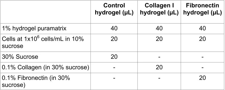
Table 1: Components of the hydrogel culture systems
Control hydrogel (µL)
Collagen I hydrogel (µL)
Fibronectin hydrogel (µL)
1% hydrogel puramatrix 40 40 40
Cells at 1x10⁶ cells/mL in 10% sucrose
20 20 20
30% Sucrose 20 - -
0.1% Collagen (in 30% sucrose) - 20 -
0.1% Fibronectin (in 30%
Figure 1
MSCs TDCsGlass
Thermanox
Collagen type I
Fibronectin
poly-L-lysine
(b)
(d) (c)
(e)
(g)
(f)
(h)
(i) (j)
(a)
100µm 100µm
100µm 100µm
100µm 100µm
100µm 100µm
(a)
(b)
(c)
(d)
(e)
(f)
(g)
(h)
MSCs TDCs
Fibronectin hydrogel
Control hydrogel
Collagen type I hydrogel
ANTs
0 10 20 30 40 50 60 70 0 25 50 75 100 Glass Thermanox Fibronectin Poly-L-lysine Collagen I (a)
Time after seeding (min)
No . Ce lls a dh er ed to s ub st ra te
0 10 20 30 40 50 60 70
0 25 50 75 100 Glass Thermanox Fibronectin Poly-L-lysine Collagen I (b)
Time after seeding (min)
No . Ce lls a dh er ed to s ub st ra te
Figure 3
(a) MSCs
(b)
0 10 20 30 40 50 60 70
0 25 50 75 100 Glass Thermanox Fibronectin Poly-L-lysine Collagen I (a)
Time after seeding (min)
No . Ce lls a dh er ed to s ub st ra te
0 10 20 30 40 50 60 70
0 25 50 75 100 Glass Thermanox Fibronectin Poly-L-lysine Collagen I (b)
Time after seeding (min)
No . Ce lls a dh er ed to s ub st ra te Glass
Thermanox Coll agen I Fibr onec tin Poly-L-lysi ne 0.0 0.1 0.2 0.3 0.4 0.5
Beta 1 blocked Control
**
(a)
**
**
**
**
Substrate Ab so rb an ce ( 65 0n m ) GlassThermanox Coll agen I Fibr onec tin Poly-L-lysi ne 0.0 0.1 0.2 0.3 0.4 0.5
Beta 1 blocked Control
(b)
*
*
*
*
*
Substrate Ab so rb an ce ( 65 0n m ) GlassThermanox Coll agen I Fibr onec tin Poly-L-lysi ne 0.0 0.1 0.2 0.3 0.4 0.5
Beta 1 blocked Control
**
(a)
**
**
**
**
Substrate Ab so rb an ce ( 65 0n m ) GlassThermanox Coll agen I Fibr onec tin Poly-L-lysi ne 0.0 0.1 0.2 0.3 0.4 0.5
Beta 1 blocked Control
(b)
*
*
*
*
*
Substrate Ab so rb an ce ( 65 0n m )(c) MSCs (d) TDCs
Figure 4
(c) 2D
(d) 3D
Figure 5
0 0.4 0.8 1.2 1.6 2Col-1 COMP Decorin Aggrecan Tenomodulin SOX-9
ge ne e xp re ssi on re la tive to GAPD H
*
*
*
0 0.4 0.8 1.2 1.6 2Col-1 COMP Decorin Aggrecan Tenomodulin SOX-9
ge ne e xp re ssi on re la tive to GAPD H MSCs TDCs Collag en I
Collag en III
Decorin Fibron
ectinCOMP
Tenom
odulinSOX9 Aggreca n Osteo pontin 0 10 20 30 Collagen I Fibronectin (a) Genes Ra ti o of g en e ex pr es si on to th e co nt ro l h yd ro gel Collag en I
Collag en III
Decor in
Fibro
nectinCOMP
Teno modu lin SOX9 Aggr ecan Osteopon tin 0 10 20 30 Collagen I Fibronectin (b) Genes Ra ti o of g en e ex pr es si on to th e co nt ro l h yd ro gel Collag en I
Collag en III
Decorin Fibron
ectinCOMP
Tenom
odulinSOX9 Aggreca n Osteo pontin 0 10 20 30 Collagen I Fibronectin (a) Genes Ra ti o of g en e ex pr es si on to th e co nt ro l h yd ro gel Collag en I
Collag en III
Decor in
Fibro
nectinCOMP
Teno modu lin SOX9 Aggr ecan Osteopon tin 0 10 20 30 Collagen I Fibronectin (b) Genes Ra ti o of g en e ex pr es si on to th e co nt ro l h yd ro gel
(a) MSCs
(b) TDCs
Figure 6
(a)
(b)
(e)
(f)
(g)
(h)
MSCs TDCs
(c)
(d)
Anti-α5 integrin Anti-α2 integrin
Anti-vinculin