28077 1 2 2 3 8
INTERSTITIAL LASER PHOTOCOAGULATION
AS A TREATMENT FOR BREAST CANCER
Thesis submitted for the degree of
Master of Surgery
in the
University of London
by
Simon Anthony Harries
M .B. B.S., F .R .C .S.(E ng.)
%YAL
HAW^SrEAD.
The National Medical Laser Centre,
Department of Surgery, University College London Medical School,
London.
ProQuest Number: U075783
All rights reserved
INFORMATION TO ALL USERS
The quality of this reproduction is dependent upon the quality of the copy submitted.
In the unlikely event that the author did not send a complete manuscript and there are missing pages, these will be noted. Also, if material had to be removed,
a note will indicate the deletion.
uest.
ProQuest U075783
Published by ProQuest LLC(2016). Copyright of the Dissertation is held by the Author.
All rights reserved.
This work is protected against unauthorized copying under Title 17, United States Code. Microform Edition © ProQuest LLC.
ProQuest LLC
789 East Eisenhower Parkway P.O. Box 1346
A bstract
Conservative surgery is a safe alternative to mastectomy for some patients with breast
cancer. A survey of surgeons in this thesis has shown that more surgeons would now
undertake conservative surgery than they have done in the past. Recently a new
technique, interstitial laser photocoagulation(ILP) has been described which is capable
of in situ tissue necrosis with safe healing. The idea of ILP takes the concept of conservative surgery for breast cancer a step further. The main purpose of this thesis
was to investigate the potential value of ILP as a future method of destroying breast
cancers in situ leaving the area to heal via resorption and fibrosis.
The aims of this thesis were to study the biology of laser interactions with breast
cancers scheduled for surgery(and not to completely destroy the tumour), to optimise
the laser parameters of power and exposure for a particular tumour and to find an
imaging technique which will accurately predict the extent of laser damage. Forty five
patients were treated with ILP prior to surgery(median 7 days). Tumour necrosis varied
from 2-25mm. No laser damage was noted in 4 patients. Two patients developed minor
complications and treatment was abandoned early due to pain in a further 4 patients.
The presence of charring within the tumour was associated with larger diameters of
necrosis than when charring was absent(median 13 vs 6 mm, p=0.002) and use of a pre
charred fibre produced similar lesions(median 14mm) which were more predictable.The
histological features in the tumour following ILP were of coagulative necrosis which
appeared to heal by the form ation of fibrous tissue. An area of heat fixed,
morphologically preserved tissue was noted within the zone of coagulative necrosis
which was thought to be non-viable.
U ltrasonography, C om puterised Tom ography(CT) and M agnetic Resonace
Imaging(MRI) were all used to monitor necrosis. Ultrasound was unable to predict the
extent of necrosis as measured in the resected specimen(r=0.3, p=N.S.) but was
reasonable at predicting tumour size(r=0.6, p=0.001). CT and MRI show some promise
This study has shown that ILP is simple and safe and when using a pre-charred fibre,
predictable. If the initial results of imaging using CT and MRI are confirmed in larger
Contents
Page
Abstract
2-3
Contents
4-10
List of tables and illustrations
11-15
Acknowledgements
16-17
Dedication
18
Chapter 1
Breast Cancer: Methods used to obtain the Diagnosis
19-29
1.1
Tissue diagnosis
1.1.1 Introduction
1.1.2 Fine needle aspiration cytology(FNAC)
1.1.3 Core biopsy
1.1.4 Excision biopsy
1.2
The Imaging of breast cancer
1.2.1 Mammography
1.2.2 Ultrasound
1.2.3 Colour doppler ultrasound
1.2.4 Computerised Tomography (CT)
1.2.5 Magnetic Resonance Imaging
1.2.6 Other techniques
Chapter 2: The treatment of localised breast cancer
30-58
2.1
Surgery
2.1.1 History: The latter part of the Nineteenth
Century onwards
2.1.2 Trials comparing mastectomy and breast
conservation
2.1.3 Breast conservation
2.1.3a Introduction and patient selection
2.1.3b Cosmetic appearence and patient acceptability
2.1.3c Local recurrence following breast
conservation and radiotherapy
2.1.4 The management of the axilla
2.2
Radiotherapy
2.3
Chemo-endocri ne therapy
2.3.1 Introduction
Chapter 3 : Prognostic factors in early breast cancer
59-74
3.1
Introduction
3.2
Patient related factors
3.2.1 Patient age
3.2.2 Pre-morbid weight
3.2.3 Breast Cancer during pregnancy
3.2.4 Timing of surgery in relationship to the menstrual
cycle
3.2.5 Psycho-social factors
3.3
Tumour related factors
3.3.1 Tumour size
3.3.2 Lymph node status
3.3.3 Histopathology sub types
3.3.4 Steroid hormone receptors
3.3.5 Cell kinetics and ploidy
3.3.6 C e r b B 2
3.3.7 Cathepsin D
Chapter 4 : Interstitial Laser Photocoagulation
75-95
4.1
Introduction
4.2
Hyperthermia
4.3
Lasers
4.4
Principles of Interstitial Laser Photocoagulation(ILP)
4.5
Light delivery systems
4.6
Laser tissue interactions
4.7
Experimental work on ILP
4.7.1 Experimental work in normal tissue
4.7.2 Experimental work in tumour models
4.7.3 Charring
4.7.4 Histological features of ILP
4.7.5 Lasers for ILP
4.8
Imaging of ILP
4.8.1 Introduction
4.8.2 Ultrasound
4.8.3 Computerised Tomography (CT)
4.8.4 Magnetic Resonance Imaging(MRI)
4.8.5 Invasive monitoring techniques
4.9
Clinical experience with ILP
4.9.1 Clinical work in liver métastasés
4.9.2 Clinical work in pancreas and prostate
4.9.3 Clinical work in breast cancer
Chapter 5:
A survey of the management of breast cancer in
England and Wales
96-109
5.1
Introduction
5.2
Aims of the study
5.3
Materials and methods
5.4
Results
5.5
Discussion
Chapter 6:
Interstitial laser photocoagulation for breast cancer:
Quantitative studies
110-135
6.1
Introduction
6.2
Aims of the study
6.3
Materials and methods
6.3.1 Patients
6.3.2 Laser
6.3.3 Equipment necessary for the procedure
6.3.4 The procedure
6.4
Statistical analysis
6.5
Results
6.5.1 Patient outcome and compl ications
6.5.2 Gross pathologic features associated with ILP
treatment in breast cancers
6.6
Discussion
Chapter 7: Histopathological features of ILP on breast cancer and
adjacent normal breast tissue
136-149
7.1
Introduction
7.2
Microscopic features of ILP induced necrosis in breast
cancers
7.3
Effect of ILP on normal breast tissue
7.4
Discussion
Chapter 8 The imaging of interstitial laser photocoagulation in breast
cancers
150-172
8.1
Ultrasound
8.2
Computerised Tomography(CT)
8.3
Magnetic Resonance Imaging(MRI)
8.4
Discussion
Chapter 9: Interstitial laser photocoagulation for breast
cancer:-General discussion, conclusions and future
research
173-182
Appendix I Copy of questionnaire from chapter 5
183-187
Publications and presentations whilst at the National Medical Laser
Centre, January 1992-March 1994
221-229
A little light relief!
230
List of tables and illustrations
Tables
Chapter 2
Table 2.1
Local recurrence rates following wide excision for
breast cancer.
Table 2.2
Randomised trials to assess efficacy of conservative surgery
compared to mastectomy.
Table 2.3
Local recurrence rates as a function of patient age, based on
the treatment of T1 and T2 tumours at the Joint Centre For
Radiation Therapy and at Marseille.
Table 2.4
Local recurrence rates correlated with the presence or absence
of ETC from 5 centres.
Table 2.5
Local recurrence rates and margin status from 13 centres
Chapter 5
Table 5.1
Regional variations in the number of surgeons
recommending mastectomy.
Chapter 6
Table 6.1
ILP patient details and results
Chapter 8
Table 8.1
CT results
Table 8.2
MRI results
Illustrations
Chapter 4
Figure 4.1
Laser light interacts with biological tissue in one of four
ways
Chapter 6
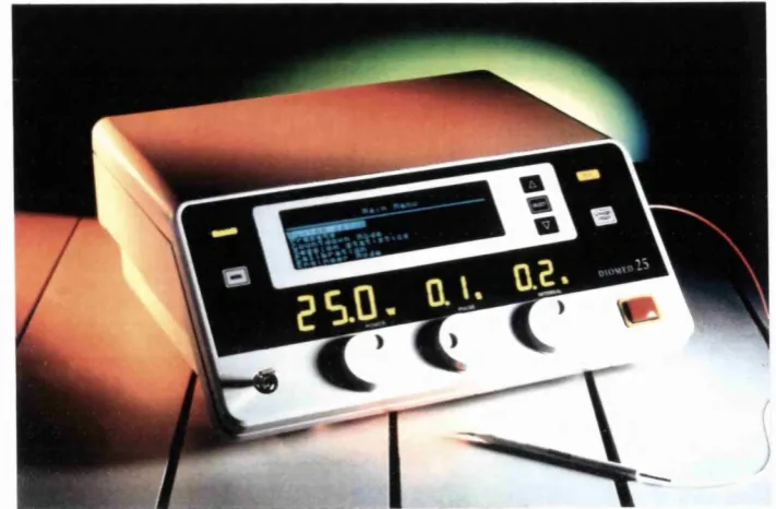
Figure 6.1
The Diomed® diode laser
Figure 6.2
Equipment necessary for the procedure of ILP
Figure 6.3
The Toshiba SAL 38B® ultrasound machine with the diode
laser
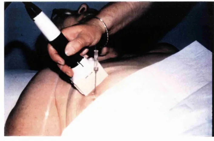
Figure 6.4 a: 14G needle inserted into the tumour under ultrasound
control using the 7.5 MHz transducer and b: patient
undergoing ILP
Figure 6.5
ILP in process using ultrasound to monitor hyperthermic
changes
Figure 6.6 a: the tumour before treatment and b: characteristic
hyperthermic changes(arrowed) seen within the tumour
during ILP treatment.
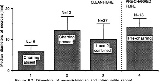
Figure 6.7
Diameters of necrosis(median and interquartile range)
Figure 6.8 The resected specimen(cut through and opened) 24 hours after
ILP and 30 minutes following surgery. The specimen has been
inked to assess the resection margins. a)The characteristic rim
of haemorrhage b)The area of necrosis c)charring present
within the tumour at the site of the laser fibre tip.
Figure 6.9 The resected specimen 3 days after ILP(2.5 watts, 500 seconds
using a clean fibre). The area of laser damage is roughly
spherical in shape. a)area of necrosis b)normal breast tissue
c)rim of haemorrhage d)cavity with charring at the site of the
fibre tip.
Figure 6.10: Low power microscopic view of a resected cancer 27 days
after ILP using a pre-charred fibre(2.5 watts, 500 seconds)
a)skin b) position of the fibre tip c) area of necrosis d) viable
tumour e) charring.
Chapter 7
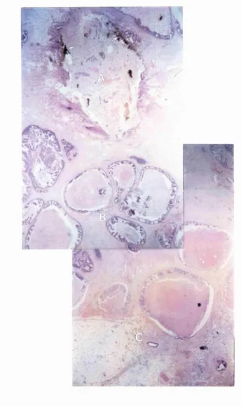
Figure 7.1 : The cavity(A) with evidence of charring lining the
cavity (arrowed),the area of
in situ
fixation(B) and surrounding
necrosis(C) seen in a tumour excised 12 days after ILP using a
clean fibre at 2 watts for 500 seconds.
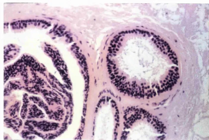
Figure 7.2: The area of
in situ
fixation in greater detail(same specimen as
figure 7.1) showing elongation of the nuclei and spindle like
cells in a patient with a pure ductal carcinoma in situ.
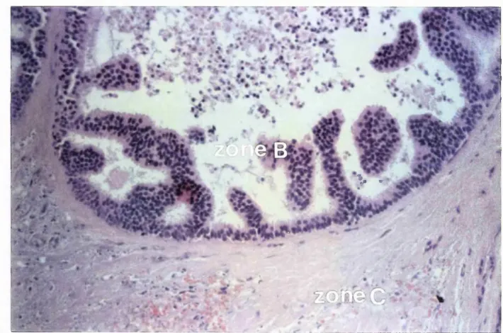
Figure 7.3: In areas, the zone of
in situ
fixation(same specimen as figure
7.1) did not differ in appearence from viable tumour tissue.
This figure shows an area of ductal carcinoma in situ within
zone B.
Figure 7.4: The junction between the
in situ
fixation and necrotic tissue
12 days post ILP, there is no evidence of inflammation.
Figure 7.5: The zone of necrosis(zone C) showing a) pyknotic cells b)
haemorrhage c) ghost like cells after karyolysis 3 days after
ILP using a clean fibre 2.5 watts for 500 seconds.
Figure 7,6: Thrombosis evident 3 days after ILP in the outer aspect of
zone C(arrowed). The laser was activated at 2.5 watts for 500
seconds using a clean fibre.
Figure 7.7: Acute inflammatory response(polymorph arrowed) at the
interface between necrotic tumour(zone C) and normal tissue
3 days after ILP using a clean fibre, 2.5 watts for 500 seconds.
Figure7.8:
Junction between zone C and normal breast tissue 7 days after
ILP(fibroblasts arrowed)
Figure 7.9: The area of granulation tissue gradually expands. A resected
specimen 27 days after ILP. a) necrosis b)granulation tissue
and c)viable tumour.
Figure 7.10: The resected tumour 94 days after ILP, a) fibrous tissue and
b) fat necrosis.
Figure 7.11: A foreign body giant cell response(arrowed) evident 27 days
after ILP.
Figure 7.12: Flow diagram of histopathological changes that occur after
ILP in breast cancers.
Figure 7.13: Effect of ILP on normal breast tissue 1 day after ILP
a) normal breast stroma b) fat necrosis with foamy
macrophages(arrowed)
Chapter 8
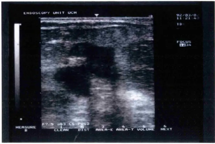
Figure 8.1
Comparison of tumour size as measured by ultrasound and
histology.
Figure 8.2
Comparison of the hyperechioc area seen on ultrasound and
histological necrosis.
Figure 8.3: Ultrasound pictures seen a)before treatment b) during
treatment(at 40 seconds) c) the hyperechoic area reached a
peak at 490 seconds d)the area seen on ultrasound correlated
poorly with the final histology when the tumour was resected
27 days after ILP and e) seen 3 minutes after the completion
of ILP, the hyperechoic area having diminished in size.
Figure 8.4: Ultrasound underestimates the final diameter of necrosis, in
this case(a papillary carcinoma) the maximum hyperechoic
area was only 3mm(top and arrowed) and yet there was 25mm
of necrosis in the final specimen(resected 6 days after
ILP)which included some damage to surrounding normal
breast tissue(bottom picture, the laser damage is within the
area marked by the black ink) which was not visible on
ultrasound.
Figure 8.5: CT scan 24 hours after ILP. The tumour enhances (a) showing
a devascularised area (b) which measures 15mm. The final
diameter of necrosis measured 11mm histologically when
resected 12 days after ILP.
Figure 8.6: CT scans seen 24 hours post ILP. 8.6a) post laser pre-contrast
l=fibre tract 2= tumour and 8.6b) post laser post contrast
l=fibre tract 2=tumour 3=laser induced necrosis.
Figure 8.7: CT scans seen in real time, no demonstrable changes were
visible.a) pre laser post contrast(tumour arrowed) b)during
ILP treatment needle(l) and tumour(2). Following treatment
no changes were visible either in the tumour or surrounding
tissue on CT scanning but 8mm of laser damage was
subsequently found in pectoralis major.
Figure 8.8: MRI scans 24 hours post ILP(patient 3), a) pre-laser pre
contrast, the tumour is arrowed, b) post laser pre-contrast(laser
fibre track arrowed) and c) Post laser post contrast an area of
reduced enhancement is evident(arrowed) which corresponded
exactly with the histology(d).
Figure 8.9: MRI scans a) pre laser pre-contrast b) pre-laser post contrast,
the tumour enhances(arrowed) c) post laser pre-contrast and d)
post laser post contrast
Acknowledgements
The work included in this thesis was undertaken at the National Medical Laser Centre,
in the Department of Surgery at University College London Medical School and in the
Department of Surgery at The Royal Surrey County Hospital, Guildford, Surrey. I am
particularly grateful to my supervisors, a special thanks goes to Professor Steve Bown,
who thought of the concept of ILP and initiated this project, for his continued support,
abounding enthusiasm and encouragement and to Mr. Mark Kissin for his support,
enthusiasm and ideas. Mark taught me a lot about breast disease in the breast clinic at
Guildford and fired my enthusiasm to pursue a general surgical career with a strong
interest in breast disease. To you both I am very grateful.
The work contained in this thesis and my salary were provided by the Guildford Laser
Appeal and I am particularly indebted to Mrs. Gillie Ross chairwoman and the Ladies
and gentleman of the appeal committee for all their extremely hard work in raising the
money to support me. I would especially like to thank the kind ladies of Guildford and
London who agreed to take part in the study armed with the knowledge that the
technique would have no direct benefit for them but possibly for women like them in
the future. It was at the suggestion of Mr.R.C.G.Russell that I was introduced to
Professor Bown and the ILP project and I am grateful for his guidance and advice
during the last 2 years and to another of my old mentors, Mr .John Scurr who also
assissted me financially. Professor Irving Taylor and Mr.Tim Davidson have
scrutinised some of the chapters contained herein and I am grateful for their help and
suggestions. Dr.Tim Mills, Dr.Giovanni Buonaccorsi and Dr.Matthew Clemence from
the medical physics department have been of invaluable assistance non the least in
teaching me some physics! Dr.Bill Lees and Dr. Julie Cooke, both consultant
radiologists were of great help and taught me how to perform breast ultrasound. I am
grateful to pathologists Dr.Martin Cook and especially to Dr.Mark Smith for their help
and enthusiasm in the project. I would also like to thank Dr Waddy Gedrojk, consultant
radiologist in the MRI unit at St. Mary's Hospital, London and G.E.medical systems,
Millwaukee who funded Dr.Clemence and the breast MRI's which were performed in
their breast MRI coil at St.Mary's Hospital. Thank you also to Dr.Tony Raven, Mr.Ilka
Mannonen and Mr.Ian Cameron from Diomed Ltd., Cambridge for loan of the laser and
for all their invaluable assistance. Sisters Pat Willgoose and Dorothy George, breast
care nurses at Guildford and London, Pat Jenkins sister in the Jarvis Breast Screening
Centre and Sister Ann Aldborough, endoscopy sister at University College Hospital
were all of enormous help. I am also grateful to Mr. Abdul Gafur and the staff of the
medical illustration department at the Middlesex Hospital for excellent photographic
assistance. A special thanks goes to Mr. Ross Scrivener, facilitator in the medical audit
department for creating the database and loading data onto the computer for chapter 5 . 1
am indebted to Dr.Secker Walker and Dr Jane Silk from the medical audit department
and Farmitalia Carlo Erba Ltd. for funding the questionnaires(chapter 5). I am also
grateful to Dr. John Bulmer of the Eastman Dental Hospital for statistical advice. Miss
Amanda J.S. Jones, secretary to Professor Bown has been of enormous help especially
with the computers. I am grateful to those I have shared an office with over the last 2
years who have been most supportive and friendly. Dr. Glen Spencer, Miss Sally
Thorpe, Dr. Zahir Amin, Dr. Hugh Roberts, Mr. Isaac Nyamecke, Dr Charles Millson,
Dr. Ian Sergeant, Mr.Ken Anson and Mr. Kenneth Port.
And finally a special thanks to my wife Tamara for all her help and support and
tolerance over the last two years. Without you all, I would not have written this thesis.
Dedication
To my wife Tamara
and to my parents
Marion and Derek
CHAPTER 1: BREAST CANCER ; METHODS USED TO
OBTAIN THE DIAGNOSIS
1.1 Tissue diagnosis
1.1.1 Introduction
1.1.2 Fine needle aspiration cytoIogy(FNAC)
1.1.3 Core biopsy
1.1.4 Excision biopsy
1.2 The imaging of breast cancer
1.2.1 Mammography
1.2.2 Ultrasound
1.2.3 Colour doppler ultrasound
1.2.4 Computerised Tomography(CT)
1.2.5 Magnetic Resonance Imaging(MRI)
1.2.6 Other techniques
1.2.7 Summary
1.1 Tissue diagnosis
L L l Introduction
The modem management of a patient with breast cancer requires that a certain amount
of tissue be obtained from the tumour not only to confirm the diagnosis of malignancy
but also to give an indication of tumour type and grade in order that appropriate
adjuvant treatment can be planned. For instance, the treatment of a patient with a pure
ductal carcinoma in situ will be different from that of a patient with an aggressive grade
III ductal carcinoma and different again from a patient with a tubular carcinoma. The
main methods available to obtain tissue from a patient with a suspected breast
carcinoma are fine needle aspiration cytology, core biopsy and excision biopsy.
1.1.2 Fine needle aspiration cytology(FNAC)
Fine needle aspiration cytology(FNAC) was initially introduced by Martin and Ellis in
1930 and is a reasonably simple technique(often deceptively so!) which requires little in
the way of equipment but a fair degree of skill in order to obtain sufficient material and
considerable skill in interpretation requiring an experienced cytologist. The use of
FNAC as a method of obtaining a tissue diagnosis has accelerated in popularity in the
past 10 years or so and some centres now have a cytologist in the clinic who not only
performs the FNAC's but also reports the results immediately. Basnett et al(1992)
showed how the use of breast FNAC increased in the period 1982-86 in a teaching and
a non-teaching hospital. The technique obviously depends upon there being sufficient
material in the aspirate to make a diagnosis and in the larger series(Ciatto et al, 1993a)
the inadequacy rate was 6.9% for cancers and 24.1% for benign lesions. Cytology can
give a definitive diagnosis of malignancy, can give an indication of tumour type and in
some cases an indication of tumour grade(Hunt et al, 1990, Ciatto, 1993b), oestrogen
and progesterone receptor status(Weintraub et al, 1987, Redard et al, 1989) and even
proliferative indices(Remvikos et al, 1991). FNAC does have its limitations, for
example, it cannot distinguish malignant cells from an in situ carcinoma from malignant
cells from an invasive carcinoma with any degree of accuracy and it is certainly not
possible at the present time to plan subsequent therapy for early breast cancer(apart
from surgery) just on the results of a positive FNAC. Fentiman(1990) has recently
published an overview of 10 studies evaluating FNAC and subsequent histology. The
number of patients in these studies ranged from 369 to 3545. The sensitivity of FNAC
was on average 87% and the specificity 99% with a positive predictive value of 97%.
A large single centre study published recently by Ciatto et al( 1993a) analysed 9533
cases of breast FNAC. The overall sensitivity was 89.5% and was dependent upon
histological type with a sensitivity of 90.1% for invasive ductal carcinomas, 79.6% for
in situ ductal cancers, 84.5% for invasive lobular carcinomas and 90.6% for special type
cancers. The specificity in this series was 98.5%. Therefore, FNAC is a reliable,
safe and simple technique but does require some skill in the performance of the
technique and interpretation of the specimens and the information gained is limited.
L I.3 Core biopsy
The technique of core biopsy involves the insertion of a larger bore needle into the
tumour consequently producing more tissue from which to make a definitive diagnosis.
Usually the Trucut® or Bioptycut® devices can easily be used under local anaesthesia
in the outpatient clinic. Fentiman(1990) has reviewed 7 studies evaluating the value of
Trucut® needle biopsies for the confirmation of malignancy in breast masses. The 7
series included between 87 and 278 breast cancers, the average sensitivity from the 7
series was 82% with a specificity of 100% and a positive predictive value of 100%.
Trucut® biopsies can give much more information about tissue type and can
differentiate between invasive and in-situ cancer, although the results have to be
interpreted with caution(the presence of in-situ disease in a biopsy does not exclude
invasive disease elsewhere in the lesion). Core biopsy can also give an indication of
tumour grade, steroid hormone receptors and tumour type. Baildam et al(1989) studied
140 patients who had trucut biopsies and subsequently underwent surgery in order to
ascertain exactly how much information can be obtained from core biopsies by
comparison with the final resected histological specimen. One hundred and thirty had
carcinomas. The sensitivity was 95% and the specificity 100%. In 93% of cases the
pathologist was able to type the tumour correctly and in 69% was able to correctly
grade the tumour but lymphatic invasion or elastosis could not be accurately predicted.
In 28% of cases it was possible to perform DNA flow cytology and in 45% of cases
steroid receptor assays. This study confirmed that core biopsies were capable of
providing a reasonable amount of information prior to surgery.
1.1.4 Excision biopsy
With the advent of FNAC and the use of core biopsies the use of excision biopsy as a
means of obtaining a tissue diagnosis has declined. Sometimes it is necessary to
perform an excision biopsy in the face of a patient with a persistent breast lump when
FNAC and core biopsies were normal and it is still necessary often for patients with
screen detected abnormalities. The use of excision biopsy as a primary method of
obtaining a diagnosis has now largely fallen into the archives of surgical history
although it is still practiced in some centres(see chapter 5). The practice of excision
biopsy results in many patients undergoing unnecessary surgery and in some cases more
than one surgical procedure.
1.2 The imaging of breast cancer
1.2.1 Mammography
The technique of mammography was first reported by Warren in 1930 and popularised
by Egan in 1960 who reported a series of 1000 cases with a sensitivity of 97%.
Mammography is an excellent technique for the detection of lesions within the breast
but it is fairly non-specific; the classical radiological signs suggestive of malignancy
being a spiculated mass with ill defined margins, the presence of architectural distortion
or skin thickening and characteristic fine microcalcifications. Mammography can
confirm the presence of malignancy diagnosed clinically and is essential for any woman
who presents with breast cancer, firstly to exclude multicentricity and multifocality and
secondly to exclude impalpable contralateral breast cancer(Dixon and Chetty, 1991).
Mammography can detect early impalpable cancer and can predict the presence of an
extensive intraduct carcinoma within an invasive carcinoma(Dixon and Chetty, 1991,
Stomper and Connolly, 1991), a known risk factor predicting local recurrence following
conservative breast cancer surgery . The sensitivity of mammography depends upon
many features probably the most important of which is patient age. The pre menopausal
dense breast is more difficult to visualise on mammography than the older, post
menopausal fatty breast and this is reflected in the sensitivity of the technique for
malignancy. The overall sensitivity of mammography is between 61-87%(Fentiman,
1990) with an average sensitivity of 70% obtained from 10 series(Fentiman, 1990) but
for women under 50 years the figure was 56% compared to 78% for those over 51
years. Mammography is said to be less accurate at diagnosing infiltrative lobular
carcinoma(Fentiman, 1990) possibly because lobular carcinoma produces masses which
are of relatively low radiologic opacity similar to normal fibro-glandular breast tissue
and in addition lobular carcinomas tend, as reported in some series, to have less in the
way of w orrisom e m icrocalcifications when com pared to invasive ductal
carcinomas(Krecke and Gisviold, 1993). Mammography is not an accurate tool for
measuring tumour size(Pain et al, 1992) tending to underestimate the size of larger
tumours and in this study it was not possible to determine the size of some of the
tumours(in 18% of cases). Another series(Report from the Yorkshire Breast Cancer
Group, 1980) found concordance between radiological(by mammography) and
pathological tumour size in 59% of 348 cancers and Fomage et al( 1987) found a
correlation coefficient of 0.72 between mammographie and pathologic tumour size in a
study of 31 patients.
7.2.2 Ultrasound
Ultrasound is now well established as an indispensable complementary investigation to
mammography in the diagnosis of breast disease. Ultrasound is not useful as a
screening modality being unable to detect fine microcalcifications(Muir et al, 1983,
Sickles et al, 1983) but is extremely useful in differentiating an impalpable cystic from
an impalpable solid lesion seen on mammography with reported accuracy’s in the range
of 96-100%(Greenstein Orel and Troupin, 1993). Ultrasound can also be useful in
differentiating solid from cystic palpable lesions which are not seen on mammography
due to a dense fibro-glandular background and can be used to guide interventional
procedures such as fine needle aspiration cytology (for example of an impalpable solid
lesion detected on mammography). Ultrasound is unable to differentiate solid lesions
which are benign from solid malignant lesions although both benign lesions such as
fibroadenomas and carcinomas are said to have characteristic echoic appearances. The
sensitivity of ultrasound varies in reported series from 68-93% with an average of 84%,
a specificity of 89% and a positive predictive value of 88%(Fentiman, 1990). Pain et
al(1992) found that ultrasound consistently underestimated tumour size as measured by
subsequent histology reporting a correlation coefficient of 0.75 and a correlation
coefficient of 0.84 was found in a similar study by Fomage et al(1987) with ultrasound
tending to overestimate tumour size.
1.23 Colour doppler ultrasound
Malignant breast tumours stimulate the growth of new blood vessels by the release of
angiogenesis factor giving rise to characteristic features on colour doppler sonography.
Cosgrove et al(1993) recently reported the use of colour doppler ultrasound in 210
patients with 222 breast lesions including 58 breast cancers. Colour doppler signals
were obtained from 57 of the 58 breast cancers with an average of 2.16 vessels per
lesion compared to colour doppler signals from 5 of 104 patients with benign breast
changes and 5 out of 36 patients with fibroadenomas and 7 out of 12 patients with
miscellaneous conditions(which included 4 cases of breast infection all of which gave
colour doppler signals). Cosgrove et al(1993) described a sensitivity of 98% and a
specificity of 89%. The technique has not been reported to be so accurate by other
workers, McNicholas et al(1993) reported a sensitivity of 87% with a specificity of
32% in a study of 54 cancers and 77 benign lesions and cite an overall sensitivity of
87%, a specificity of 70% and a positive predictive value of 82 % from 4 series reported
in the literature.
1.2 A Computerised Tomography(CT)
CT scanning of the breast has not been shown to be of value in the routine detection of
breast cancers. The early enthusiasm for the technique in the late 1970's and early
1980’s was based on the work of Chang et al who found that carcinomas enhanced with
the use of iodonated contrast but other workers have found that reliable differentiation
between benign and malignant tumours could not be achieved despite the administration
of intravenous contrast(Greenstein Oriel and Troupin, 1993). In addition the use of CT
results in a higher dose of radiation than mammography both to the breast and to the
thorax and the technique is considerably time consuming and relatively expensive.
Hence breast CT has not found a place in the routine imaging of breast cancer.
1.2.5 Magnetic Resonance Imaging(MRl)
Magnetic Resonance Imaging allows visualisation of the breast without radiation, in
fact, some of the earliest clinical work done on MRI was on the breast. The principle
of MRI is based on the fact that some nuclei(with unpaired electrons) behave like small
magnets. Water is the principle constituent of most body tissues and the hydrogen
nuclei are therefore present in large numbers. The use of a strong external magnet will
force the nuclei to align in a new magnetic axis, a pulse of radiowaves then displaces
the magnetised nuclei from their new alignment releasing the energy they absorbed as a
radiosignal and this signal is detected by the coil used for excitation. The signal is then
converted by computer into an image. The magnetic fields used in clinical practice
range from 0.15 to 1.5 Tesla(or 1500 to 15000 Gauss where the earth’s magnetic field is
0.5 Gauss). The main radiofrequency pulse sequences used are called saturation
recovery, inversion recovery and spin echo. Inversion recovery sequences(Tl weighted
images) show better anatomical detail whilst spin echo(T2 weighted images) sequences
are better at imaging pathological changes. MRJ has good soft tissue resolution and has
been investigated by several workers as a potential imaging device for the breast.
The early reports of the use of breast MRI were encouraging(Greenstein Orel and
Troupin, 1993) but large slice thickness with long acquisition times and initially only
moderate sensitivities diminished the initial enthusiasm for breast MRI and its future
place in the diagnosis of breast cancer was uncertain. However, more recent
developments including the use of breast surface coils instead of body coils, improved
signal to noise ratios, the use of intravenous gadolinium to enhance the tumour and fat
suppression sequences have all significantly increased the sensitivity of breast MRI and
another surge of enthusiasm has resulted. For MRI to have a major role in breast cancer
diagnostic management it must, a) have high resolution for detection of small cancers
(this requires intravenous gadolinium to enhance the tumour to increase the sensitivity),
b) employ fat suppression sequences for differentiating enhanced tumours from breast
fat and c) have rapid acquisition times(less than 6 minutes) for differentiation of
enhancing tumours from breast parenchyma(Harms et al, 1993). MRI has much higher
sensitivities for malignancy than mammography, Kaiser(1993) reported a 98.4%
sensitivity for detection of 63 carcinomas and Harms et al(1993) a 94% sensitivity for
the detection of 47 carcinomas. Harms et al(1993) also found a good correlation
between tumour size as measured by MR and subsequent histology. It appears that
intravenous administration of the paramagnetic agent gadolinium increases sensitivity.
In one study 20% of cancers w ere observed only after adm inistration of
gadolinium(Kaiser and Zeitler, 1989) and in this series 8 cancers were visualised on
MRI which were not seen on mammography. When tumours do enhance they
sometimes appear isointense with the surrounding breast fat but fat suppression
sequences have now been developed to overcome this problem(Pierce et al, 1991). The
third problem is that enhancement of breast cancers with gadolinium is due to the
difference in vasculature between tumour tissue and normal breast tissue, essentially
benign lesions such as fibroadenomas can enhance and in time the surrounding normal
tissue will enhance as well. Hence the need for rapid acquisition sequences as benign
tissue enhances much more slowly than tumour tissue. The fact that benign lesions and
in time breast parenchyma will enhance with gadolinium results in breast MRI having a
high false positive rate varying from 18-40%(Hussman et al, 1993).
As well as in primary diagnosis MRI probably has a place in the detection of local
recurrence in the breast(Lewis-Jones et al, 1991 )folio wing breast conserving surgery and
radiotherapy where it may be difficult on mammography, especially in the
premenopausal dense breast, to differentiate between local recurrence and post
operative scar tissue, as scar tissue does not enhance with gadolinium, although 1 report
suggested that scar tissue will enhance in the first 6 months after surgery(Cohen, 1993).
It may also be possible in the near future to perform stereotactic needle localisation
under MRI control or even minimally invasive procedures following the recent
description by Hussman et al(1993) of early work with a MRI stereotactic localisation
device for the breast.
MRI then has potential as a future imaging device for breast disease, it has greater
sensitivity than mammography if gadolinium is given to enhance the tumour but has a
lower specificity and in addition it is more time consuming, more expensive and less
readily available than mammography. Current work in progress suggests that a
stereotactic device could soon be available to allow biopsy or interventional procedures
under MRI control but the future role of breast MRI has yet to be fully determined.
1.2.6 Other techniques
A variety of other techniques have either been investigated and abandoned or are
currently under investigation seeking to find a role to play in diagnostic breast
radiology. Thermography comes under the auspices of the first category(i.e.it has
largely been abandoned). Greenstein Orel and Troupin(1993) reviewed the published
results of thermography and found that it was associated with an unacceptably high
false negative rate(49.5% of cancers were missed in one study!) and there is no current
data to support its use. Similarly they reviewed the place of transillumination, a light
scanning technique again associated with a high false negative rate(transillumination
only detected 19% of tumours less than 1cm compared to mammography which
detected 90% in 1 series).
Whilst these techniques are not in current use digital mammography is a new technique
which has been described as "the evolving technology with the greatest potential impact
on breast cancer detection and diagnosis"(Greenstein Orel and Troupin, 1993). Digital
mammography is thought to have great potential in detection of lesions in the
mammographically dense breast an area where mammography has only moderate
sensitivity. However, it is still at an early stage of development and clinical evaluation.
Another technique. Positron emission tomography(PET) scanning has been evaluated
and is able to detect breast cancers with the aid of FDG(2-[F-18]-Fluro-2-deoxy-D-
glucose) and F-18-ES (16-[F-18]-fluroestradiol-17). PET scanning may be able to
monitor changes in the primary tumour following primary chemotherapy(Greenstein
Orel and Troupin, 1993) and it appears that PET with F18ES may be able to predict the
oestrogen receptor status of a tumour(Mintum et al, 1988). PET is at a very early stage
of development in breast disease and has yet to find a role.
Summary
It is important to assess the patient with a suspected breast cancer thoroughly in order to
plan subsequent treatment appropriately. A tissue diagnosis of malignancy can be
reliably achieved by the use of fine needle aspiration cytology(FNAC) which has
largely replaced excision biopsy for this task, the information gained from FNAC is
limited and more information can be achieved by means of a core biopsy.
Mammography is currently the standard imaging technique for the breast and is
essential for all women who present with breast cancer. It has good sensitivity for the
detection of malignancy but the sensitivity is lower in younger women. Ultrasound is a
useful adjunct to mammography, its main value being to differentiate between a solid
and cystic lesion. Colour doppler is currently under evaluation as is digital
mammography. MRI shows great potential as an imaging tool for the breast but its
exact role has yet to be defined. Breast CT, thermography and transillumination are now
largely obsolete and PET scanning is at a very early stage of development.
CHAPTER 2 ; THE TREATMENT OF LOCALISED BREAST CANCER
2.1 Surgery
2.1.1 History : The latter part of the Nineteenth Century onwards.
2.1.2 Trials comparing mastectomy and breast conservation.
2.1.3 Breast conservation.
2.1.3a Introduction
2.1.3b Cosmesis and patient acceptability
2.1.3c Local recurrence
2.1.4 The treatment of the axilla
2.2. Radiotherapy
2.3 Chemo-endocrine therapy
2.1 Surgery
2.1.1 History : The latter part of the Nineteenth Century onwards
Towards the latter part of the nineteenth century the standard treatment for breast cancer
was mainly by local excision. The published results of this form of treatment revealed
unacceptably high local recurrence rates (see Table 2.1)
Author time period no.cases local recurrence
Bergmann 1882-1887 114 51-60%
Billroth 1867-1876 170 82%
Czerny 1877-1886 102 62%
Fischer 1871-1878 147 75%
Konig 1875-1885 152 58-62%
Kuster 1871-1885 228 60%
Von-Volkman 1874-1878 131 60%
Table 2.1 ; Local recurrence rates following wide excision for breast cancer.
(from Harris, Heilman, Henderson and Kinne, 1991)
Charles Hewitt Moore( 1821-1870) a surgeon at St. Luke’s and the Middlesex Hospital
in 1867 was one of the first surgeons to encourage more extensive surgery for breast
cancer. Believing that local recurrences were due to a continuation of the growth of
fragments of the primary tumour, he advised excision of the primary tumour along with
any tissues likely to be affected- including skin, fat, muscle and lymphatic tissue. His
ideas laid the foundation for the development of more radical surgical procedures which
were to revolutionise the treatment of the disease at the end of the nineteenth and for
much of the twentieth century.
William Stewart Halsted in 1894 reported his results with the operation of radical
mastectomy which he first performed in 1882 and described a local recurrence rate of
only 6% in the 50 patients that he operated on(Halsted, 1894). These results were in
stark contrast to the published results of conservative treatment(Table 2.1) and were
achieved even though all patients in his series had involved axillary lymph nodes and
many had advanced disease at presentation. An era had began of radical surgery for
breast cancer and surgery was to become even more radical in the early and mid parts
of the twentieth century. Halsted extended his operation to include a supraclavicular
node dissection in combination with the radical mastectomy but abandoned this due to
an increased operative morbidity and the observation that there was no survival
advantage. Urban(1952) described an en-bloc (or in-continuity) resection of the internal
mammary chain in combination with the radical mastectomy and Wangensteen(1949)
described the supra-radical mastectomy combining radical mastectomy with supra
clavicular node dissection, median sternotomy and resection of both internal mammary
chains. However, both these procedures were associated with an increased morbidity
without improvement in survival.
The Halsted radical mastectomy became the "gold standard" treatment for breast cancer
for much of the early and mid-part of this century. This radical approach to breast
cancer treatment was based on the thought that breast cancer metastasised by means of
the lymphatic system and that eradication of that system not only gave an improved
local control but an improvement in overall survival. This theory was really an over
simplification and paid little emphasis to the problem of venous spread. Although some
surgeons such as Urban at Memorial Sloan Kettering in New York advocated
operations such as the extended radical mastectomy many became disillusioned by
radical surgery and reports appeared of less radical forms of mastectomy.
In 1948 David Patey, a surgeon at the Middlesex Hospital, London published his results
of a lesser form of mastectomy than the Halsted operation which became known as the
Patey mastectomy(Patey and Dyson, 1948). In this procedure pectoralis major was left
behind and the pectoralis minor muscle excised in order that a full axillary dissection
could be performed. The local relapse free survival for the radical mastectomy was 76%
compared to 82% for the modified operation. The operation became modified further by
Madden(Madden et al 1972) who advised leaving both pectoralis major and minor
muscles behind in the so called modified radical mastectomy. Surgical practice
gradually changed with a move away from the Halsted operation toward the Patey or
the modified radical mastectomy as described by Madden. A survey of American
Surgeons conducted by The American College of Surgeons in 1982(Wilson et al, 1984)
and compared to a similar survey carried out in 1977 found an increase in the use of the
modified radical operation(from 55.6% in 1977 to 78.2% in 1982) and a move against
Halsted's operation(from 27.5% in 1977 to only 3.4% in 1982).
Whilst extending the radical mastectomy to include the supra clavicular or internal
mammary nodal areas was felt to be acceptable surgical practice and the less radical
Patey mastectomy became fashionable any lesser procedure was felt to be most
unacceptable and indeed negligent. A leading article in the British Medical Journal in
October 1953(Anon) summed up the current feeling on the subject
"So firmly established had the practice of radical mastectomy become that even to question its value smacked of lese majeste , and those who had the courage to look for other methods had to face, not only the barriers o f reasoned resistance, but an array o f prejudice which lifted the discussion out o f the cool atmosphere o f scientific argument and plunged it into a cauldron seething with emotion".
Even before 1953 there were people who seriously questioned the value of the radical
mastectomy operation. The absolute cure rate was low and there was definite morbidity
in the form of arm oedema and limitation of movement at the shoulder associated with
the procedure. An early sceptic was Professor George Gask at St. Bartholomew's
Hospital, London who in 1922 initially instigated Mr.Geoffrey Keynes to investigate
the use of radium needles to treat recurrent breast cancer. Encouraged by the results he
obtained he extended the use of radiotherapy to patients with advanced breast cancer
and then to treat early breast cancer. He even undertook simple tumour excision and
followed this with breast radiotherapy. Another early advocate of the use of
conservative surgery in combination with radiotherapy was M. Vera Peters a
radiotherapist at St. Margarets Hospital in Toronto who started this form of treatment
as far back as 1939.
Apart from Keynes and Peters few people practised conservative surgery for breast
cancer. However, a few reports started to emerge in the 1950’s to support the use of a
more conservative approach. Mustakallio(1954) reported an 84% five year survival in
127 patients with clinical stage one breast cancers treated by local excision and
radiotherapy and Sir Arthur Porritt(1964) from St. Mary's Hospital, London described a
personal series of patients treated by conservative surgery and radiotherapy with
promising results. These reports were largely anecdotal arising from non-randomised
retrospective studies and it was not until 1972 that the safety of a more conservative
surgical approach combined with radiotherapy was reported by Atkins et al at Guy's
Hospital.
2.7.2 Trials comparing mastectomy and breast conservation
In 1955 Sir Hedley Atkins et al considered the hypothesis that radical mastectomy was
no more effective in preserving life in cases of early breast cancer than simple
lumpectomy. The first prospective randomised trial comparing radical mastectomy and
wide excision followed by radiotherapy(in this paper wide excision was referred to as
extended tylectomy which was defined as removal of the tumour with a 3cm rim of
surrounding normal breast tissue) began to accrue patients in 1961 at Guy's Hospital.
Three hundred and seventy six patients were entered and randomised to radical
mastectomy and post operative radiotherapy or to extended tylectomy without axillary
dissection followed by post operative radiotherapy both to the breast and nodal areas.
Both groups were well matched for age(all were over 60 years old) tumour size etc. By
the time the first paper appeared in (Atkins et al, 1972) it was evident that there were
serious problems with the tylectomy group. Statistical analysis revealed that in the
clinical stage II group radical mastectomy gave significantly better survival than
extended tylectomy(60% vs 28% at lOyears), however in the clinical stage I group there
was no significant difference in survival. Local recurrence was significantly better for
the radical mastectomy group for clinical stage I and II patients. Clear interpretation of
the results was difficult, the radical mastectomy group had received axillary surgery and
axillary radiotherapy whereas the tylectomy group received only axillary radiotherapy,
furthermore the radiotherapy doses given were certainly inadequate by today's
standards. However, it emerged from the data that the survival in the clinical stage I
group was similar and this then encouraged a second study comparing radical
mastectomy and tylectomy in the clinical stage I group only. A further 252 patients
were accrued between 1971 and 1975 but surprisingly the group treated by conservation
fared worse both in terms of relapse free and overall survival(Hayward 1985).
The differences in the results between the two trials were at first difficult to explain.
Hayward and Caleffi(1987) in a subsequent analysis were able in part to explain the
differences by the observation that there were a greater number of smaller (Ti)tumours
in the second series and that the T% group in the second series fared similarly in terms of
local control and overall survival. It was in the Ti group in the second series that the
major differences emerged, the patients in this group treated by mastectomy had
significantly better relapse free and overall survival than the tylectomy group. The
lessons concluded from the Guy's studies were that surgery was capable of cure for
small lesions and that inadequate local treatment led to an increased incidence of local
relapse and an inferior overall survival. For lesions larger than 2cms(i.e.T2 cancers) the
inference was that loco-regional therapy might not be as important as systemic therapy
in affecting long term overall survival.
Two other prospective randomised clinical trials began in 1972 and in 1973. The first
which started in 1972 was organised by the Institut Gustave-Roussy, Villejuif,
France(Sarrazin et al, 1984) and accrued 179 patients between 1972 and 1980. Criteria
for entry to the trial were patients with unilateral breast cancers 2cm or less on
macroscopic examination, aged less than 70 years. Pregnant patients with breast cancer
were excluded. Random isation was to simple m astectom y or local tum our
excision(tumour excision plus a margin of 2cms of glandular tissue) plus breast
irradiation. Both groups underwent a low axillary dissection and those with positive
nodes underwent a full axillary dissection. This latter group were then randomised
further to receive postoperative nodal irradiation or no nodal irradiation. One hundred
and seventy nine patients were entered into the first trial, 88 randomised to
tum ourectom y and 91 to mastectomy. When m astectomy was compared to
tumourectomy there was no significant difference in overall or relapse free survival.
These results were confirmed at 10 years of follow up(Sarrazin et al, 1989), furthermore
92% of patients treated by tumourectomy had either an excellent or a good cosmetic
result.
In 1973 another trial was set up by the Istituto Tumori in Milan under the auspices of
Professor Umberto Veronesi. Seven hundred and one patients with breast cancers less
than 2cms in diameter without palpable axillary nodes were randomised to either
Halsted radical mastectomy or to quadrantectomy, axillary dissection and breast
radiotherapy. Patient accrual took place between 1973 and 1980 and those found to
have positive nodes after 1976 received CMF adjuvant chemotherapy. At 5 years of
follow up(Veronesi et al, 1981) there was no difference in disease free or overall
survival. Only one patient in the quadrantectomy group developed a local recurrence.
The balance in breast conservation surgery lies in the "trade off" between performing a
wide excision leading to a low local recurrence rate and an inferior cosmetic result
versus a narrower excision, a better cosmetic result but a higher local failure rate.
Veronesi et al(1981)achieved a low local recurrence rate but almost 1 in 3 of his
patients had an inferior cosmetic result(Fentiman, 1990). At a median follow up of 13
years (Veronesi et al, 1990) overall and disease free survival remained identical.
Probably the most referenced paper confirming the safety and efficacy of breast
conservation as an alternative to mastectomy are the published results of the National
Surgical Adjuvant Breast Project, a multi-centre trial known as the NSABP B06
trial(Fisher et al, 1985). Patients with breast cancer, up to 4cm in diameter were
randomised to one of three treatment groups:- total mastectomy or segmental
mastectomy with or without breast irradiation. All patients underwent an axillary
dissection and patients with positive lymph nodes received systemic adjuvant
chemotherapy. Those who received breast irradiation were given a dose of 50 Gy
without a boost to the excision site. Patients treated by segmental mastectomy who had
positive tumour margins on histological examination were converted to total
mastectomy but remained in their original randomisation group when comparisons were
made of survival i.e. the randomisation was an intention to treat rather than actual
treatment. Over eighteen hundred patients were entered into the trial between 1976 and
1984. The mean follow up was only 39 months(range 5-99) and the disease free
survival, distant disease free survival and overall survival were similar in all three
groups. The local recurrence rate in the breast of those patients treated by segmental
mastectomy and irradiation was 8% at 5 years compared to 30% for those treated by
segmental mastectomy alone and in this latter group local recurrence rose to 36% in
those with positive axillary nodes. Breast irradiation significantly reduced the incidence
of local recurrence(pcO.OOl). Logical assumption would therefore suggest that disease
free survival would be reduced in the segmental mastectomy group but the protocol of
the study
stated:-’T /ie occurrence o f tumour in the breast after segmental mastectomy was not considered an event in the disease free survival o f a patient, since patients who underwent total mastectomy as their initial operation were not at risk for the occurrence of a breast tumour”.
Therefore according to the protocol of the NSABP B06 trial it was possible to develop a
local recurrence in the ipsilateral treated breast but this was not considered to constitute
a reduction in the disease free survival. Other features worth noting were that 10% of
patients in the segmental mastectomy group had positive tumour margins and were
converted to mastectomy. The follow up at eight years confirmed the 5 year
findings(Fisher et al, 1989) and further follow up(Fisher et al,1991) showed that by 9
years the local recurrence rate had risen in the segmental mastectomy and irradiation
group from 8% to 12% and from 30% to 43% in the non-irradiated group. At nine years
of follow up(Fisher et al, 1991) even though a high proportion of those treated by
segmental mastectomy without irradiation had developed an isolated breast recurrence
their distant disease free survival was no different from the segmental mastectomy and
irradiation or from the total mastectomy group. But a Cox's regression analysis of the
lumpectomy group revealed that those patients who developed a local recurrence were
at 3.41 times greater risk of developing distant disease and also, the development of an
early local recurrence carried a less favourable distant disease free survival. They
concluded that the development of an isolated breast recurrence was a marker of an
increased risk of, but not a cause of, distant métastasés. Local recurrence will be
discussed in greater detail below.
The clear messages from the NSABP B06 trial were that segmental mastectomy with
clear pathological margins, axillary clearance and breast irradiation was a safe
alternative to mastectomy for tumours of 4 cms or less and that segmental mastectomy
alone leads to an unacceptably high local recurrence rate. Other prospective studies
have confirmed the safety of breast conservation and an overview of these studies and
the studies presented in this chapter are outlined in table 2.2
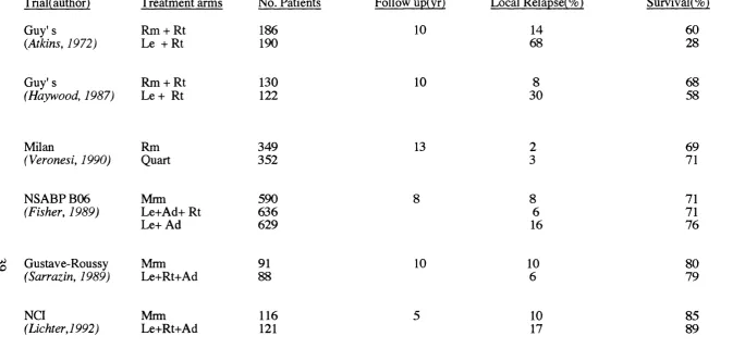
TriaKauthor)
Guy's
{Atkins, 1972)
Treatment arms
Rm + Rt Le + Rt
No. Patients 186 190 Follow up(yr) 10 Local Relapse(%) 14 68 Survival(%) 60 28 Guy's
(Haywood, 1987)
Rm + Rt Le + Rt
130 122 10 8 30 68 58
Milan Rm
(Veronesi, 1990) Quart
349 352
13 69
71
NSABP B06 (Fisher, 1989)
Mrm Le+Ad+ R.t Le+ Ad 590 636 629 8 8 6 16 71 71 76
^ Gustave-Roussy (Sarrazin, 1989)
Mrm Le+Rt+A d 91 88 10 10 6 80 79 NCI (Lichter,1992) Mrm Le+Rt+Ad 116 121 10 17 85 89
Table 2.2:- Randomised trials to assess efficacy of conservative surgery compared to mastectomy(from Sacks and Baum, 1993)
Rm=Radical mastectomy, Mrm= Modified radical mastectomy, Rt=Radiotherapy, Le=Local excision, Ad= Axillary dissection.
To conclude, the work reviewed in this chapter so far has shown how the treatment of
breast cancer has essentially done an about turn in the last 100 years having moved
away from an era of treating breast cancer conservatively in the late 1800's through a
radical era and now into an era where several studies with good statistical correlation
have shown that conservative surgery combined with radiotherapy in appropriately
selected patients is a safe alternative to mastectomy.
2.13 Breast conservation
2.1.3a Introduction and patient selection
The above trials have outlined the safety of breast conservation, but the technique is
only of proven efficacy for selected patients e.g. those with tumours less than 4 cms in
diameter. It would be worthwhile at this point identifying which patients are and which
are not suitable for breast conservation.
The upper size limit considered suitable for breast conservation is 4cms, without signs
of local advancement, extensive nodal involvement or distant métastasés. Many patients
with smaller tumours would not be suitable for conservation because of small breast
size resulting in a poor cosmetic result and are best treated by mastectomy possibly with
immediate reconstruction. Central tumours were not initially considered suitable but
are not a complete contra-indication to conservation, the nipple is lost but the general
shape remains. Multi-focality is considered a contra-indication to conservation as it
leads to a high incidence of local recurrence(Kurtz et al, 1990). Breast conservation has
been shown to be safe for tumours other than invasive ductal carcinomas(Kurtz et al,
1989) but is not suitable for the treatment of inflammatory carcinomas.
The safety of this technique for treating early carcinomas such as Ductal Carcinoma in
Situ(DCIS) has yet to be fully established and is currently the subject of a nationwide
U.K. study. Early results from an NSABP Study(Fisher et al, 1993) comparing 818
patients with DCIS randomised to segmental mastectomy with or without irradiation
have found a significant improvement in "event free survival"(subsequent tumour at a
local, regional or distant site after the initial operation) in the irradiated breast group
even at 43 months of follow up. The presence of an extensive in-situ component in
association with an invasive ductal carcinoma appears to predispose patients to an
increased risk of local recurrence and these patients are possibly not suitable for
conservation(see below). Age is not a contra-indication to conservation but young
patients(<35 years) appear to have an increased incidence of local recurrence(Recht et
al ,1988). Elderly patients are suitable for conservation provided that they are treated in
a conventional manner by lumpectomy, radiotherapy and tamoxifen(Dixon, 1992).
Pregnant women with breast cancer are best treated either by modified radical
mastectomy or if the pregnancy is terminated by conservation followed by radiotherapy.
Patients with connective tissue disorders are not suitable. The final barrier to breast
conservation is patient choice. Some patients opt for mastectomy rather than
conservation even after counselling(Wilson et al, 1988) The reasons why some patients
choose mastectomy rather than conservation are varied. In addition, some patients are
advised to undergo mastectomy by their surgeons even if they have small
carcinomas(see chapter 5). In the U.K. there is a regional variation in the treatment of
breast cancer with mastectomy being advised more frequently in certain parts of
England(Harries et al, 1993).
2.1.3b Cosmetic appearance
The cosmetic appearance following conservative surgery and radiotherapy is an
important consideration. The art of successful breast conservation is to achieve a good
cosmetic result without compromising local control. Large resections especially in a
small breast lead to poor cosmesis. The Milan group(Veronesi et al, 1981) were able to
produce a low local recurrence rate but the procedure of quadrantectomy lead to a poor
cosmetic result in nearly 1 in 3 patients(Fentiman, 1990).
Sarrazin et al(1984) used the operation of tumourectomy combined with axillary
dissection and radiotherapy, local relapse was 6% at 10 years and 92% had a
satisfactory cosmetic appearance. Radial incisions in the breast give an inferior