R E S E A R C H
Open Access
Differential expression of microRNAs in
GH-secreting pituitary adenomas
Zhi-Gang Mao
1, Dong-Sheng He
1, Jing Zhou
2, Bin Yao
3, Wei-Wei Xiao
2, Chun-Hua Chen
2,
Yong-Hong Zhu
2*, Hai-Jun Wang
1*Abstract
Background:The purpose of this study was to (1) identify specific miRNAs in growth hormones (GH)-secreting pituitary adenomas; (2) determine the relationship between the expression of these miRNAs and tumor size, somatostatin analogs treatment, and responsiveness to somatostatin analogs (SSA).
Methods:Fifteen GH-secreting adenomas patients were treated with lanreotide for 4 months before surgery. Patients with 50% reduction of GH secretion by lanreotide were considered as SSA responders, while patients with less than 50% of GH reduction were considered as SSA nonresponders. We analyzed the miRNAs in 21 GH-secreting pituitary adenomas and 6 normal pituitaries by miRCURY™LNA array and some differentially expressed miRNAs were validated by quantitative real-time PCR.
Results:Fifty-two miRNAs were differentially expressed between GH-secreting pituitary adenomas and normal pituitaries. Differential expression of 9 miRNAs was observed between micro- and macro-adenomas. Thirteen miRNAs were differentially expressed between tumor samples from lanreotide-treated patients and those from lanreotide-untreated patients. Seven miRNAs were differentially expressed between SSA responders or GH
nonresponders. Several identified miRNAs may be involved in cell proliferation, apoptosis, cancer development and progression.
Conclusions:Our results indicate that altered miRNAs expression is involved in GH-secreting pituitary adenomas transformation, which will shed light on the mechanisms for the treatment of acromegaly by SSA. Identification and characterization of the targets of altered miRNAs genes may elucidate molecular mechanisms involved in the pathogenesis of pituitary adenoma.
Background
MicroRNAs (miRNAs) are a class of non-coding RNAs that post-transcriptionally regulate the expression of
downstream mRNAs by targeting the 3’ untranslated
regions [1,2]. Since the discovery that miRNAs are a class of conserved genes, hundreds of miRNA genes have been identified. More than 6000 miRNAs encoded by virus, plant and animal species have been recorded in miRBase bank [3,4]. miRNAs are a huge class of nega-tive gene regulators controlling a wide range of biologi-cal functions such as cell proliferation, differentiation,
signaling pathways, apoptosis and metabolism [5,6]. Recently, it has been shown that several human cancers, e.g. breast, colon, lung, brain, thyroid, and hematologic malignancies are associated with altered miRNAs expression [7]. In addition, more and more evidences suggested that some miRNAs might have oncogenic or tumor suppressor functions [8], and play an important role in tumorigenesis [9]. Previous studies have shown that expression of miR-15a and miR-16-1 in pituitary adenomas is lower than that in the normal pituitary tis-sues. Further more, the expression level of miR-15a and miR-16-1 is inversely correlated with tumor diameter and directly correlated with the secretion of the anti-neoplastic cytokine p43 [10]. Further studies have demonstrated that several identified miRNAs are involved in cell proliferation, apoptosis and cortico-trophic tumorigenesis, suggesting that deregulation of
* Correspondence: zhuyongh@mail.sysu.edu.cn; mmh222111@yahoo.com.cn 1
Department of Neurosurgery and Pituitary Tumor Center, The First Affiliated Hospital, Sun Yat-sen University, Guangzhou 510080, China
2
Department of Histology and Embryology, Zhongshan School of Medicine, Sun Yat-sen University, Guangzhou 510080, China
Full list of author information is available at the end of the article
miRNAs expression may be involved in pituitary tumori-genesis [11,12]. Predictive miRNAs could be potentially useful diagnostic markers, improving the classification of pituitary adenomas. Nevertheless, the role of transcrip-tional regulation of miRNAs and their target genes in the pathogenesis of pituitary adenomas remains largely unknown. Development of acromegaly is caused by the proliferation of somatotrophs and oversecretion of the hormone. A cascade of transcription factors and genetic elements normally determine the ability of somatotroph cells to synthesize and secrete growth hormone [13]. In this study, we compared the miRNAs expression between GH-secreting pituitary adenomas samples and
normal pituitary samples by miRCURY™Locked Nucleic
Acid Array in order to identify miRNAs that are specifi-cally associated with GH-secreting pituitary adenomas. The possible role of these identified miRNAs was also discussed.
Materials and methods Patient information
The study was approved by the Institutional Review Board of the First Affiliated Hospital, Sun Yat-sen Uni-versity (Guanzhou, China). The local ethical committee approved the pre-surgical medical treatment and all par-ticipants had informed written consent. The study is registered at ClinicalTrials.gov (NCT00993356). Tissue samples were collected in accordance with the guide-lines of local committee on human research. The biolo-gical diagnosis of acromegaly was based on the criteria that (1) plasma GH concentration is higher than 1μg/l after oral administration of 75 g of glucose (oral glucose tolerance test, OGTT); (2) insulin-like growth factor 1 (IGF-1) concentration is increased compared to the nor-mal population in the same age and sex; and (3) relevant clinical features associated with acromegaly occurred and pituitary adenoma appeared on the magnetic reso-nance imaging examination.
Experimental design
We examined 21 GH-secreting pituitary adenoma sam-ples and found that 3 samsam-ples belonged to micro-adeno-mas (maximum diameter <10 mm), while the other 18 samples were macro-adenomas (maximum diameter >10 mm) [14]. Fifteen patients were treated with lanreotide (Somatuline Autogel, Beaufour Ipsen, Paris, France) for 4 months before surgery. Six patients did not receive any pre-surgical medical treatments. Previous conven-tional or stereotaxic (Gamma knife) radiotherapy was not performed for all the patients. Patients treated with lanreotide presented no evidence of previous cholecysto-lithiasis or any other abnormalities. As reported by Maiza, the initial dosage of lanreotide was 60 mg/28 days [15]. Patients with >50% reduction of GH secretion
after treatment with lanreotide were considered as SSA responders, while patients with <50% GH secretion were considered as SSA nonresponders [16]. We assessed the responsiveness of GH secretion after the second injec-tion because of the slow-release of lanreotide formula-tions. The dosage of lanreotide was adjusted according to the hormone response. In the case of nonresponders, the dosage of lanreotide was increased to 90 mg/28 days, and the dosage of lanreotide was maintained at 60 mg/28 days in the case of responders. SSA nonre-sponding patients received the treatment of lanreotide continuously because it ameliorated the clinical symp-toms including headache, fatigue and peripheral soft-tissue swelling. All patients were well tolerated by lanreotide and they all completed the study. Twelve patients experienced bowel cramps and diarrhoea for several days after the first injection of lanreotide and the symptoms were resolved after the third injection. No adverse events were reported during the study. Magnetic resonance imaging scan of the pituitary, detection of plasma GH and IGF-1 concentration were performed after treatment.
Detection of miRNAs expression and data analysis Pituitary tumor samples were obtained during transphe-noidal surgery. Tumor samples were divided two parts: one was used for pathologic analysis including hematox-ylin eosin (H&E) and immunohistochemical staining and another was used for miRNAs expression. Tumor
samples were snapped-frozen and stored at -80Ω°C.
(biospec). Total RNAs were isolated by using TRIzol reagent (Invitrogen, Carlabad, CA) and RNeasy mini kit (Qiagen, Valencia, CA) according to the manufacturer’s instruction. The quality and quantity of RNA were mea-sured using a NanoDrop ND-1000 (NanoDrop). The ratio of OD260/280for pure RNA should be close to 2.0
(ratios between 1.8 and 2.1 are acceptable). The ratio of OD260/230should be larger than 1.8.
After the measurement of RNA quantity and quality, the samples are labeled using the miRCURY™Hy3™/Hy5™ Power labeling kit (Exiqon, Denmark) according to the manufacturer’s instruction. The Hy3™/Hy5™-labeled RNA
molecules were hybridized on the miRCURY™ LNA
Array (Exiqon, Denmark). The latest version of the array (v.10.0) consists of control probes, mismatched probes and more than 1200 capture probes, which cover all human, mouse and rat miRNAs sequences annotated in miRBase 10.0 at The Wellcome Trust Sanger Institute. Scanning was performed with the Axon GenePix 4000B microarray scanner. GenePix Pro 6.0 software (Axon Instruments, Union City, CA) was used to analyze the raw intensity of the images. Each sample was hybridized
with miRCURY LNA™Arrays in triplicate with three
independent samples. The intensity of green signal was calculated after background subtraction and four repli-cated spots of each probe on the same slide were aver-aged. We used median normalization method to obtain “normalized data”based on the following formula: nor-malized data = (foreground-background)/median. The median is 50 percent quantile of the miRNA intensity, which was larger than 50 in all samples after background correction. After normalization,student t-test was used to identity the miRNAs that were differentially expressed between tumor and normal samples. Unsupervised hier-archical clustering and correlation analyses were per-formed for the miRNAs data. The differential expression of miRNAs was based on the criteria that (1) up- or down-regulated miRNAs were fold change >2.00 or <0.50, respectively; (2) the threshold value of false discov-ery rate (FDR) <0.10.
Quantitative Real Time PCR
For quantitative real time PCR (qRT-PCR), total RNAs were extracted from pituitary adenomas with TRIzol reagent, digested with DNase I and reverse-transcribed into cDNA using applied biosystems 9700 Thermocycler (Applied Biosystems). The reverse transcription
con-tained 1 μg of purified total RNAs, 2 μl of dNTP
(2.5 mM each), 2μl of RT Buffer (Epicentre), 1μl of RT Prime (1μM), 2 μl of MMLV reverse transcriptase and 0.3μl of RNase inhibitor mix in a 20μl volume. Reverse transcription was performed at 16°C for 30 min, 42°C for 30 min and 85°C for 5 min. All reverse transcription reactions were run in duplicate. qRT-PCR was
performed using a standard TaqMan PCR kit protocol on the applied biosystems 7700 sequence detection sys-tem (Applied Biosyssys-tems). The 25 μl PCR included 2μl of RT product, 1×TaqMan Universal PCR Master Mix (Applied Biosystems) according to the manufacturer’s instruction. The reactions were incubated in a 96-well plate at 95°C for 5 min, followed by 40 cycles of 95°C for 10 sec, 60°C for 20 sec and 72°C for 20 sec. All reac-tions were run in triplicates for each data point, and data analysis was performed by using Sequence Detec-tion System 1.9.1 software (Applied Biosystems). Primers used for qRT-PCR are listed in Additional file 1: Table S1. U6 was used as endogenous control to nor-malize the expression level of target genes because preli-minary experiments showed that U6 expression level was very constant in all samples. Data was normalized by the endogenous control U6 Ct-median expression
and calibrated by ΔCt-median value obtained from all
sample tissues. Relative quantification of miRNA expres-sion was calculated using the 2-ΔΔCTmethod [17].
We selected these miRNAs, such as miR-125a-5p, miR-125b, miR-524-5p etc, for qRT-PCR verification because these miRNAs were involved in the regulation of pituitary tumor-transforming gene (PTTG), insulin-like growth factor-binding protein 3 (IGFBP-3) and insulin-like growth factor-binding protein complex acid labile subunit chain precursor (IGFALS) based on the miRBase targets bank. We were particularly interested in the function of these miRNAs and their targets in the future research. Another reason that we selected these miRNAs for qRT-PCR was that some of these miRNAs were also reported to be differentially expressed in pre-vious studies [11,12].
Results
The diagnosis of GH-secreting pituitary adenoma was confirmed by H&E and immunohistochemical staining for all the samples (Additional file 2: Figure 1). In the 15 patients treated with lanreotide, 12 patients were considered as SSA responders and the remaining 3 patients were SSA nonresponders. The GH, IGF-1 level and the volume of tumors before and after lanreo-tide treatment were list in Additional file 1: Table S2. Significant shrinkage of the tumor volume was observed in two patients after treatment, we have published it in European Journal of Endocrinology [18].
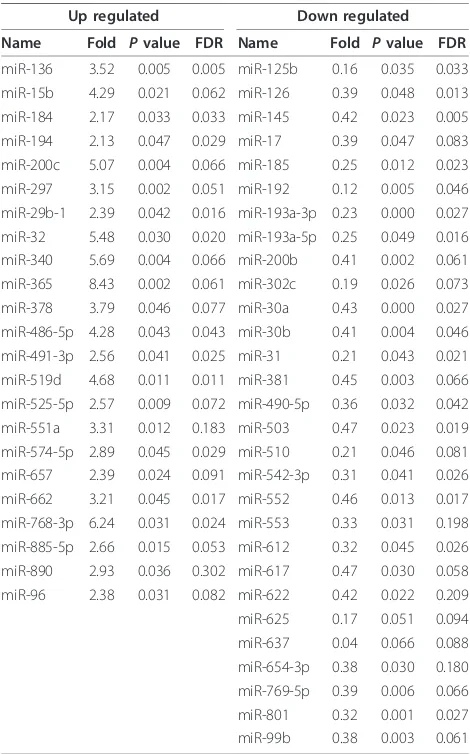
A total of 52 miRNAs were differentially expressed between GH-secreting pituitary adenomas and normal pituitaries. Twenty-three miRNAs were up-regulated and twenty-nine miRNAs were down-regulated in GH-secret-ing pituitary adenomas (Table 1). Although, the fold change of some up- and down-regulated miRNAs was >2.00 or <0.50, the miRNAs are not listed in Table 1
P= 0.12, FDR = 0.021; miR-222, up, 2.87,P= 0.23, FDR = 0.023; miR-516b, up, 2.48,P= 0.16, FDR = 0.065; miR-601, up, 2.67,P= 0.11, FDR = 0.094; miR-629, up, 2.71, P= 0.31, FDR = 0.212; miR-886-5p, up, 3.49,P = 0.09, FDR = 0.025; miR-766, 2.53,P= 0.18, FDR = 0.033;
miR-124, down, 0.29,P= 0.27, FDR = 0.005; miR-125a-5p,
down, 0.41,P= 0.16, FDR = 0.029; miR-15a, down, 0.32,
P= 0.21, FDR = 0.076; miR-198, down, 0.36, P= 0.25,
FDR = 0.005; miR-630, down, 0.39,P= 0.26, FDR = 0.095;
miR-744, down, 0.43, P= 0.24, FDR = 0.072; miR-765,
down, 0.37, P = 0.14, FDR = 0.173). Cluster analysis
showed a clear distinction between pituitary adenomas and normal pituitary (Figure 1). When adding the thresh-old value of FDR <0.10 to the differentially expressed cri-teria, eight miRNAs lost their status of differentially expressed (five in Table 1, two in Table 2 and one in Table 3).
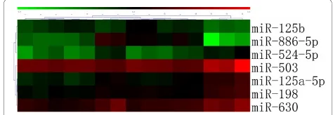
A total of 9 miRNAs were differentially expressed between macro- and micro-adenomas (Table 3). Cluster analysis based on these differentially expressed miRNAs showed that macro- and micro-adenomas belonged to two distinct groups (Figure 2). A total of 13 miRNAs were differentially expressed between the GH-secreting pituitary adenomas with lanreotide-treated patients and those without lanreotide-treatment (Table 2). We also found that 7 miRNAs were differentially expressed between SSA responders and SSA nonresponders (Table 4). Cluster analysis based on these differentially expressed miRNAs showed a clear distinction between SSA responders and SSA nonresponders (Figure 3).
To verify the results obtained by microarray analysis, we performed qRT-PCR for some of the differentially expressed miRNAs. qRT-PCR results for the expression of miR-124, miR-125a, miR-126, miR-223, miR-381, miR-503, miR-524-5p, miR-525-5p, and miR-886-5p were consistent with the results obtained from microar-ray analysis (Table 5). In supplementary experiments, we performed qRT-PCR for analyzing more miRNAs
Figure 1Cluster analysis shows a clear distinction between GH-secreting pituitary adenomas and normal pituitary. GH-secreting pituitary adenomas: 1-21, Normal pituitary: 22-27.
Table 1 miRNAs differentially expressed in GH-secreting pituitary adenomas vs. normal pituitaries
Up regulated Down regulated
Name Fold Pvalue FDR Name Fold Pvalue FDR
miR-136 3.52 0.005 0.005 miR-125b 0.16 0.035 0.033 miR-15b 4.29 0.021 0.062 miR-126 0.39 0.048 0.013 miR-184 2.17 0.033 0.033 miR-145 0.42 0.023 0.005 miR-194 2.13 0.047 0.029 miR-17 0.39 0.047 0.083 miR-200c 5.07 0.004 0.066 miR-185 0.25 0.012 0.023 miR-297 3.15 0.002 0.051 miR-192 0.12 0.005 0.046 miR-29b-1 2.39 0.042 0.016 miR-193a-3p 0.23 0.000 0.027 miR-32 5.48 0.030 0.020 miR-193a-5p 0.25 0.049 0.016 miR-340 5.69 0.004 0.066 miR-200b 0.41 0.002 0.061 miR-365 8.43 0.002 0.061 miR-302c 0.19 0.026 0.073 miR-378 3.79 0.046 0.077 miR-30a 0.43 0.000 0.027 miR-486-5p 4.28 0.043 0.043 miR-30b 0.41 0.004 0.046 miR-491-3p 2.56 0.041 0.025 miR-31 0.21 0.043 0.021 miR-519d 4.68 0.011 0.011 miR-381 0.45 0.003 0.066 miR-525-5p 2.57 0.009 0.072 miR-490-5p 0.36 0.032 0.042 miR-551a 3.31 0.012 0.183 miR-503 0.47 0.023 0.019 miR-574-5p 2.89 0.045 0.029 miR-510 0.21 0.046 0.081 miR-657 2.39 0.024 0.091 miR-542-3p 0.31 0.041 0.026 miR-662 3.21 0.045 0.017 miR-552 0.46 0.013 0.017 miR-768-3p 6.24 0.031 0.024 miR-553 0.33 0.031 0.198 miR-885-5p 2.66 0.015 0.053 miR-612 0.32 0.045 0.026 miR-890 2.93 0.036 0.302 miR-617 0.47 0.030 0.058 miR-96 2.38 0.031 0.082 miR-622 0.42 0.022 0.209 miR-625 0.17 0.051 0.094 miR-637 0.04 0.066 0.088 miR-654-3p 0.38 0.030 0.180 miR-769-5p 0.39 0.006 0.066 miR-801 0.32 0.001 0.027 miR-99b 0.38 0.003 0.061
Note: The threshold value used to screen up- and down-regulated miRNAs was fold change >2.00 or fold change <0.50.
Table 2 miRNAs differentially expressed with vs. without lanreotide treatment
Up regulated Down regulated
Name Fold Pvalue FDR Name Fold Pvalue FDR
miR-183 2.47 0.043 0.021 miR-124 0.25 0.024 0.005 miR-193a-5p 2.82 0.026 0.025 miR-32 0.41 0.031 0.082 miR-222 2.96 0.035 0.049 miR-574-5p 0.42 0.041 0.073 miR-516b 2.31 0.033 0.065 miR-744 0.31 0.025 0.158 miR-524-5p 2.49 0.021 0.011 miR-96 0.30 0.017 0.051 miR-601 2.58 0.030 0.094
miR-629 2.85 0.050 0.212 miR-99b 2.29 0.032 0.034
expression (Table 5). As described by Bottoni [11], pre-diction of miRNAs target genes can be analyzed using four algorithms: TargetScan, PicTar, miRanda and miR-Base Targets http://microrna.sanger.ac.uk/targets/. Using these algorithms, we performed prediction analysis for the target genes of the differentially expressed miRNAs identified in microarray (124, 125a-5p, 125b, 126, 145, 151-3p, 524-5p, miR-516b, miR-744 and miR-96). We also analyzed the target genes of qRT-PCR confirmed miRNAs (223, miR-381, miR-503, miR-525-5p and miR-886-5p).
Discussion
GH-secreting pituitary adenoma and normal pituitary Among the differentially expressed miRNAs between GH-secreting pituitary adenomas and normal pituitary samples, some are involved in cell growth, apoptosis, cell proliferation and tumor development, as reported by the previous studies [19-21]. Yu et al. [19] found that ectopic overexpression of miR-184 resulted in a marked increase in apoptosis and cell death. Meanwhile, Hino et al. [20] demonstrated that miR-194 was involved in the differentiation of intestinal epithelial cells. Ferretti et al. [21] found that down-regulation of miR-125b caused the proliferation of tumor cells. In our study, we found that miR-126 and miR-381 were down-regulated in GH-secreting pituitary adenomas compared to normal pitui-tary. Previous study has shown that miR-126 is located in the chromosome region of 9q34.3 and regulates phos-phatidylinositol 3-kinase (PI3K) signaling by targeting
the PI3K regulatory subunit beta (p85b). Guo et al. [22] reported that miR-126 modulated the activity of PI3K at the level of signal initiation by limiting p85b levels in the normal colon epithelium. Loss of miR-126 during tumorigenesis would eliminate this check point and facilitate the amplification of PI3K signal, which may provide a growth advantage during colon carcinogenesis. The target of miR-126 and miR-381 is PTTG protein 1, which is involved in multiple cellular pathways, includ-ing proliferation, DNA repair, transformation, angiogen-esis induction, invasion, and the induction of genetic instability. PTTG is overexpressed in most pituitary ade-nomas and is correlated to the recurrence and angiogen-esis [23]. Our results, therefore, indicated that altered expression of miR-126 genes may play an important role in the development of GH-secreting pituitary adenomas.
Tumor size
Among 9 differentially expressed miRNAs between micro- and macro- GH-secreting pituitary adenomas, the expression of miR-15a was down-regulated. There was no correlation between the reduced expression of miR-15a and tumor size (miR-15a, down-regulated, 0.32, P= 0.21, FDR = 0.076, AP value >0.05, the data wasn’t shown in the Table 1). This finding was supported by previous study using the samples of corticotropinoma [12]. Bottoni et al. [10] demonstrated an inverse correla-tion between the tumor diameter and the expression level of miR-15a and miR-16 in samples of GH- or PRL-secreting pituitary adenomas, which was different from Table 3 miRNAs differentially expressed in macro- vs.
micro- GH-secreting pituitary adenomas
Up regulated Down regulated
Name Fold Pvalue FDR Name Fold Pvalue FDR
miR-184 2.68 0.043 0.029 miR-124 0.43 0.031 0.005 miR-524-5p 2.85 0.021 0.027 miR-222 0.36 0.022 0.023 miR-629 2.49 0.015 0.061 miR-32 0.47 0.011 0.016 miR-766 2.17 0.031 0.033 miR-744 0.42 0.020 0.072 miR-765 0.41 0.042 0.173
Note: The threshold value used to screen up- and down-regulated miRNAs was fold change >2.00 or fold change <0.50.
Figure 2Cluster analysis shows a clear distinction between macro- and microadenomas in GH-secreting pituitary
adenomas. Macroadenomas:1-18, Microadenomas:19-21.
Table 4 miRNAs differentially expressed in treatment with GH responders vs. GH noresponders
Up regulated Down regulated
Name Fold Pvalue FDR Name Fold Pvalue FDR
miR-125b 2.73 0.034 0.013 miR-125a-5p 0.42 0.043 0.029 miR-886-5p 12.41 0.015 0.025 miR-198 0.46 0.012 0.005 miR-503 0.31 0.031 0.077 miR-524-5p 0.41 0.036 0.033 miR-630 0.42 0.022 0.095
Note: The threshold value used to screen up- and down-regulated miRNAs was fold change >2.00 or fold change <0.50.
our findings. In short, these findings suggested a role of reduced expression of miR-15a and miR-16 in the pathogenesis of pituitary tumors. Consistent with these results, enforced expression of the miR-222 can induce the thyroid papillary carcinoma cell line to progress to the S phase of the cell cycle, indicating that miR-222 negatively regulates p27Kip1 protein expression and cell cycle [24]. Amaral et al. [12] found that the expression of miR-21, miR-141 and miR-150 was reduced in corti-cotropinomas. Patients with lower expression of miR-141 had a higher chance of remission after transphenoi-dal surgery, suggesting a possible role of the miR-141 in the regulation of pituitary genes involved in tumor growth and tumor local invasion. However, in this study, we did not observe significant difference of miR-21 and miR-141 expression between micro- and macro-GH-secreting pituitary adenomas. Further studies are
needed to elucidate the pathogenesis of different subtype pituitary tumors including GH-secreting pituitary adenomas, PRL-secreting pituitary adenomas, and corticotropinoma.
Lanreotide treatment and SSA responders or nonresponders
Among the putative targets of miRNAs, miR-125a-5p, miR-125b and miR-524-5p are associated with IGFBP-3 and IGFALS chain precursor, which are involved in pro-tein binding, receptor binding, cell communication and regulation of growth. The role of IGFALS is associated with the regulation of the bioavailability of IGFs during postnatal growth. Up to 90% of circulating IGF-I and IGF-II are carried by binding to either IGFBP-3 or IGFBP-5. ALS, in the form of tertiary complexes, can extend their circulating half-life of IGF-I and IGF-II [25]. Moreover, miR-524-5p targets matrix metallopro-teinase-9, which is involved in metabolism, ion binding and extracellular matrix. However, miR-524-5p was down-regulated in SSA responders as compared to SSA nonresponders. In contrast, miR-524-5p was up-regu-lated in the lanreotide-treated patients as compared to the untreated patients. The possible reason of miR-524-5p up and down regulation in different groups is due to the different responsiveness to SSA treatment in various cases. The response to SSA depends on the presence of a sufficiently high number of somatostatin receptors (SSRs) (subtypes, sst1-5) on the tumor cells. GH secre-tion is regulated through ligand binding of somatostatin to both sst2 and sst5, whereas SSA binds preferentially sst2 [26]. The GH-lowering effect of SSA is positively correlated with the level of sst2 mRNA expression [27-29]. Thus, in GH-responder cases, the level of growth factor is high. Lanreotide is a type of inhibitory growth factor. It is possible that miR-524-5p is nega-tively correlated with growth factors, e.g., lanreotide pro-motes miR-524-5p up regulation and inhibits the growth factors. Another possible reason is that miR-524-5p is inhibited itself by growth factor. When the inhibition is removed (lanreotide treatment), the level of expression of miR-524-5p is up-regulated. The similar reason may explain the opposite phenomenon occurred with the miR-193a-5p, miR-574-5p, miR-96 and miR-99b in Table 2 and Table 5. However, further studies are needed to elucidate the function and mechanisms of altered expression of miR-524-5p.
Further analyses showed that miR-516b and miR-96 target IGFBP-7, miR-744 targets IGFBP-6, and miR-99b targets homeobox protein prophet of Pit-1. All these target genes are involved in the regulation of organ development, nucleic acid binding and membrane-bound organelles. Visvanathanet et al. [30] demonstrated that SCP1 (small C-terminal domain phosphatase Table 5 Some microarray data validation by qRT-PCR
Different miRNAs U6
Microarray Real time Microarray Real time
miR-124 0.54 0.39 1 1
miR-125a 1.26 1.95 1 1
miR-126 0.39 0.42 1 1
miR-223 2.78 1.42 1 1
miR-381 0.86 0.96 1 1
miR-503 0.22 0.37 1 1
miR-524-5P 0.28 0.34 1 1
miR-525-5p 5.14 4.05 1 1
miR-886-5P 3.71 1.51 1 1
miR-125b 2.85 1.92 1 1
miR-145 1.60 1.76 1 1
miR-151-3p 1.16 0.91 1 1
miR-183 1.20 1.59 1 1
miR-184 0.63 0.77 1 1
miR-193a-5p 0.62 0.73 1 1
miR-194 1.11 0.84 1 1
miR-198 0.46 0.49 1 1
miR-222 1.12 1.42 1 1
miR-30b 0.49 1.11 1 1
miR-32 1.64 1.03 1 1
miR-516b 0.64 0.87 1 1
miR-574-5p 1.10 1.15 1 1
miR-601 0.53 0.79 1 1
miR-629 0.45 0.31 1 1
miR-630 0.38 0.48 1 1
miR-744 2.03 1.74 1 1
miR-765 1.49 2.11 1 1
miR-766 0.57 0.47 1 1
miR-96 1.85 2.23 1 1
miR-99b 2.53 1.19 1 1
1) played an anti-neural role during CNS development. miR-124 can inhibit SCP1 expression by directly target-ing SCP1-3’ untranslated region (UTR). It is suggested that during CNS development, timely down-regulation of SCP1 is critical for induction of neurogenesis. Contri-bution of miR-124 to this process is at least partially through down-regulation of SCP1 expression. These results also implied that establishing a novel evolutiona-rily conserved strategy to keep the balance between miRNAs and their transcriptional regulatory programs is necessary.
miR-145 was down-regulated in GH-secreting pitui-tary adenomas as compared to normal pituipitui-tary. Amaral et al. [12] observed that miR-145 was down-regulated in 11 samples of corticotropinomas, suggesting a possible role of miR-145 in carcinogenesis. The potential target genes of miR-145 encode oncogenic proteins, such as myc, kras, fos, yes, fli, cyclin D2, and MAPK transduc-tion proteins [31]. miR-145 targets the insulin receptor substrate-1 (IRS-1) and miR-151-3p targets the insulin receptor substrate-4 (IRS-4), which regulated cell com-munication, receptor and membrane activities. Further-more, Shi et al. [32] demonstrated experimentally that miR145 targeted IRS-1 and had a profound biological effect on human colon cancer cells. IRS-1, a docking protein for both the type 1 insulin-like growth factor receptor (IGF-IR) and the insulin receptor, is known to transmit a mitogenic, anti-apoptotic, and anti-differen-tiation signal.
Some of the miRNAs in our study, such as miR-769-5p, miR-885-miR-769-5p, miR-886-5p and miR-890 are newly discovered in pituitary adenomas samples because the new array contains more capture probes and their func-tions are unknown [33]. The differentially expressed miRNAs are correlated with adenoma characteristics. In this study, the sample size is relatively low, and 4 months of treatment is relatively short. However, it must be emphasized that our studies represent promis-ing preliminary results. Therefore, further studies are needed to predict up-regulated or down-regulated miR-NAs target genes and their correlation with GH-secret-ing pituitary adenomas characteristics.
In conclusion, our results indicated that altered miR-NAs expression may be involved in GH-secreting pitui-tary adenomas transformation. Furthermore, some differentially expressed miRNAs are associated with tumor diameter, lanreotide treatment, and responsive-ness to SSA. These results will facilitate our understand-ing on mechanism of SSA treatment for acromegaly. Further studies are needed to predict the up-regulated
or down-regulated miRNAs’ targets and their partner
factors in pituitary adenomas. Studying the targets of deregulated miRNAs may elucidate molecular mechan-isms involved in pituitary adenoma pathogenesis.
Additional material
Additional file 1: Table S1 and S2. Supplemental tables
Additional file 2: Figure S1. Diagnosis of GH-secreting pituitary adenoma with H&E and immunohistochemical staining. Panel A indicated the microphotographs with H&E staining for growth hormone pituitary adenoma sections. A strong and diffuse acidophilic staining was shown in the cytoplasm of tumor cells. (original magnification, 200×). Panel B indicated microphotographs of immunohistochemically stained tumor sections, the tumor shows strong and diffuse staining for growth hormone antigen (original magnification, 400×).
Acknowledgements
This study was supported by National natural science foundation of China (No: 30971538), and Natural science foundation of Guangdong Province (No: 9151140004000001 and No: 10151008901000176).
Author details
1Department of Neurosurgery and Pituitary Tumor Center, The First Affiliated Hospital, Sun Yat-sen University, Guangzhou 510080, China.2Department of Histology and Embryology, Zhongshan School of Medicine, Sun Yat-sen University, Guangzhou 510080, China.3Department of Endocrinology and Pituitary Tumor Center, The First Affiliated Hospital, Sun Yat-sen University, Guangzhou 510080, China.
Authors’contributions
ZGM carried out the research studies, participated in the drafting of the manuscript. DSH and JZ carried out the immunohistochemical studies and detection of miRNAs expression and data analysis. BY participated in the design of the study. YHZ and HJW conceived of the study, helped draft the manuscript and participated in its design and coordination. All authors read and approved the final manuscript.
Competing interests
The authors declare that they have no competing interests.
Received: 7 September 2010 Accepted: 7 December 2010 Published: 7 December 2010
References
1. Nilsen TW:Mechanisms of microRNA-mediated gene regulation in animal cells.Trends Genet2007,23:243-249.
2. Stefani G, Slack FJ:Small non-coding RNAs in animal development.Nat Rev Mol Cell Biol2008,9:219-230.
3. Reinhart BJ, Slack FJ, Basson M, Pasquinelli AE, Bettinger JC, Rougvie AE, Horvitz HR, Ruvkun G:The 21-nucleotide let-7 RNA regulates developmental timing in caenorhabditis elegans.Nature2000,
403:901-906.
4. Pasquinelli AE, Reinhart BJ, Slack F, Martindale MQ, Kuroda MI, Maller B, Hayward DC, Ball EE, Degnan B, Muller P, Spring J, Srinivasan A, Fishman M, Finnerty J, Corbo J, Levine M, Leahy P, Davidson E, Ruvkun G:Conservation of the sequence and temporal expression of let-7 heterochronic regulatory RNA.Nature2000,408:86-89.
5. Esquela-Kerscher A, Slack FJ:Oncomirs–microRNAs with a role in cancer.
Nature Reviews Cancer2006,6:259-269.
6. Kloosterman WP, Plasterk RH:The diverse functions of microRNAs in animal development and disease.Dev Cell2006,11:441-450. 7. Johnson CD, Esquela-Kerscher A, Stefani G, Byrom M, Kelnar K,
Ovcharenko D, Wilson M, Wang XW, Shelton J, Shingara J, Chin L, Brown D, Slack FJ:The let-7 microRNA represses cell proliferation pathways in human cells.Cancer Res2007,67:7713-7722.
8. Gartel AL, Kandel ES:miRNAs: Little known mediators of oncogenesis.
Semin Cancer Biol2008,18:103-110.
10. Bottoni A, Piccin D, Tagliati F, Luchin A, Zatelli MC, degli Uberti EC:miR-15a and miR-16-1 downregulation in pituitary adenomas.J Cell Physiol2005,
204:280-285.
11. Bottoni A, Zatelli MC, Ferracin M, Tagliati F, Piccin D, Vignali C, Calin GA, Negrini M, Croce CM, Degli Uberti EC:Identification of differentially expressed microRNAs by microarray: a possible role for microrna genes in pituitary adenomas.J Cell Physiol2007,210:370-377.
12. Amaral FC, Torres N, Saggioro F, Neder L, Machado HR, Silva WA, Moreira AC, Castro M:MicroRNAs differentially expressed in ACTH-secreting pituitary tumors.J Clin Endocrinol Metab2009,94:320-323. 13. Melmed S:Medical progress: Acromegaly.N Engl J Med2006,
355:2558-2573.
14. Shimon I, Cohen ZR, Ram Z, Hadani M:Transsphenoidal surgery for acromegaly: endocrinological follow-up of 98 patients.Neurosurgery2001,
48:1239-1245.
15. Maiza JC, Vezzosi D, Matta M, Donadille F, Loubes-Lacroix F, Cournot M, Bennet A, Caron P:Long-term (up to 18 years) effects on GH/IGF-1 hypersecretion and tumour size of primary somatostatin analogue (SSTa) therapy in patients with GH-secreting pituitary adenoma responsive to SSTa.Clin Endocrinol2007,67:282-289.
16. Plöckinger U, Albrecht S, Mawrin C, Saeger W, Buchfelder M, Petersenn S, Schulz S:Selective loss of somatostatin receptor 2 in octreotide resistant growth hormone secreting adenomas.J Clin Endocrinol Metab2008,
93:1203-1210.
17. Pfaffl MW:A new mathematical model for relative quantification in real-time RT-PCR.Nucleic Acids Res2001,29:2002-2007.
18. Mao ZG, Zhu YH, Tang HL, Wang DY, Zhou J, He DS, Lan H, Luo BN, Wang HJ:Preoperative lanreotide treatment in acromegalic patients with macroadenomas increases short-term postoperative cure rates: a prospective, randomized trial.Eur J Endocrinol2010,162:661-666. 19. Yu J, Ryan DG, Getsios S, Oliveira-Fernandes M, Fatima A, Lavker RM:
MicroRNA-184 antagonizes microRNA-205 to maintain SHIP2 levels in epithelia.PNAS2008,105:19300-19305.
20. Hino K, Tsuchiya K, Fukao T, Kiga K, Okamoto R, Kanai T, Watanabe M:
Inducible expression of microRNA-194 is regulated by HNF-1a during intestinal epithelial cell differentiation.RNA2008,14:1433-1442. 21. Ferretti E, Smaele ED, Miele E, Laneve P, Po A, Pelloni M, Paganelli A,
Marcotullio LD, Caffarelli E, Screpanti I, Bozzoni I, Gulino A:Concerted microRNA control of hedgehog signalling in cerebellar neuronal progenitor and tumour cells.J EMBO2008,27:2616-2627. 22. Guo CG, Sah JF, Beard L, Willson JKV, Markowitz SD, Guda K:The
noncoding RNA, mir-126, suppresses the growth of neoplastic cells by targeting phosphatidylinositol 3-kinase signaling and is frequently lost in colon cancers.Gene Chromosome Canc2008,47:939-946.
23. Salehi F, Kovacs K, Scheithauer BW, Lloyd RV, Cusimano M:Pituitary tumor-transforming gene in endocrine and other neoplasms: a review and update.Endocrine-Related Cancer2008,15:721-743.
24. Visone R, Russo L, Pallante P, Martino ID, Ferraro A, Leone V, Borbone E, Petrocca F, Alder H, Croce CM, Fusco A:MicroRNAs (miR)-221 and miR-222, both overexpressed in human thyroid papillary carcinomas, regulate p27Kip1 protein levels and cell cycle.Endocrine-Related Cancer 2007,14:791-798.
25. Heath KE, Argente J, Barrios V, Pozo J, Díaz-González F, Martos-Moreno GA, Caimari M, Gracia R, Campos-Barros Á:Primary acid-labile subunit deficiency due to recessiveIGFALSmutations results in postnatal growth deficit associated with low circulating insulin growth factor (IGF)-I, IGF binding protein-3 levels, and hyperinsulinemia.J Clin Endocrinol Metab 2008,93:1616-1624.
26. Moller LN, Stidsen CE, Hartmann B, Holst JJ:Somatostatin receptors.
Biochim Biophys Acta2003,1616:1-84.
27. Saveanu A, Gunz G, Dufour H, Caron P, Fina F, Ouafik L, Culler MD, Moreau JP, Enjalbert A, Jaquet P:Bim-23244, a somatostatin receptor subtype 2- and 5-selective analog with enhanced efficacy in suppressing growth hormone (GH) from octreotide-resistant human GH-secreting adenomas.J Clin Endocrinol Metab2001,86:140-145.
28. Hofland LJ, van der Hoek J, van Koetsveld PM, de Herder WW, Waaijers M, Sprij-Mooij D, Bruns C, Weckbecker G, Feelders R, van der Lely AJ, Beckers A, Lamberts SW:The novel somatostatin analog SOM230 is a potent inhibitor of hormone release by growth hormone- and prolactin-secreting pituitary adenomas in vitro.J Clin Endocrinol Metab2004,
89:1577-1585.
29. Taboada GF, Luque RM, Bastos W, Guimaraes RF, Marcondes JB, Chimelli LM, Fontes R, Mata PJ, Filho PN, Carvalho DP, Kineman RD, Gadelha MR:Quantitative analysis of somatostatin receptor subtype (SSTR1-5) gene expression levels in somatotropinomas and non-functioning pituitary adenomas.Eur J Endocrinol2007,156:65-74. 30. Visvanathan J, Lee S, Lee B, Lee JW, Lee SK:The microRNA miR-124
antagonizes the anti-neural REST/SCP1 pathway during embryonic CNS development.Genes & Dev2007,21:744-749.
31. Iorio MV, Ferracin M, Liu CG, Veronese A, Spizzo R, Sabbioni S, Magri E, Pedriali M, Fabbri M, Campiglio M, Menard S, Palazzo JP, Rosenberg A, Musiani P, Volinia S, Nenci I, Calin GA, Querzoli P, Negrini M, Croce CM:
MicroRNA gene expression deregulation in human breast cancer.Cancer Res2005,65:7065-7070.
32. Shi B, Sepp-Lorenzino L, Prisco M, Linsley Peter , deAngelis T, Baserga R:
Micro RNA 145 targets the insulin receptor substrate-1 and inhibits the growth of colon cancer cells.J Biol Chem2007,282:32582-32590. 33. Landgraf P, Rusu M, Sheridan R, Sewer Al, Iovino N, Aravin A, Pfeffer S,
Rice A, Kamphorst AO, Landthaler M, Lin C, Socci ND, Hermida L, Fulci V, Chiaretti S, Foa’R, Schliwka J, Fuchs U, Novosel A, Müller RU, Schermer B, Bissels U, Inman J, Phan Q, Chien M, Weir DB, Choksi R, Vita GD, Frezzetti D, Trompeter HI, Hornung V, Teng G, Hartmann G, Palkovits M, Lauro RD, Wernet P, Macino G, Rogler CE, Nagle JW, Ju J, Papavasiliou NF, Benzing T, Lichter P, Tam W, Brownstein MJ, Bosio A, Borkhardt A, Russo JJ, Sander C, Zavolan M, Tuschl T:A mammalian microRNA expression atlas based on small RNA library sequencing.Cell2007,129:1401-1414.
doi:10.1186/1746-1596-5-79
Cite this article as:Maoet al.:Differential expression of microRNAs in GH-secreting pituitary adenomas.Diagnostic Pathology20105:79.
Submit your next manuscript to BioMed Central and take full advantage of:
• Convenient online submission
• Thorough peer review
• No space constraints or color figure charges
• Immediate publication on acceptance
• Inclusion in PubMed, CAS, Scopus and Google Scholar
• Research which is freely available for redistribution