R E S E A R C H
Open Access
Change in diaphragm and intercostal
muscle thickness in mechanically ventilated
patients: a prospective observational
ultrasonography study
Nobuto Nakanishi
1*, Jun Oto
1, Yoshitoyo Ueno
1, Emiko Nakataki
2, Taiga Itagaki
1and Masaji Nishimura
2Abstract
Background:Diaphragm atrophy is observed in mechanically ventilated patients. However, the atrophy is not
investigated in other respiratory muscles. Therefore, we conducted a two-center prospective observational study to evaluate changes in diaphragm and intercostal muscle thickness in mechanically ventilated patients.
Methods:Consecutive adult patients who were expected to be mechanically ventilated longer than 48 h in the ICU
were enrolled. Diaphragm and intercostal muscle thickness were measured on days 1, 3, 5, and 7 with ultrasonography. The primary outcome was the direction of change in muscle thickness, and the secondary outcomes were the relationship of changes in muscle thickness with patient characteristics.
Results:Eighty patients (54 males and 26 females; mean age, 68 ± 14 years) were enrolled. Diaphragm muscle thickness decreased, increased, and remained unchanged in 50 (63%), 15 (19%), and 15 (19%) patients, respectively. Intercostal muscle thickness decreased, increased, and remained unchanged in 48 (60%), 15 (19%), and 17 (21%) patients, respectively. Decreased diaphragm or intercostal muscle thickness was associated with prolonged
mechanical ventilation (median difference (MD), 3 days; 95% CI (confidence interval), 1–7 and MD, 3 days; 95% CI, 1– 7, respectively) and length of ICU stay (MD, 3 days; 95% CI, 1–7 and MD, 3 days; 95% CI, 1–7, respectively) compared with the unchanged group. After adjusting for sex, age, and APACHE II score, they were still associated with prolonged mechanical ventilation (hazard ratio (HR), 4.19; 95% CI, 2.14–7.93 and HR, 2.87; 95% CI, 1.53–5.21, respectively) and length of ICU stay (HR, 3.44; 95% CI, 1.77–6.45 and HR, 2.58; 95% CI, 1.39–4.63, respectively) compared with the unchanged group.
Conclusions:Decreased diaphragm and intercostal muscle thickness were frequently seen in patients under
mechanical ventilation. They were associated with prolonged mechanical ventilation and length of ICU stay.
Trial registration:UMIN000031316. Registered on 15 February 2018
Keywords:Diaphragm, Intercostal muscle, Atrophy, Ultrasonography
© The Author(s). 2019Open AccessThis article is distributed under the terms of the Creative Commons Attribution 4.0 International License (http://creativecommons.org/licenses/by/4.0/), which permits unrestricted use, distribution, and reproduction in any medium, provided you give appropriate credit to the original author(s) and the source, provide a link to the Creative Commons license, and indicate if changes were made. The Creative Commons Public Domain Dedication waiver (http://creativecommons.org/publicdomain/zero/1.0/) applies to the data made available in this article, unless otherwise stated. * Correspondence:nakanishi.nobuto@tokushima-u.ac.jp
1Emergency and Critical Care Medicine, Tokushima University Hospital, 2-50-1 Kuramoto, Tokushima 770-8503, Japan
Background
Critically ill patients experience atrophy of various mus-cles including respiratory musmus-cles, with diaphragm muscle atrophy garnering increasing attention. Patients undergoing prolonged mechanical ventilation experience sustained diaphragm muscle loss, which leads to worse clinical outcomes [1].
Mechanical ventilation can affect not only the dia-phragm but also the intercostal muscles, as demon-strated in animal studies [2]. Although diaphragm is the main respiratory muscle, several other respiratory mus-cles contribute to the respiratory effort as well. Specific-ally, when respiratory workload increases, other respiratory muscles are more active than the diaphragm [3]. Among these, intercostal muscles are one of the most important respiratory muscles in manipulating the movement of the rib cage [4].
Although diaphragm atrophy was reported to affect clinical outcomes in mechanical ventilation [1, 5], changes in other respiratory muscles during mechanical ventilation are not well investigated. Since diaphragm and intercostal muscles play an important role in critic-ally ill patients, it is reasonable to hypothesize that the thickness of both diaphragm and intercostal muscles could change over the mechanical ventilation. Therefore, we evaluated changes in diaphragm and intercostal muscle thickness and the potential relationship with pa-tient characteristics, medications, ventilator mode, dur-ation of mechanical ventildur-ation, and length of intensive care unit (ICU) stay in patients undergoing mechanical ventilation.
Methods Study design
This two-center prospective observational study was conducted in the mixed medical/surgical ICUs of Tokushima University Hospital and Tokushima Prefec-tural Central Hospital between June 2016 and June 2018 (Additional file1: Table S1). This study was approved by both clinical research ethics committees at Tokushima University Hospital (approval number 2593) and Tokushima Prefectural Central Hospital (approval num-ber 1739). This trial was registered on a clinical trial (UMIN-Clinical Trials Registry: 000031316). At the time of enrollment, written informed consent was obtained from patients or their authorized surrogate decision makers.
Study population
Consecutive adult patients who were expected to be mechanically ventilated longer than 48 h were enrolled in this study. Patients were recruited prospectively within 24 h following ICU admission. Patients who met the following criteria were excluded: age under 18 years,
trauma or chest tube at the measurement point, diagno-sis of primary neuromuscular disease.
Measurements of diaphragm and intercostal muscle thickness
Imaging was performed with B-mode ultrasound using liner transducers. Diaphragm and intercostal muscle thickness were evaluated with serial ultrasound measure-ments on days 1, 3, 5, and 7. Recordings were discontin-ued at extubation, discharge from the ICU, or death of the patient, whichever occurs first.
All measurements were performed with patients in su-pine anatomical position, reclining in bed at a 30-degree angle. The transducer was perpendicularly placed on the right chest wall at the zones of apposition: between the eighth and tenth intercostal spaces, between the antero-axillary and the midantero-axillary lines, and 0.5–2 cm below the costophrenic sinus, as previously reported (Add-itional file 1: Figure S1) [6]. A mark was drawn on the patient’s skin to ensure consistency. Diaphragm and intercostal muscle thickness were measured three times at end expiration, and the mean value was recorded for evaluation. In this area, the diaphragm is observed as a three-layered structure, with the hypoechogenic muscu-lar layer bordered by echogenic layers—the peritoneum and the diaphragmatic pleurae (Additional file 1: Figure S2). Thickening fraction of the diaphragm was calculated as [thickness at inspiration − thickness at expiration]/ [thickness at expiration] × 100 [7]. As intercostal mus-cles comprise internal and external intercostal muscle fi-bers that are difficult to discriminate by ultrasonography, intercostal muscles including the in-ternal and exin-ternal intercostal muscles were measured at the zone of apposition. In order to blind the data ana-lysis from the patient status, the image was stored in ultrasound machine at the bedside and then, the same investigator measured the muscle thickness which was blinded from patients’name and measurement days. Be-fore commencing the study, intra- and inter-observer re-producibility were 0.92 and 0.96 for the diaphragm and 0.92 and 0.90 for the intercostal muscles, as assessed by two ICU physicians (Additional file1: Figures S3-S6).
Definitions
10% decrease, and the remaining patients were catego-rized into the unchanged group.
Outcomes
The primary outcome was direction and rate of changes in the diaphragm and intercostal muscle thickness. The thickness change was defined as percent variation in muscle thickness compared with the admission day. The secondary outcomes were the relationship of changes in muscle thickness with patient characteristics; ventilator mode; duration of mechanical ventilation; length of ICU and hospital stay; reintubation and tracheostomy; the use of high-flow nasal cannula (HFNC) and noninvasive positive pressure ventilation (NPPV) after extubation; and medications including analgesics, sedatives, cate-cholamines, steroids, and muscle relaxants.
Statistical analysis
No articles reporting on intercostal muscle atrophy in the ICU were found by the literature search. Therefore, a feas-ible sample size of 80 patients was planned for enrollment based on two studies on diaphragm atrophy (the average of 54 and 107 patients in these studies) [5,8]. Continuous data were presented as means ± standard deviation or medians (interquartile range (IQR)), whereas categorical data were expressed as numbers (%). Changes in muscle thickness over time were assessed using a linear mixed model for re-peated measures: statistical significance of changes in muscle thickness at each time point was also tested using 95% confidence intervals (CIs), with intervals not including zero considered as statistically significant. Kappa statistics was used to evaluate the relationship between changes in diaphragm and intercostal muscle thickness. Variables were compared using one-way analysis of variance or the Kruskal-Wallis test. Post hoc correction for multiple com-parisons was performed with Dunnett’s or Steel’s test to compare decreased or increased thickness group versus un-changed group. Continuous outcomes were evaluated using Cox regression analysis, adjusted for age, sex, and APACHE II score. We presented cumulative incidence of liberation from mechanical ventilation, in which death was treated as a competing risk. Data were compared using Gray’s test with Bonferroni correction for two pairwise comparisons (significant at p < 0.025 vs. unchanged group). Data ana-lyses were conducted using JMP 13.1.0 and SAS 9.4 (SAS Institute, Cary, NC). All statistical tests were two-tailed and apvalue < 0.05 was regarded as statistically significant.
Results
Patient characteristics
The patient characteristics are shown in Table1. Among a total of 84 patients, 80, 49, and 32 patients remained in the study on days 3, 5, and 7, respectively. Four pa-tients were excluded due to only one measurement,
leaving 80 patients in analysis. The mean age was 68 ± 14 years, and 54 patients were male. The median Acute Physi-ology and Chronic Health Evaluation II score was 24 (IQR, 19–30). The causes for admission were respiratory failure (28%), post-cardiac surgery (19%), and heart failure (10%).
Main outcomes
The diaphragm muscle thickness decreased, increased, and remained unchanged in 50 (63%), 15 (19%), and 15 (19%) patients, respectively. In the decreased thickness group, it decreased by 11.3% (95% CI, 7.6–15.0%), 13.0% (95% CI, 8.7–17.4%), and 16.3% (95% CI, 10.7–21.9%) on days 3, 5, and 7 (p< 0.01; Fig.1). In the increased thick-ness group, it increased by 26.7% (95% CI, 19.1–34.2%), 10.6% (95% CI, 1.4–19.8%), and 29.4% (95% CI, 19.2– 39.7%), on days 3, 5, and 7 (p< 0.01). Finally, in the un-changed group, it un-changed by 0.5% (95% CI, − 1.7– 2.8%), − 2.0% (95% CI, − 7.7–3.7%), and −0.02% (95% CI,−6.9–6.9%) on days 3, 5, and 7 (p= 0.86).
Throughout the study period, the intercostal muscle thickness decreased, increased, and remained unchanged in 48 (60%), 15 (19%), and 17 (21%) patients, respect-ively. In the decreased thickness group, it decreased by 15.4% (95% CI, 11.5–19.2%), 20.9% (95% CI, 16.5– 25.4%), and 19.8% (95% CI, 14.6–25.0%) on days 3, 5, and 7 (p< 0.01). In the increased thickness group, it in-creased by 25.7% (95% CI, 15.8–35.6%), 28.4% (95% CI, 16.5–40.3%), and 28.6% (95% CI, 10.4–46.8%) on days 3, 5, and 7 (p< 0.01). Finally, in the unchanged group, it changed by 0.5% (95% CI, −2.2–1.2%), 2.9% (95% CI,−
Table 1Patient characteristics
Characteristics Overall (n= 80)
Age, mean ±SD, year 68 ± 14
Male/female 54/26
Body mass index, mean ± SD, kg/m2 24 ± 4
APACHE II score 24 (19–30)
SOFA, mean in the first 3 days 9 (5–12)
Sepsis (sepsis-3 criteria),n(%) 31 (39) ICU admission reasons,n(%)
Respiratory failure 22 (28)
Post-cardiac surgery 15 (19)
Heart failure 8 (10)
Sepsis, nonrespiratory 7 (9)
Stroke 7 (9)
Cardiac arrest 5 (6)
Traumas 2 (3)
Others 14 (18)
Data were expressed as median (IQR) unless otherwise indicated
0.5–6.4%), and− 1.3% (95% CI, −5.2–2.7%) on days 3, 5, and 7 (p= 0.26).
The relationship between the diaphragm and the intercostal muscle
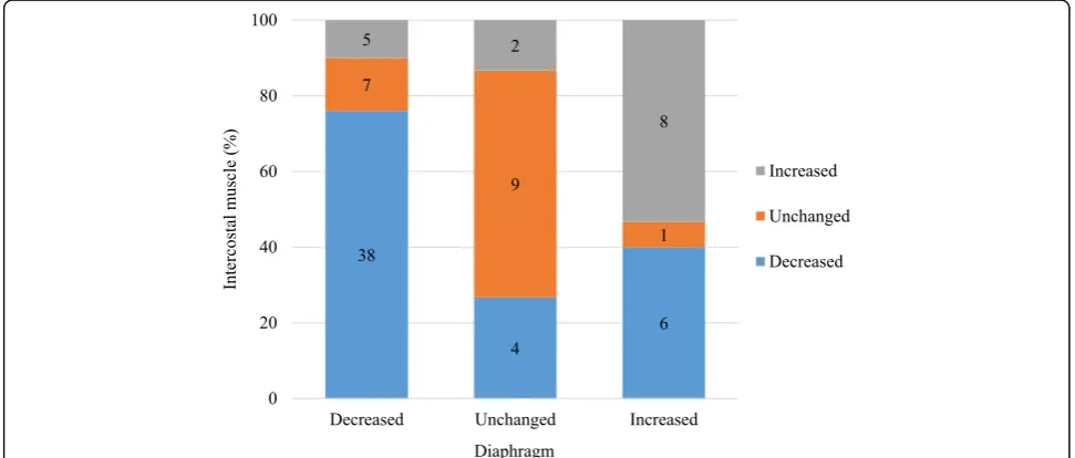
Out of fifty patients with the decreased diaphragm thick-ness, 38 patients (76%) exhibited decreased intercostal muscle thickness (Fig. 2). In the diaphragm unchanged
group, 9 patients (60%) had no change in intercostal muscle thickness, whereas 8 patients (53%) in the in-creased diaphragm thickness group also had inin-creased intercostal muscle thickness. In total, 55 patients (69%) exhibited muscle thickness changes in the same direc-tion for both the diaphragm and intercostal muscle (de-creased in 38 patients (48%), unchanged in 9 patients (11%), increased in 8 patients (10%)). The change in Fig. 1Time course of the diaphragm and intercostal muscle thickness. Time course for the measurement of the diaphragm and intercostal muscle thickness over the first 7 days of mechanical ventilation. The horizontal line represents the time from admission to the intensive care unit (ICU), and the vertical line represents the change in diaphragm and intercostal muscle thickness. Solid lines represent the changes in diaphragm muscle thickness, and dotted lines represent the changes in intercostal muscle thickness. Data are expressed as means and 95% confidence intervals
diaphragm thickness was associated with the change in intercostal muscle thickness, with the kappa value of 0.28 (95% CI, 0.14–0.41, p < 0.001), suggesting a poor association.
The relationship of changes in muscle thickness with ventilator mode
Over the first 3 days, 75 patients (94%) were in assist-control ventilation mode (ACV), and all patients were controlled by pressure-control ventilation (Table2). The thickening fraction of diaphragm was 7.1% (IQR, 4.3– 13.1%) in decreased thickness, 8.7% (IQR, 5.8–11.9%) in increased thickness, and 8.7% (IQR, 6.5–16.1%) in un-changed (not in Table2). There were no significant as-sociations between thickening fraction and changes in diaphragm thickness (p= 0.65). Set inspiratory pressure above positive end-expiratory pressure was higher in the decreased and increased intercostal muscle thickness groups than the unchanged group (p= 0.02), but the in-spiratory pressure was not significantly associated with the changes in diaphragm thickness (p= 0.28).
Patient outcomes
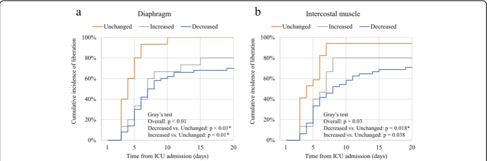
The duration of mechanical ventilation and the length of ICU stay were different among three groups (p < 0.01; Table3). The duration of mechanical ventilation was lon-ger in decreased diaphragm and intercostal muscle thick-ness group (median difference (MD), 3 days; 95% CI, 1–7 and MD, 3 days; 95% CI, 1–7, respectively). Moreover, it was longer in the increased diaphragm thickness group (MD, 2 days; 95% CI, 0–5,p= 0.04). Similarly, the length of ICU stay was longer in the decreased diaphragm and intercostal muscle thickness group (MD, 3 days; 95% CI, 1–7 and MD, 3 days; 95% CI, 1–7). In Cox regression ana-lysis, the duration of mechanical ventilation and the length of ICU stay were also associated with decreased dia-phragm and intercostal muscle thickness with the com-parison to unchanged (p< 0.01; Table 4). Moreover, the duration of mechanical ventilation was associated with in-creased diaphragm thickness with the comparison to un-changed (p = 0.03). Compared with unchanged, those with decreased or increased diaphragm thickness during the first week had a lower cumulative incidence of liber-ation from mechanical ventilliber-ation (p< 0.01 in both group; Fig.3). On the other hand, those with decreased intercos-tal muscle thickness had a lower cumulative incidence of liberation from mechanical ventilation (p= 0.018), and in-creased intercostal muscle thickness did not have the sig-nificant difference to unchanged (p = 0.038, significant level atp< 0.025 by Bonferroni correction).
Discussion
This prospective observational ultrasonography study in-vestigated the time course of change in diaphragm and
intercostal muscle thickness daily for up to 7 days in mechanically ventilated patients. The diaphragm muscle thickness can decrease, increase, or remain unchanged during mechanical ventilation. A similar change was ob-served in intercostal muscles, which are also essential re-spiratory muscles. Our study found that atrophy of diaphragm or intercostal muscles was associated with prolonged mechanical ventilation and length of ICU stay.
While the diaphragm is the main respiratory muscle, accounting for 60–80% of the inspiratory work [9], other respiratory muscles also play an important role in mov-ing the rib cage, especially in cases where the diaphragm function deteriorates or work associated with breathing increases [3]. Several studies detected rib cage muscle activity at high inspiratory efforts by electromyography [10, 11]. In one study, the activity of rib cage muscles exceeded that of the diaphragm during increased in-spiratory workload [9]. Rib cage muscles comprise inter-costal, sternocleidomastoid, scalene, pectoralis, serratus, oblique, transverse abdominis, and rectus abdominis muscles. Among these, the intercostal muscles are the most important respiratory muscles that significantly contribute to the movement of the rib cage [9]. In pa-tients with chronic obstructive pulmonary disease, inter-costal muscle atrophy occurs in advanced stages [12], and the decreased mass of intercostal muscles is associ-ated with exacerbation [13]. Our findings highlight the important role of intercostal muscles in critically ill pa-tients and suggest a possible association of their atrophy with difficult weaning from mechanical ventilation and prolonged ICU stay.
Several studies reported on rib cage muscle atrophy under mechanical ventilation. In one study, mechanical ventilation led to intercostal muscle atrophy as well as diaphragm muscle in animals [2]. Capdevila et al. sug-gested that 48 h of mechanical ventilation led to a 29% decrease in intercostal muscle mass in rabbits [2]. An-other study by Levine et al. investigating atrophy of the pectoralis major muscle and the diaphragm in mechanic-ally ventilated patients [14] found that the muscle fibers of the pectoralis major muscle did not decrease, al-though the cross-sectional area of the diaphragm muscle fibers was decreased. However, that study compared a limited number of 22 patients with different pathologies. In contrast, Nakanishi et al. reported that biceps brachii, which can also function to expand rib cage, showed sig-nificant progressive atrophy in critically ill patients [15]. Thus, rib cage muscles can undergo atrophy in critically ill patients.
potential explanations. First, majority of the patients in the current study were controlled by ACV, which was previously shown to be associated with excessive respira-tory support and neglected spontaneous breathing [16, 17]. Therefore, an average thickening fraction was 8% in the current study, which should be approximately 25% in individuals with normal breathings. Second, the mean patient age was higher in the current study compared with previous studies (68 vs. 59–60 years), which is asso-ciated with prominent muscle atrophy and decreased functional status [18]. As previously reported, we also found that decreased diaphragm thickness was
associated with the duration of mechanical ventilation and the length of ICU stay, and diaphragm thickness de-creased quickly in the early phase after mechanical ven-tilation [1,8].
The increased diaphragm thickness was observed in 19% of the patients and remained unchanged in another 19%, which were previously reported as 12–24% and 35–44%, respectively [1, 5]. Increased diaphragm thick-ness was associated with prolonged mechanical ventila-tion. Increased diaphragm thickness has been garnering increased interest based on a study by Goligher et al. who recently reported that increased diaphragm
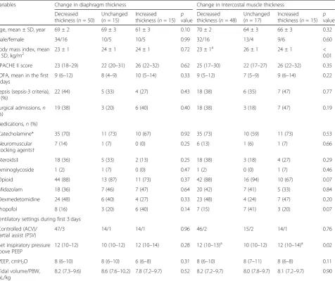
Table 2The relationship of changes in diaphragm and intercostal muscle thickness with patient characteristics, medications, and ventilator mode
Variables Change in diaphragm thickness Change in intercostal muscle thickness
Decreased thickness (n= 50)
Unchanged (n= 15)
Increased thickness (n= 15)
p
value
Decreased thickness (n= 48)
Unchanged (n= 17)
Increased thickness (n= 15)
p
value
Age, mean ± SD, year 69 ± 2 69 ± 3 61 ± 3 0.10 70 ± 2 64 ± 3 66 ± 3 0.32
Male/female 34/16 10/5 10/5 0.99 32/16 13/4 9/6 0.60
Body mass index, mean ± SD, kg/m2
23 ± 1 24 ± 1 24 ± 1 0.72 23 ± 1a 26 ± 1 24 ± 1 <
0.01
APACHE II score 23 (18–29) 22 (20–31) 26 (22–32) 0.62 25 (17–30) 22 (17–27) 26 (22–32) 0.35
SOFA, mean in the first 3 days
9 (6–12) 8 (4–9) 10 (5–14) 0.33 9 (5–12) 7 (5–9) 9 (6–14) 0.22
Sepsis (sepsis-3 criteria),
n(%)
22 (44) 5 (33) 4 (27) 0.43 18 (38) 6 (35) 7 (47) 0.77
Surgical admissions,n (%)
19 (38) 3 (20) 6 (40) 0.40 18 (38) 3 (18) 7 (47) 0.19
Medications,n(%)
Catecholamine* 35 (70) 11 (73) 10 (67) 0.92 35 (73) 10 (59) 11 (73) 0.53
Neuromuscular blocking agents†
7 (14) 1 (7) 0 (0) 0.25 6 (13) 1 (6) 1 (7) 0.66
Steroids‡ 18 (36) 5 (33) 2 (13) 0.25 18 (38) 3 (18) 4 (27) 0.29
Aminoglycoside 1 (2) 1 (7) 0 (0) 0.47 1 (2) 0 (0) 1 (7) 0.46
Opioid 44 (88) 13 (87) 11 (73) 0.37 42 (88) 16 (94) 10 (67) 0.07
Midazolam 18 (36) 7 (46) 7 (47) 0.64 20 (42) 7 (41) 5 (33) 0.84
Dexmedetomidine 24 (48) 6 (40) 4 (27) 0.33 23 (48) 4 (24) 7 (47) 0.20
Propofol 8 (16) 3 (20) 6 (40) 0.14 7 (15) 7 (41) 3 (20) 0.07
Ventilatory settings during first 3 days
Controlled (ACV)/ partial assist (PSV)
47/3 14/1 14/1 0.96 46/2 15/2 14/1 0.76
Set inspiratory pressure above PEEP
12 (10–12) 10 (10–12) 12 (10–14) 0.28 12 (10–13)a 10 (10–12) 12 (10–14)a 0.02
PEEP, cmH2O 8 (6–10) 8 (6–10) 6 (6–8) 0.31 8 (6–10) 8 (7–11) 8 (6–8) 0.11
Tidal volume/PBW, mL/kg
8.2 (7.3–9.6) 8.6 (7.6–10.2) 7.8 (7.2–9.7) 0.52 8.2 (7.2–9.7) 8.0 (7.8–9.7) 8.1 (7.2–9.7) 0.90
Data were expressed as median (IQR) unless otherwise indicated.pvalues were obtained using one-way analysis of variance (ANOVA) or the Kruskal-Wallis test
SDstandard deviation,APACHEAcute Physiology and Chronic Health Evaluation,SOFASequential Organ Failure Assessment,ACVassist-control ventilation,PSV pressure-support ventilation,PEEPpositive end-expiratory pressure,PBWpredicted body weight
a
Significant atp< 0.05 vs. Unchanged by post hoc Dunnett’s or Steel’s test *Catecholamine (dopamine, dobutamine, noradrenaline, or adrenaline) †Neuromuscular blockers with continuous use
thickness predicted prolonged mechanical ventilation (OR, 1.38; 95% CI, 1.00–1.90). The rate of increased dia-phragm thickness in the current study is comparable with those reported in previous studies [1, 5]. However, increased diaphragm thickness was not significantly as-sociated with excessive thickening fraction as previously reported [5]. The potential reason is the limited number of high thickening fraction, defined as more than 25% thickening (2.5% in our study population vs. 12.3% in Goligher’s study) [1]. Despite the relatively lower thick-ening fraction in the current study, increased thickness was observed in 15 patients, suggesting increased dia-phragm thickness is likely multifactorial. The clinical sig-nificance of increased diaphragm thickness is unclear in this research because of the relatively lower number of increased thickness in the study cohort (15 with in-creased thickness vs. 50 patients with atrophy). Further-more, most studies on diaphragm focuses on atrophy and not increased thickness [8, 17, 19, 20], reflected on
the paucity of data on its increased thickness [1,5]. In-creased diaphragm thickness needs further investigation.
The changes in the diaphragm and intercostal muscle thickness showed a poor association (κ= 0.27,p< 0.01). Although the association was poor, the atrophy of both the diaphragm and intercostal muscles was related to prolonged mechanical ventilation. This result shows both of the diaphragm and intercostal muscles are im-portant for critically ill patients, but different factors may influence the atrophy. The extent of disuse may be different because diaphragm and rib cage muscles con-tribute to respiration differently [9]. In spontaneous breathing patients, mechanical ventilation can support some part of diaphragm function and may disuse inter-costal muscles. On the other hand, interinter-costal muscles may exceed the diaphragm function in increased respira-tory workload [3]. Therefore, the relationship was lim-ited to a poor association between the diaphragm and the intercostal muscles.
Table 3Outcomes
Outcomes Change in diaphragm thickness Change in intercostal muscle thickness
Decreased thickness (n= 50)
Unchanged (n= 15)
Increased
thickness (n= 15) pvalue
Decreased thickness (n= 48)
Unchanged (n= 17)
Increased
thickness (n= 15) pvalue Duration of mechanical
ventilation, day
7 (5–15)a 4 (3–5) 7 (4–12)a < 0.01
8 (5–17)a 4 (3–6) 6 (5–8) < 0.01
Length of ICU stay, day 10 (6–16)a 5 (5–7) 8 (5–15) < 0.01
10 (6–18)a 5 (4–9) 7 (5–10) < 0.01
Length of hospital stay, day
34 (22–54) 32 (13–118) 27 (11–65) 0.71 34 (22–72) 29 (13–46) 34 (16–65) 0.37
Reintubation,n(%) 9 (18) 1 (7) 1 (7) 0.36 10 (21) 1 (6) 0 (0) 0.07
Tracheostomy,n(%) 12 (24) 1 (7) 2 (13) 0.27 12 (25) 1 (6) 2 (13) 0.19
The use of HFNC,n(%) 29 (58) 8 (53) 7 (47) 0.73 27 (56) 9 (53) 8 (53) 0.96
The use of NPPV,n(%) 3 (6) 0 (0) 0 (0) 0.39 2 (4) 1 (6) 0 (0) 0.66
Mortality in the ICU,n (%)
10 (20) 0 (0) 1 (7) 0.10 8 (17) 0 (0) 3 (20) 0.17
Mortality in the hospital,
n(%)
18 (36) 1 (7) 5 (33) 0.09 17 (35) 1 (6) 6 (40) 0.048
Data were expressed as median (IQR) unless otherwise indicated.pvalues were obtained using one-way analysis of variance (ANOVA) or the Kruskal-Wallis test
ICUintensive care unit,HFNChigh-flow nasal cannula,NPPVnoninvasive positive pressure ventilation a
Significant atp< 0.05 vs. unchanged by post hoc Dunnett’s or Steel’s test
Table 4Outcomes by multivariate analysis
Outcomes Change in diaphragm thickness Change in intercostal muscle thickness
Decreased thickness vs. unchanged
Increased thickness vs. unchanged
Decreased thickness vs. unchanged
Increased thickness vs. unchanged
Duration of mechanical ventilation, day
4.19 (2.14–7.93)* 2.38 (1.08–5.29)† 2.87 (1.53–5.21)* 1.71 (0.79–3.81)
Length of ICU stay, day 3.44 (1.77–6.45)* 1.99 (0.92–4.39) 2.58 (1.39–4.63)* 1.43 (0.66–3.16)
Length of hospital stay, day 1.34 (0.66–2.60) 0.89 (0.36–2.28) 2.04 (1.06–3.81)† 2.21 (0.94–5.61)
Data are expressed as hazard ratio (95% confidence interval) with intervals not including zero considered as statistically significant. Cox regression analysis was used for the analysis, adjusted for age, sex, and APACHE II score
ICUintensive care unit
Ventilator settings caused contradictory results in dia-phragm and intercostal muscle thickness. We did not find a significant difference in the inspiratory pressure between the patients with changed and preserved dia-phragm thickness. The limited range of set inspiratory pressure (IQR, 12–14 cmH2O) may explain our results,
since a previous study compared patients with a set in-spiratory pressure of≥12 cmH2O to those with a set
in-spiratory pressure of 5–12 cmH2O [20]. Conversely,
high inspiratory pressure was associated with both the decreased and increased thickness of intercostal muscles. Changes in thickness were easier to monitor for inter-costal muscles than the diaphragm because of the bigger muscle size (2.1 mm for diaphragm vs. 4.2 mm for inter-costal muscles). Therefore, the impact of pressure was likely detected in the intercostal muscles. As excessive inspiratory support causes atrophy of the disused muscle, excessive pressure might cause injury to respira-tory muscles and structural changes with gradual in-creased thickness. Since set inspiratory pressure does not reflect the actual pressure loaded to respiratory mus-cles, transpulmonary pressure should be measured to elucidate its precise influence. Similarly, the significance of the observed association between low body mass index (BMI) and intercostal muscle atrophy is unclear. It is possible that this result reflects the association of low BMI with malnutrition and increased respiratory work-load. Further elucidation of this aspect is limited due to the small study sample size that was not powered to de-tect the significant differences.
Changes in respiratory muscle thickness occur fre-quently in critically ill patients. As reported for the dia-phragm, intercostal muscles exhibit significant changes
in these patients, and the underlying causes are multifac-torial. The severity of the clinical status, medications, and ventilatory settings should be further investigated as potential factors. Compared with immobilized limb mus-cles, respiratory muscles are permanently activated by the ventilator [15]. Therefore, the duration and settings of mechanical ventilation may be the key factors to pre-serve respiratory muscles. Whether prolonged mechan-ical ventilation led to respiratory muscle atrophy or respiratory muscle atrophy induced prolonged mechan-ical ventilation remains to be elucidated. Weaning pa-tients from mechanical ventilation may be essential to prevent or minimize respiratory muscle changes. It is suggested that maintaining and improving respiratory muscles thickness is essential in order to do fast-track extubation in the ICU. Further studies are needed to de-termine approaches to maintain respiratory muscles. Taken together, the current findings emphasize that it is essential to monitor changes in diaphragm and intercos-tal muscles.
Limitations
depend on the specific intercostal muscle that was measured.
Conclusion
This two-center prospective observational study evaluat-ing changes in diaphragm and intercostal muscle thick-ness in mechanically ventilated patients measured with ultrasound revealed that decreased diaphragm and inter-costal muscle thickness were frequently seen in patients under mechanical ventilation. The decreased diaphragm and intercostal muscle thickness were associated with prolonged mechanical ventilation and length of ICU stay.
Supplementary information
Supplementary informationaccompanies this paper athttps://doi.org/10. 1186/s40560-019-0410-4.
Additional file 1: Table S1.Facility and equipment in this two-center prospective observational study.Figure S1.Anatomical structures at the zone of apposition.Figure S2.Ultrasound image of the diaphragm and intercostal muscles from the intercostal view.Figure S3.Intra-observer reproducibility of diaphragm.Figure S4.Inter-observer reproducibility of diaphragm.Figure S5.Intra-observer reproducibility of intercostal muscle. Figure S6.Inter-observer reproducibility of intercostal muscle.
Abbreviations
ACV:Assist-control ventilation; BMI: Body mass index; CI: Confidence interval; HFNC: High-flow nasal cannula; ICU: Intensive care unit; IQR: Interquartile range; NPPV: Noninvasive positive pressure ventilation; VS: Versus
Acknowledgements
The authors thank Yoshihiro Okayama of the Clinical Trial Center for Development Therapeutics of the Tokushima University Hospital for his statistical support.
Authors’contributions
NN was involved in study design, analysis, and interpretation of the data, and drafting of the manuscript. OJ took part in study design, analysis, and drafting of the manuscript. UY was involved in acquisition of the data. NE and IT did analyze and interpreted the data. NM took part in study concept, interpretation of the data, and critical revision of the manuscript. All authors read and approved the final manuscript.
Funding
This research did not receive any specific grant from funding agencies in the public, commercial, or not-for-profit sectors.
Availability of data and materials
The datasets used and/or analyzed during the current study are available from the corresponding author on reasonable request.
Ethics approval and consent to participate
Ethics approval was obtained from the clinical research ethics committee at Tokushima University Hospital (approval number 2593) and Tokushima Prefectural Central Hospital (approval number 1739). Informed consent to participate in the study was also obtained from patients or from an authorized surrogate.
Consent for publication Not applicable
Competing interests
The authors declare that they have no competing interests
Author details
1Emergency and Critical Care Medicine, Tokushima University Hospital, 2-50-1 Kuramoto, Tokushima 770-8503, Japan.2Tokushima Prefectural Central Hospital, 1-10-3 Kuramoto, Tokushima 770-8539, Japan.
Received: 10 June 2019 Accepted: 17 October 2019
References
1. Goligher EC, Dres M, Fan E, Rubenfeld GD, Scales DC, Herridge MS, Vorona S, Sklar MC, Rittayamai N, Lanys A, et al. Mechanical ventilation-induced diaphragm atrophy strongly impacts clinical outcomes. Am J Respir Crit Care Med. 2018;197(2):204–13.
2. Capdevila X, Lopez S, Bernard N, Rabischong E, Ramonatxo M, Martinazzo G, Prefaut C. Effects of controlled mechanical ventilation on respiratory muscle contractile properties in rabbits. Intensive Care Med. 2003;29(1):103–10. 3. Hershenson MB, Kikuchi Y, Tzelepis GE, McCool FD. Preferential fatigue of
the rib cage muscles during inspiratory resistive loaded ventilation. J Appl Physiol. 1989;66(2):750–4.
4. Ratnovsky A, Elad D, Halpern P. Mechanics of respiratory muscles. Respir Physiol Neurobiol. 2008;163(1-3):82–9.
5. Goligher EC, Fan E, Herridge MS, Murray A, Vorona S, Brace D, Rittayamai N, Lanys A, Tomlinson G, Singh JM, et al. Evolution of diaphragm thickness during mechanical ventilation. Impact of inspiratory effort. Am J Respir Crit Care Med. 2015;192(9):1080–8.
6. Bunnell A, Ney J, Gellhorn A, Hough CL. Quantitative neuromuscular ultrasound in intensive care unit-acquired weakness: a systematic review. Muscle Nerve. 2015;52(5):701–8.
7. Umbrello M, Formenti P, Longhi D, Galimberti A, Piva I, Pezzi A, Mistraletti G, Marini JJ, Iapichino G. Diaphragm ultrasound as indicator of respiratory effort in critically ill patients undergoing assisted mechanical ventilation: a pilot clinical study. Crit Care. 2015;19:161.
8. Schepens T, Verbrugghe W, Dams K, Corthouts B, Parizel PM, Jorens PG. The course of diaphragm atrophy in ventilated patients assessed with ultrasound: a longitudinal cohort study. Crit Care. 2015;19:422.
9. Ratnovsky A, Elad D. Anatomical model of the human trunk for analysis of respiratory muscles mechanics. Respir Physiol Neurobiol. 2005;148(3):245–62. 10. Costa D, Vitti M, de Oliveira TD, Costa RP. Participation of the
sternocleidomastoid muscle on deep inspiration in man. An electromyographic study. Electromyogr Clin Neurophysiol. 1994;34(5): 315–20.
11. Breslin EH, Garoutte BC, Kohlman-Carrieri V, Celli BR. Correlations between dyspnea, diaphragm and sternomastoid recruitment during inspiratory resistance breathing in normal subjects. Chest. 1990;98(2):298–302. 12. Ju S, Lee SJ, Park MJ, Cho YJ, Jeong YY, Jeon KN, Bae K, Lee JD, Kim HC.
Clinical importance of cross-sectional area of intercostal muscles in patients with chronic obstructive pulmonary disease. Clin Respir J. 2018;12(3):939–47. 13. Guerri R, Gayete A, Balcells E, Ramirez-Sarmiento A, Vollmer I,
Garcia-Aymerich J, Gea J, Orozco-Levi M. Mass of intercostal muscles associates with risk of multiple exacerbations in COPD. Respir Med. 2010;104(3):378–88. 14. Levine S, Nguyen T, Taylor N, Friscia ME, Budak MT, Rothenberg P, Zhu
J, Sachdeva R, Sonnad S, Kaiser LR, et al. Rapid disuse atrophy of diaphragm fibers in mechanically ventilated humans. N Engl J Med. 2008;358(13):1327–35.
15. Nakanishi N, Oto J, Tsutsumi R, Iuchi M, Onodera M, Nishimura M. Upper and lower limb muscle atrophy in critically ill patients: an observational ultrasonography study. Intensive Care Med. 2018;44(2):263–4. 16. Zambon M, Greco M, Bocchino S, Cabrini L, Beccaria PF, Zangrillo A.
Assessment of diaphragmatic dysfunction in the critically ill patient with ultrasound: a systematic review. Intensive Care Med. 2017;43(1):29–38. 17. Glau CL, Conlon TW, Himebauch AS, Yehya N, Weiss SL, Berg RA, Nishisaki A.
Progressive diaphragm atrophy in pediatric acute respiratory failure. Pediatr Crit Care Med. 2018;19(5):406–11.
18. Sacanella E, Perez-Castejon JM, Nicolas JM, Masanes F, Navarro M, Castro P, Lopez-Soto A. Functional status and quality of life 12 months after discharge from a medical ICU in healthy elderly patients: a prospective observational study. Crit Care. 2011;15(2):R105.
20. Zambon M, Beccaria P, Matsuno J, Gemma M, Frati E, Colombo S, Cabrini L, Landoni G, Zangrillo A. Mechanical ventilation and diaphragmatic atrophy in critically ill patients: an ultrasound study. Crit Care Med. 2016;44(7):1347– 52.
Publisher’s Note