Mycobacterium bovis
as a Significant Cause of Tuberculosis in
Children Residing Along the United States–Mexico Border
in the Baja California Region
Wayne M. Dankner, MD*, and Charles E. Davis, MD‡§
ABSTRACT. Objective. To determine the role of My-cobacterium bovisin active pediatric tuberculosis (TB) in a United States–Mexico border region.
Method. We reviewed all new cases of pediatric (<15 years old) TB presenting to San Diego hospitals and clinics from 1980 to 1997. Patients were categorized by age, ethnicity, country of origin, culture results, and dis-ease manifestations. Case definitions were similar to those used by the Centers for Disease Control and Pre-vention.M boviswas distinguished fromMycobacterium tuberculosisby standard biochemical tests.
Results. The median age of the 563 identified patients was 4.1 years old. The yearly incidence began rising in 1989 and peaked in the mid-1990s. Hispanics constituted 78.9% of the patients, but they were less likely to be foreign-born (21.6%) than were black children and Asian/ Pacific Islanders. Overall,M boviscaused 10.8% of all TB during this period. Of the 180 patients with positive culture results, however, M bovis accounted for 33.9% and M tuberculosis 66.1%. This high percentage of M bovisinfections was largely attributable to its contribu-tion to extrapulmonary TB (55.2% of all culture-positive specimens).M bovispatients were also even more likely to be Hispanic (90.2%), to present with extrapulmonary disease (95.1%), and to be older than 12 months (96.8%). Conclusion. These data demonstrate the dramatic im-pact of this underappreciated cause of zoonotic TB on US children at the Mexican border and underscore the need for cross-collaboration to enforce existing Mexican pas-teurization laws. Pediatrics 2000;105(6). URL: http:// www.pediatrics.org/cgi/content/full/105/6/e79; pediatric tuberculosis, Mycobacterium bovis, Mycobacterium tuber-culosis, zoonotic tubertuber-culosis, extrapulmonary tuberculo-sis, United States–Mexico border area.
ABBREVIATIONS. HIV, human immunodeficiency virus; TB, tu-berculosis; CDC, Centers for Disease Control and Prevention; PPD, purified protein derivative; INH, isoniazid; AFB, acid-fast bacillus.
B
eginning in the mid-1980s, a sharp increase in the incidence and prevalence of tuberculous disease in the United States reversed the steady decline of the last several decades. This increase, which persisted through the early- to mid-1990s, wasfueled by the human immunodeficiency virus (HIV) epidemic and by an increasing proportion of active disease in foreign-born individuals.1,2Although most
of the attention was directed toward the significant rise of disease in adults, there was an alarming in-crease in the pediatric-aged population as well.3,4
Much of the pediatric disease was identified in mi-nority populations, especially in children from large cities, mirroring the increasing epidemic in adults.3
Traditional teaching has stressed that children with tuberculous disease are sentinel events for active adult disease and that preadolescents usually ac-quire disease from close contact with a family mem-ber or other adult household contact.4Because
aero-sols are the route of transmission to these pediatric patients,Mycobacterium tuberculosisis usually the pri-mary organism considered to be responsible for in-fection and disease in this age group. As we reported previously, however,Mycobacterium bovis causes tu-berculous disease in both adults and children in the San Diego/Baja California region.5Because the
infec-tions in children likely represented recent acquisi-tion, we sought to determine the actual contribution of this mycobacterial species to the current preva-lence of pediatric tuberculous disease in this unique crossborder region.
METHODS
New cases of tuberculous disease in children⬍15 years of age for the years 1980 –1997 were identified through investigation of multiple sources. These included medical chart review of both inpatient and outpatient records using theInternational Classifica-tion of Diseases, Ninth Revisioncodes 010-018 inclusive and review of tuberculosis (TB) culture logs at the 2 primary pediatric medical facilities in San Diego (University of California, San Diego Medical Center and Children’s Hospital of San Diego). In addition, we reviewed yearly reports of pediatric TB cases reported to the Department of Health Services and the San Diego County Public Health Laboratory to identify unique cases seen at other facilities in San Diego. Patients were included regardless of whether they resided in San Diego County. The majority of patients who resided outside San Diego County came from neighboring counties (Im-perial County, Orange County, and Los Angeles County) and Tijuana, Mexico. The inclusion of patients from outside the San Diego County area precluded performing population-based inci-dence rates.
Case definitions were similar to those used by the Centers for Disease Control and Prevention (CDC)3except that we considered
children who presented with isolated hilar adenopathy (intratho-racic lymph nodes) on chest radiograph to have pulmonary TB instead of lymphatic TB.4Children were considered foreign-born
if their country of origin was outside of the 50 states, District of Columbia, Puerto Rico, or any of the US territories. For purposes of categorizing patients as to the source of their tuberculous in-fection, we counted patients with a well-documented history of
From the Departments of *Pediatrics, ‡Pathology, and §Medicine, Univer-sity of California, San Diego, San Diego, California.
Received for publication Oct 4, 1999; accepted Jan 5, 2000.
Reprint requests to (W.M.D.) University of California, San Diego Medical Center, 200 W Arbor Dr, 8468, San Diego, CA 92103. E-mail: wdankner@ ucsd.edu
contact to a source case (with an identifiedM tuberculosisisolate) to be in theM tuberculosiscategory and not to have disease caused byM bovis.
BecauseM bovisandM tuberculosisare both homologous to the nucleic acid probes used to identify species of theM tuberculosis complex, M boviswas distinguished fromM tuberculosisby stan-dard biochemical tests, including negative niacin and nitratase results; sensitivity to thiophene-2-carboxylic acid hydrazide; and in the first 14 years of surveillance, by a positive pyrazinamidase test result; and within the last 3 years, by pyrazinamide resistance in the Bactec system (Johnston Laboratories Inc, Towson, MD).6
All cultures were either performed or confirmed at University of California, San Diego Medical Center microbiology laboratory or the County Public Health Laboratory.M bovis–BCG strains were identified by phage typing or by high-performance liquid chro-matography7and patients infected with these isolates were
sub-sequently eliminated from the data analysis.
Statistical Analysis
Differences between demographic groups and clinical manifes-tations of TB caused byM bovisandM tuberculosiswere compared by the2test or Fisher’s exact test, where appropriate using Winks
statistical program (TexaSoft, Cedar Hill, TX). Statistical signifi-cance was set atP⬍.05.
RESULTS Demographic Information
During the 18 study-years, 563 children were di-agnosed with TB. The median age was 4.1 years old with a range of 1 month to 14.8 years of age (Fig 1). The number of children with tuberculous disease first began to rise in 1989 and peaked between 1991 and 1994 before beginning a gradual decline (Fig 2). The ethnic distribution revealed 444 (78.9%) His-panic children, 56 (9.9%) Asian/Pacific Islanders, 41 (7.3%) black non-Hispanics, 15 (2.7%) whites, and 7 (1.2%) Native Americans. Although there were no age differences among these ethnic groups, black children and Asian/Pacific Islanders were more likely to be foreign-born (63.4% and 78.2%, respec-tively) than children of Hispanic origin (21.6%;P ⬍
.001; Fig 3). The vast majority of the foreign-born Hispanic children (95.8%) were from Mexico with the remainder coming from Central American coun-tries. Only 7 children had conditions that could have predisposed them to develop tuberculous disease, including 4 with malignancies (3 with leukemia and 1 with neuroblastoma), 1 older child with systemic lupus erythematous on steroid therapy, a child with trisomy 21, and a neonate who was diagnosed with congenital TB. However, 30 children had evidence of prior or recent tuberculous infection or disease.
Eigh-teen had a history of purified protein derivative (PPD) reactivity, 9 had a history of tuberculous dis-ease, and 3 were reportedly receiving isoniazid (INH) preventive therapy for reactive PPD tests at the time that they were diagnosed with active TB. Twenty-three children with active tuberculous dis-ease had close adult contacts that wereM tuberculosis
culture-positive and believed to be the source cases for these children.
Overall, 325 (57.7%) patients were diagnosed with pulmonary or pleural-based disease and 238 (42.3%) with extrapulmonary disease (Table 1). Of those with extrapulmonary disease, 201 had only extrapulmo-nary disease and 373 had evidence of concomitant pulmonary disease (abnormal chest radiographs). The most common extrapulmonary site was lym-phatic (excluding intrathoracic lymph node involve-ment as described in “Methods”), followed by the central nervous system and abdominal foci. Only 3 patients with lymph node involvement had disease outside of the cervical or submandibular regions (2 had disease in an axillary node and 1 had disease in an inguinal node). Excluding patients with M bovis
infection (which contributed significantly to ex-trapulmonary manifestations and also was heavily represented in children of Hispanic origin), Hispanic patients were still more likely to have extrapulmo-nary disease than the other ethnic groups (40.6% vs 19.5%;P⬍.001).
Only 74 (22.8%) of the 325 patients with primary pulmonary or pleural-based disease had a positive acid-fast bacillus (AFB) culture result or were an intimate contact of a culture-positive source case. The majority of the patients with pulmonary disease were diagnosed by clinical criteria, ie, positive PPD, abnormal chest radiograph on presentation, and de-cision to treat with antituberculous therapy by the primary or consulting physician. In contrast, 105 (44.1%) of the 238 patients with extrapulmonary dis-ease had positive AFB culture results. The diagnosis was established on most, 106 (79.7%), of the others by histopathologic identification of granulomas with or without a positive AFB smear, by abnormal CSF findings and radiologic findings consistent with cen-tral nervous system tuberculous, or by radiograph appearance consistent with extrapulmonary tubercu-lous disease in conjunction with a positive PPD.
Thus, within the entire study population, 179
tients (31.8%) were culture-positive forM tuberculosis
complex strains, including the 23 intimate contacts of
M tuberculosis culture-positive adults. Of these 179 patients, 115 (64.2%) were positive forM tuberculosis, 61 (34.1%) forM bovis, and 2 (1.1%) forM africanum. One other isolate was identified as M tuberculosis
complex by DNA probe but could not be speciated further because of contamination. Seven children had isolates identified asM bovis-BCG; and as stated in “Methods,” these patients were eliminated from the study population and data analysis.
All 61 M bovisstrains were sensitive to the first line antituberculous drugs, excluding
pyrazin-amide to whichM bovisis innately resistant. Of the
M tuberculosisstrains, full sensitivity profiles were available for 108, including 19 from the close adult contacts. Of the M tuberculosisstrains, 16.7% were resistant to 1 or more antituberculous drugs and the INH resistance rate was 8.3%. The INH-resis-tant strains included 5 (5.2%) resisINH-resis-tant to INH alone; 2 (2.1%) resistant to streptomycin and INH; 1 (1.0%) resistant to INH, streptomycin, and ethambutol; and only 1 (1.0%) resistant to both INH and rifampin. There were no differences in the sensitivity results from the strains isolated from the infected close adult contacts, compared Fig 2. Yearly distribution of tuberculous disease
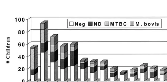
from 1980 through 1997. The Neg category includes patients with negative culture results. The ND cate-gory includes those who did not have cultures performed. TheM tuberculosiscomplex category in-cludes all patients with culture-provenM tuberculo-sis, the 2 patients infected withM africanum, the 1 patient whose isolate could not be speciated further than the stage of MTB complex, and the intimate contacts of documentedM tuberculosisadult source cases. The total number of infections is indicated by the height of the bars. The number of infections byM tuberculosis,M bovis, and those that were either cul-ture-negative or were not cultured for mycobacteria are depicted by the differences in shading, as indi-cated in the figure.
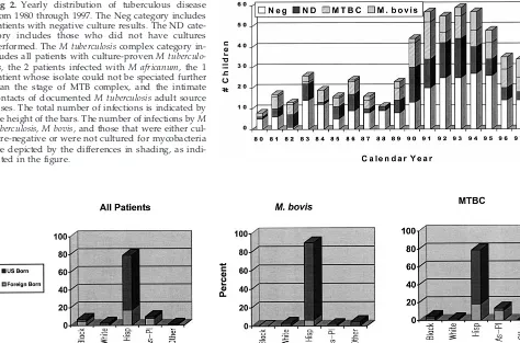
Fig 3. Ethnic distribution of children with tuberculous disease. The first panel (from left to right) includes all children. The second panel includes only those children with M bovis culture-proven disease. The third panel includes those children with culture-proven M tuberculosisdisease, the 2 patients infected withM africanum, the 1 patient whose isolate could not be speciated further than the stage of M tuberculosiscomplex, and the intimate contacts of documentedM tuberculosisadult source cases.
TABLE 1. Distribution of Tuberculous Disease Categories by Culture Isolation Results
Disease Manifestation
M tuberculosis* M bovis Culture Negative
Culture Not Done†
Total
Pulmonary 66 3 130 105 304
Pleurisy 6 0 15 0 21
Adenitis 13 31 67 17 128
Central nervous system 16 3 16 0 35
Abdominal 1 12 9 8 30
Bone/joint 4 5 11 2 22
Miliary 9 5 1 0 15
Other‡ 4 2 1 1 7
Total 119 61 250 133 563
* Also includes 2 patients withM africanum, 1 patient withM tuberculosiscomplex (not speciated), and 23 patients who were intimate contacts ofM tuberculosisculture–positive adults.
with the strains isolated directly from the infected children.
M bovis-Related Disease
As noted above, there were 61 children (25 of whom had been described previously)5with culture
provenM bovistuberculous disease identified during this 18-year period. The median age of these children (3.7 years old) was not statistically different from those who were not culture-positive for this myco-bacterial species. However, more M bovis-infected children were greater than 12 months of age at the time of their diagnosis than children with M tuber-culosis-proven disease (96.8% vs 76.1%;P⬍ .001). In addition, there were other statistically significant dif-ferences in the ethnic distribution and disease man-ifestations between those with and without docu-mentedM bovis-related disease (Fig 3). Children with
M bovis disease were more likely to be of Hispanic origin than were those without documentation of this bacterium (90.2% vs 77.5%;P⫽.022), but, some-what unexpectedly, less likely to be foreign-born (7.6% vs 20.7%; P ⫽ .006). Additionally, children withM bovisproven extrapulmonary disease seemed no less likely to have a positive chest radiograph than children with M tuberculosis-proven extrapul-monary disease (9.3% vs 18.7%;P⫽.10). This differ-ence becomes statistically significant if the children with miliary disease, who by definition invariably have abnormal chest radiographs, are eliminated from the comparison (2.0% vs 13.7%;P⫽ .019).
Children withM bovis-proven infection were also more likely to present with extrapulmonary disease than their counterparts withoutM bovisisolated from culture (95.1% vs 35.8%; P ⬍ .001), thus M bovis
proven disease accounted for 24.4% of all extrapul-monary manifestations seen over the 18-year period of surveillance. Furthermore, as demonstrated in Ta-ble 1, when only culture-proven cases are consid-ered, M bovis contributed even more significantly (55.2%) to the overall prevalence of extrapulmonary TB and individually for 92.3% of abdominal infec-tions, 72.1% of lymphadenitis, 55.6% of bone/joint infections, 35.7% of miliary infections, and 15.8% of central nervous system infections.
DISCUSSION
The alarming rise in tuberculous disease in the adult US population between the mid- to late-1980s and the early- to mid-1990s has now begun to de-cline.1,2A recent study from the CDC demonstrates a
similar trend in the pediatric population, lagging in time slightly behind the adult experience in both the increase and the subsequent decrease.3This
phenom-enon was predictable by virtue of the expected route of transmission from adults to children and the pre-dilection of young children to manifest TB as active disease.4The increasing influx of foreign-born
indi-viduals from TB endemic regions and the coepidemic of HIV infection with its risk for a higher rate of TB reactivation in the previously infected adult popula-tion have contributed to this resurgence.1–3 At first
glance, our data would seem to mirror that described in the CDC report, with an increase in pediatric TB
cases beginning in 1989, continuing without a defi-nite decline until 1995, and decreasing more signifi-cantly in the last 2 years. Additionally, foreign-born children contributed significantly in our series to the prevalence of TB in this area and our rate of 29.3% was similar to that of 19% to 23% reported by the CDC for the overall national experience.3Although
minorities also figured prominently in the preva-lence of tuberculous disease at the national level (80%– 86%) and in our own region (97.3%), there are significant differences between our series and the CDC report. Some of these differences could influ-ence the nature of TB control measures and resource allocation.
The first and most important difference that im-pacts both the prevalence and presentation of pedi-atric tuberculous disease in our region is the pre-dominance of children of Hispanic origin. They accounted for 78.9% of all the pediatric TB infections identified in this series, although Hispanics presently make up only⬃25% of the population in San Diego County.8Although we included children in our
se-ries that resided outside the county, the San Diego County statistics are a reasonable reflection of the ethnic distribution in the surrounding counties. Only 21.6% are foreign-born, with the vast majority com-ing from Mexico, but this figure may be misleadcom-ing in terms of how and where the US-born Hispanic children were exposed to TB. As pointed out in the CDC report, these young children may be exposed to recently immigrated parents from TB endemic re-gions, such as Mexico.3Exposure to extended
fami-lies, friends, and others with TB during frequent travel across the border is equally important. How-ever, our data also identify a unique epidemiologic situation associated with this crossborder phenome-non, namely exposure to a zoonotic source of TB. This is supported not only by the data presented in this report, but also by 2 recent studies performed by Besser et al9,10in San Diego County, which explored
the potential source cases for children with either tuberculous infection or tuberculous disease. In these studies, the only risk factor for TB in approximately one third of the children was ingestion of dairy prod-ucts, likely derived from raw, unpasteurized milk. Very few were exposed to adults with TB.9,10The role
of unpasteurized milk products is also strongly sug-gested in our series by the near absence of culture proven M bovis infections in the age group ⬍12 months of age, in stark contrast to the number of culture-proven infections with M tuberculosis. As shown in Fig 1, there were only 2 patients in this age group with cultures positive forM bovis, compared with 34 with M tuberculosis. This disproportionate ratio did not occur at any other age. These data strongly implicate unpasteurized milk products in the transmission ofM bovisbecause it correlates per-fectly with the practice of breastfeeding and the use of commercial infant formulas, which delay expo-sure to bovine dairy products until 6 to 12 months of age. Additionally, the uncommon isolation ofM bovis
cul-ture-proven pulmonary disease in adults in San Di-ego County (unpublished data) argues against respi-ratory transmission of this organism to the pediatric population.
Another important difference between the experi-ence in San Diego and that described for the remain-der of the United States is the rate of extrapulmonary disease in our population of children with TB.3,4
Pre-viously, we reported the unusually high number of children with abdominal TB in our region and de-scribed the manifestations and management of this form of TB.11In the present report, we analyzed all
forms of extrapulmonary disease to determine the contribution ofM bovisto the unusually high rate of extrapulmonary TB in children along the border of California with Baja. First, we found that the rate of extrapulmonary disease among non-Hispanic chil-dren was similar to that recently reported by the CDC, but significantly greater in Hispanic children, the group with the greatest risk for exposure to un-pasteurized dairy products from Mexico.3 We then
compared the isolation rates of M bovisto M tuber-culosis for each category of extrapulmonary disease and found that M boviswas contributing heavily to the burden of disease in our region. It is also possible that we underestimated the burden of disease caused byM bovisbecause its dysgonic growth characteris-tics and the absence of pyruvic acid supplements in many primary media may be disadvantageous in terms of primary isolation from culture material.12
Nevertheless,M bovisstill accounted for 55.2% of all culture-positive patients with extrapulmonary dis-ease. The strong predominance for gastrointestinal related organs without any associated pulmonary foci observed on chest radiograph, cervical lymph-adenopathy, and abdominal disease highlights the oral route of transmission for this pathogen. These figures are reminiscent of the experience with this species as a cause of childhood TB in the early 1900s. In our earlier review of the historical significance of this organism,M bovis was heavily associated with extrapulmonary disease in children ⬍14 years of age.5These similarities between the historical
docu-mentation of M bovis disease in previous endemic regions of the world, the virtual eradication of this pathogen from the milk supply in the United States for the last several decades, and the data reported here demonstrate the continued presence of this or-ganism in unpasteurized dairy products from the Baja region of northern Mexico. Its impact on the expression of tuberculous disease in the pediatric population of the San Diego region has been substan-tial.
Although we did not formally evaluate health care costs associated with M bovis-related disease, the predominance of extrapulmonary disease would be expected to have a major impact on the use of health
care resources, including hospitalizations, surgical services, treatment, and potential long-term morbid-ity. Although history has taught us that many factors must be considered in the elimination ofM bovisas a zoonotic disease in children,5,13public health
educa-tion efforts directed at high-risk populaeduca-tions and cross-border collaborative efforts to enhance the ad-herence to pasteurization laws already in effect in Mexico would be a major step toward the eradication of this pathogen from the pediatric population along the San Diego–Mexico border.
ACKNOWLEDGMENTS
We thank Dr Kathy Moser from Department of Health Services and Dr Chris Peter of the San Diego County Public Health Labo-ratory for their assistance with the identification of pediatric TB cases.
Additionally, we thank the laboratory staff in the microbiology laboratories at University of California, San Diego Medical Center, the Childrens Hospital of San Diego, and the Public Health labo-ratory for their skill in cultivation of mycobacterial strains from pediatric patients.
REFERENCES
1. McKenna MT, McCray E, Onorato I. The epidemiology of tuberculosis among foreign-born persons in the United States, 1986 to 1993.N Engl J Med. 1995;332:1071–1076
2. McCray E, Weinbaum CM, Braden CR, Onorato IM. The epidemiology of tuberculosis in the United States.Clin Chest Med. 1997;18:99 –113 3. Ussery XT, Valway SE, McKenna M, et al. Epidemiology of tuberculosis
among children in the United States: 1985 to 1994.Pediatr Infect Dis J. 1996;15:697–704
4. Starke JR, Smith MHD. Tuberculosis. In: Feigin RD, Cherry JD, eds.
Textbook of Pediatric Infectious Diseases. 4th ed. Philadelphia, PA: WB Saunders Company; 1998:1196 –1239
5. Dankner WM, Waecker NJ, Essey MA, et al.Mycobacterium bovis infec-tions in San Diego: a clinico-epidemiologic study of 73 patients and a historical review of a forgotten pathogen.Medicine. 1993;72:11–37 6. Nolte FS, Metchock B.Mycobacterium. In: Murray PR, Baron EJ, Pfaller
MA, Tenover FC, Yolken RH, eds.Manual of Clinical Microbiology. 6th ed. Washington DC: ASM Press; 1995:400 – 437
7. Floyd MM, Silcox VA, Jones WD, Butler WR, Kilburn JO. Separation of
Mycobacterium bovisBCG fromMycobacterium tuberculosisand Mycobac-terium bovisby using high-performance liquid chromatography of my-colic acids.J Clin Microbiol. 1992;30:1327–1330
8. San Diego Association of Governments.Population Estimates by Age, Sex and Ethnic Group. San Diego, CA: San Diego Association of Governments; 1995
9. Besser R, Pakiz B, Haverkamp D, et al. Source case investigations of children with tuberculosis disease in San Diego. In:Programs and Ab-stracts of the 38th Interscience Conference on Antimicrobial Agents and Chemotherapy. San Diego, CA: American Society for Microbiology; 1999; 586. Abstract L-127
10. Besser R, Pakiz B, Haverkamp D, et al. Source case investigations of children with tuberculosis infection in San Diego, CA. In:Programs and Abstracts of the 38th Interscience Conference on Antimicrobial Agents and Chemotherapy. San Diego, CA: American Society for Microbiology; 1999: 586. Abstract L-127a
11. Veeragandham RS, Lynch FP, Canty TG, Dankner WM. Abdominal tuberculosis in children: Review of 26 cases.J Pediatr Surg. 1996;31: 170 –176
12. Dixon JMS, Cuthbert EH. Isolation of tubercle bacilli from uncentri-fuged sputum on pyruvic acid medium.Am Rev Respir Dis. 1967;96: 119 –122
DOI: 10.1542/peds.105.6.e79
2000;105;e79
Pediatrics
Wayne M. Dankner and Charles E. Davis
Mexico Border in the Baja California Region
−
Along the United States
as a Significant Cause of Tuberculosis in Children Residing
Mycobacterium bovis
Services
Updated Information &
http://pediatrics.aappublications.org/content/105/6/e79
including high resolution figures, can be found at:
References
http://pediatrics.aappublications.org/content/105/6/e79#BIBL
This article cites 8 articles, 1 of which you can access for free at:
Subspecialty Collections
b
http://www.aappublications.org/cgi/collection/infectious_diseases_su
Infectious Disease
following collection(s):
This article, along with others on similar topics, appears in the
Permissions & Licensing
http://www.aappublications.org/site/misc/Permissions.xhtml
in its entirety can be found online at:
Information about reproducing this article in parts (figures, tables) or
Reprints
http://www.aappublications.org/site/misc/reprints.xhtml
DOI: 10.1542/peds.105.6.e79
2000;105;e79
Pediatrics
Wayne M. Dankner and Charles E. Davis
Mexico Border in the Baja California Region
−
Along the United States
as a Significant Cause of Tuberculosis in Children Residing
Mycobacterium bovis
http://pediatrics.aappublications.org/content/105/6/e79
located on the World Wide Web at:
The online version of this article, along with updated information and services, is
by the American Academy of Pediatrics. All rights reserved. Print ISSN: 1073-0397.