during kératinocyte terminal
differentiation and
Immortalisation
PhD Thes s
Kairbaan Jimmy Hodivala (BSc Hons)
April 1994
Internal supervisor Prof. Lewis Wolpert
University College London
All rights reserved
INFORMATION TO ALL USERS
The quality of this reproduction is dependent upon the quality of the copy submitted.
In the unlikely event that the author did not send a complete manuscript and there are missing pages, these will be noted. Also, if material had to be removed,
a note will indicate the deletion.
uest.
ProQuest 10046126
Published by ProQuest LLC(2016). Copyright of the Dissertation is held by the Author.
All rights reserved.
This work is protected against unauthorized copying under Title 17, United States Code. Microform Edition © ProQuest LLC.
ProQuest LLC
789 East Eisenhower Parkway P.O. Box 1346
Integrins are a family of cell-extracellular matrix adhesion molecuies that reguiate keratinocyte terminal differentiation. In stratified squamous epithelia integrins are normaliy expressed by the cells in the basai layer and are lost from the cell surface during terminal differentiation. In my thesis I have examined two situations in which this pattern of expression is perturbed: the presence of human papiiloma virus and in cultures of kératinocytes grown in low extracellular calcium concentrations.
I have compared two HPV-16 immortalised keratinocyte ceii iines up and vp with their normai parentai strains of normal kératinocytes from which they were derived u and v, and also with upr which was obtained by viral-Harvey-ras transfection of the up ceil lines. The HPV-16 immortaiised ceil lines displayed reduced levels of integrin protein and mRNA and changes in adhesion and motlity. The upr cell line was not tumorigenic in nude mice and had iittle effect on integrin ievels or celi motiiity when compared to the up; the oniy difference between the up and upr was that upr showed slight increased adhesion to fibronectin. Eight cervical biopsies with various grades of cervicai intraepithélial neoplasia showed evidence of HPV infection. Integrin leveis were either reduced or discontinuous in the most severe lesions and integrins were expressed in suprabasai layers in the less severe lesions. Thus, the impaired differentiation that is associated with the presence of HPV genes in vivo and in cuiture is correlated with abnormal integrin expression.
LIST OF DIAGRAMS, FIGURES AND TABLES... 1
CHAPTER 1
... 5
GENERAL INTRODUCTION... 5
1 ) S tru c tu re o f th e s k in ...5
1a) Subcutaneous fa t...6
1 b) Dermis...6
1c) Basement membrane... 7
1 d) Hair, sweat glands and sebaceous glands...8
1e) Epidermis...8
2 ) Markers fo r keratinocyte term inai d iffe re n tia tio n ... 1 1 2a) Keratins... 11
2b) Protein precursors of the comified envelope...11
2c) Filaggrin... 12
2d) Lipids... 12
2 e ) Peanut ie c tin -b in d in g g iy c o p ro te in s ...1 3 3) p ro iife ra tive disord ers o f the e p id e rm is...1 3 4 ) in v itro keratinocyte c u itu re ...1 4 5 ) I n te g r in s ...1 5 5a) The integrin family, structure and function...15
5a(i) p-subunits... 17
5a(ii) a-subunit... 18
Sa(iii) Integrins and signalling...19
5b) Keratinocyte integrins... 20
5c) Aberrant integrin expression in abnormal epidermis... 22
6) C a d h e r in s ...2 4 6a) The cadherin family, structure and function... 24
6b) Keratinocyte cadherins... 27
6c) Cadherin expression and cancer... 29
C H A P T E R 2 ...
31
MATERIALS AND METHODS...31
1 ) TISSUE CULTURE MEDIA AND SOLUTIONS...31
1a) Phosphate-buffered saline (PBS)... 31
1b) Versene... 31
1c) Trypsin...31
1 d) Standard FAD medium (FAD+FCS+HICE)... 32
1 e) Low Ca2+ FAD medium (-Ca2+ FAD+FCS+HICE)... 32
1 f) Cheiex treatment of FCS to remove free calcium and magnesium ions... 32
1 g) DMEM (E4 medium)...33
1 h) Mitomycin C... 33
2 ) C E L L C U L T U R E ... 3 3 2a) 3T3 feeder ceii layer routine cuiture... 33
2b) Primary keratinocyte isolation...33
2c) Keratinocyte subculture... 34
2c) (i) Standard calcium culture... 34
2c) (ii) Low calcium cultures... 34
2c) (ill) Culturing up, upr, and vp ceil lines...34
2d) SP2 culture... 35
2e) Freezing down celi stocks...35
2f) Thawing celi stocks... 35
3 ) IMMUNOFLUORESCENCE M ETHODS...3 6 3a) Dispase treatment... 36
3b) Preparation of tissue for cryosectioning...36
3c) Immunofluorescence staining protocol for surface integrins...36
3d) Double Immunofluorescence staining... 37
3d) (i) Tissue sections...37
3d) (ii) Whole ceiis... 37
3g) Filamentous Actin staining... 38
3h) Tubulin staining...38
3h) Geivatol recipe... 39
4 ) FLOW CYTOMETRY... 3 9 5 ) TIME-LAPSE CINEMICROSCOPY... 4 0 6) E L IS A ...4 0 7 ) GEL ELECTROPHORESIS... 41
7a) Polyacrylamide gel electrophoresis (PAGE) of proteins...41
7b) Coomassie staining...42
8) RADIOACTIVE LABELLING IMMUNOPRECIPITATION AND WESTERN BLOTTING...4 2 8a) Surface iodination... 42
8a) (i) NP40 lysis buffer recipe... 43
8b) Metabolic labelling...43
8b) (i) Overnight labelling... 43
8b (ii)Pulse labelling... 43
8c) TCA (Tri-chloro acetic acid) precipitation... 44
8d) Immunoprécipitation... 44
8e) Western blotting...45
9) ADHESION ASSAYS AND ADHESION BLOCKING A S S A Y S ...4 6 9a) Adhesion assays...46
9b) Adhesion blocking assays...48
1 0 ) TERMINAL DIFFERENTIATION A S S A Y ... 4 9 1 Da) Preparation of methyl cellulose... 49
10b) Preparation of PolyHEMA...49
1 Go) Induction of keratinocyte differentiation by suspension in methyl cellulose...49
1 1 ) RNA ISOLATION AND NORTHERN BLOTTING... 5 0 11a) Probes used for Northern blotting...50
11a) (ii) Probe preparation...51
11 b) RNA extraction (see Recipe sheet 2 for solution recipes)...51
11c) Northern blotting (see Recipe Sheet 2 for solution recipes)... 51
l i d ) Prehybridisation (see Recipe Sheet 2 for solution details)... 52
l i e ) Hybridisation... 52
I l f ) Washing the blot...52
11 g) Stripping the blot...53
1 2 ) IN SITU HYBRIDISATION... 5 3 12a) Fixation...53
12b) Probes for in situ hybridisation... 53
12c) Prehybridisation treatment (see Recipe sheet 2) ...54
12d) Hybridisation... 55
12e) Post hybridisation washes... 56
12f) Autoradiography... 56
12g) Development... 57
13) MONOCLONAL ANTIBODY PRODUCTION...5 8 13a Immunization... 58
13a) (i) Preparation of cells for immunization...58
13a) (ii) Primary immunization with basal kératinocytes... 59
13a) (iii) Boost immunizations... 59
13a) (iv) Test bleed... 59
13a) (v)F inal boost immunization... 60
13b) Fusion...60
13c) Screen 1... 61
13d) Single cell cloning, expanding and freezing clones... 62
13d) (i) Expanding clones (96 well plateÆ 24 well plate)... 62
13d) (ii) Single cell cloning...62
13d) (iii) Expanding clones 24 well platesÆ 6 well plates... 62
13d) (iv) Freezing and thawing clones... 63
13d) (v) Expanding clones (6-well plate to 10cm diameter petri ... dish and further)...63
13e) Screen 2 ...63
13f) Purification of antibodies... 64
13f) (i) Antibody (IgGI) purification on a Protein G column... 64
13f) (ii) Antibody (IgM) purification on a Con A-Sepharose column... 65
13f) (iii) Quantitating the protein content of the eluted fractions... 65
13g) Screen 3 ...6 6 13h) Immunoprécipitation and Western blot analysis of antigens recognised by monoclonal antibodies...6 6 14) PRODUCTION OF POLYCLONAL ANTIBODIES... 6 6 14a) Purifying immunoglobulins from rabbit anti-sera... 69
15) MICROINJECTIONS...6 9 16) ANTIBODY AND LECTIN LIST...71
16a) Anti-integrin antibodies... 71
16b) Anti-involucrin antibodies... 72
16c) Anti-cadherin antibodies... 72
16d) Tubulin...72
16e) Filamentous Actin... 72
16f) PNA-FITC...73
16g) Secondary antibodies... 73
16h) Negative control antibodies... 73
16i) Immunoprécipitation bridging antibodies...73
17) MICROSCOPES...7 3
RECIPE SHEET 1...
7 4
SDS-PAGE for Proteins... 7 4RECIPE SHEET 2... 75
CHAPTER 3 ... 78
ATTEMPT TO PRODUCE FUNCTION BLOCKING ANTI-INTEGRIN MONOCLONAL ANTIBODIES... 7 8 1) INTRODUCTION... 7 8 2 ) RESULTS... 7 8 2a) Fusions... 78
2b) immunofluorescence assay...79
2c) Adhesion blocking assay...79
2d) Differentiation blocking assay...80
2e) Immunoprécipitation and Westem blotting... 80
3 ) D ISC USS IO N ... 86
CHAPTER 4 ... 88
INTEGRIN EXPRESSION AND FUNCTION IN HUMAN KERATINOCYTES TRANSFORMED WITH HPV16, WITH OR WITHOUT v-Ha-ras. COMPARISON WITH CERVICAL INTRAEPITHELIAL NEOPLASIA... 8 8 1) INTRODUCTION 8 8 2 ) RESULTS...8 9 2a) Integrin expression...89
2b) Cell adhesion... 91
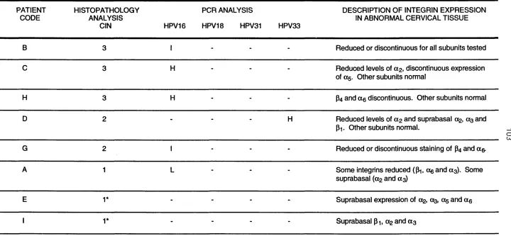
2c) Integrin expression in cervical intraepithélial neoplasia (GIN)... 92
3 ) D ISC USS IO N ...103
CHAPTER 5 ... 107
EVIDENCE THAT CADHERINS PLAY A ROLE IN THE DOWNREGULATION OF INTEGRIN EXPRESSION THAT OCCURS DURING KERATINOCYTE TERMINAL DIFFERENTIATION... 1 0 7 1) INTRODUCTION... 1 0 7 2 ) RESULTS... 10 9 2a) Selective loss of integrins from differentiating cells during calcium induced stratification... 109
2b) Integrin synthesis in low calcium and standard medium... 110
2c) Integrin synthesis by terminally differentiating kératinocytes in low calcium medium...1 1 2 2d) Role of cadherins in calcium-induced stratification...113
INVESTIGATING THE NATURE OF INTEGRIN-CADHERIN
•CROSS-TALK'...
125
1) INTRODUCTION... 1 2 5 2 ) R ESULTS... 1 2 62a) The role of protein synthesis in the ioss of integrins during calcium
induced stratification... 126 2b) Involvement of the actin cytoskeieton in the ioss of integrin from terminaiiy
differentiating kératinocytes during caicium induced stratification...127 2c) The effect of cytochaiasin D and coichicine on keratinocyte migration
and aggregation... 128 2d) Microinjections... 129 3 ) D ISC USS IO N ...1 3 9
C H A P T E R 7 ... 143
GENERAL DISCUSSION...1 4 31 ) iNTEGRiNS AND HPV... 143 2) iNTEGRIN-CADHERiN iNTERACTiONS...144 3) CONCLUSIONS...146
ACKNOWLEDGEMENTS...1 47
T
a b l e s
Chapter 1
Diagram 1
Diagram 2
Diagram 3
Diagram 4
Diagrammatic reprsentation of the structures identifiable in the skin
Diagrammatic representation of a cross-section through human epidermis
A schematic representation of an integrin complex
Schematic representation of the classical cadherin complex
Chapter 2
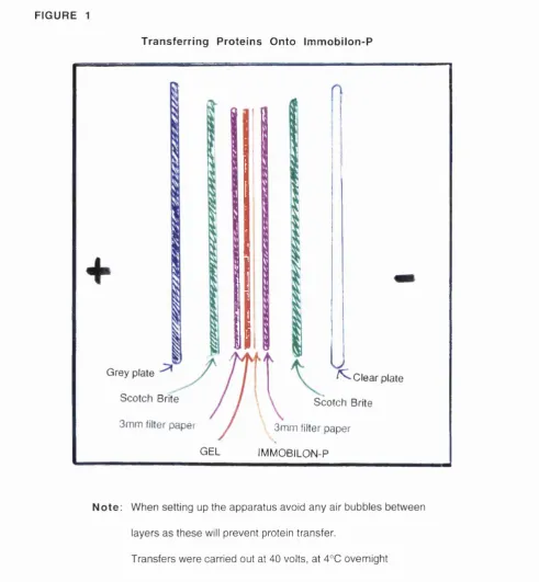
Figure 1
Tabie 1
Transferring proteins onto immobiion-P
Details of a-catenin, b-catenin, plakoglobin and bi-cytoplasmic tail peptides used for polyclonal antibody production
Chapter 3
Figure 1
Figure 2
Figure 3
Figure 4
Indirect immunofluorescence staining with monoclonal antibodies 4E2 and 2A11.
Adhesion blocking assay
Differentiation blocking assays
Figure 1 Indirect immunofluorescence staining of u, up, upr for integrins
Figure 2 Indirect immunofluorescence staining of v and vp for integrins
Figure 3 Flow cytometry of u, up, upr, v and vp
Figure 4 Immunoprécipitation of integrins from surface iodinated u, up, v and vp
Figure 5 Integrin p1 subunit mRNA levels in u, up, v , vp and upr
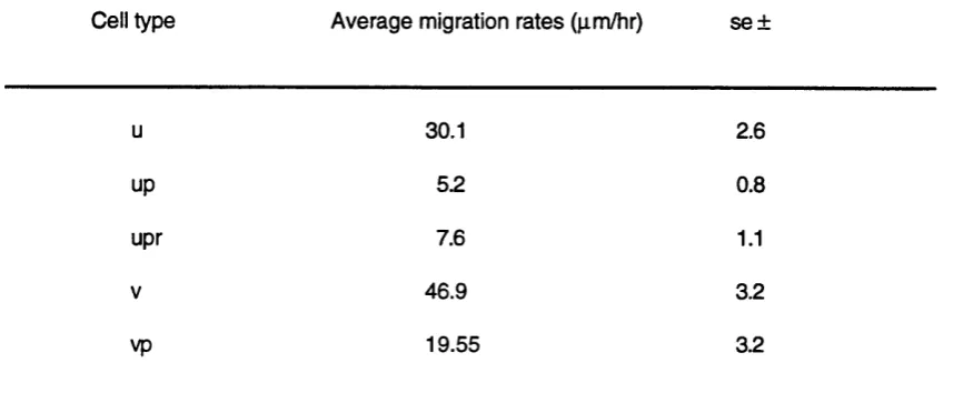
Figure 6 Adhesion of u, up, upr, v and vp to extracellular matrix proteins in vitro Figure 7 Traces of the migration paths of v, vp, u, up and upr
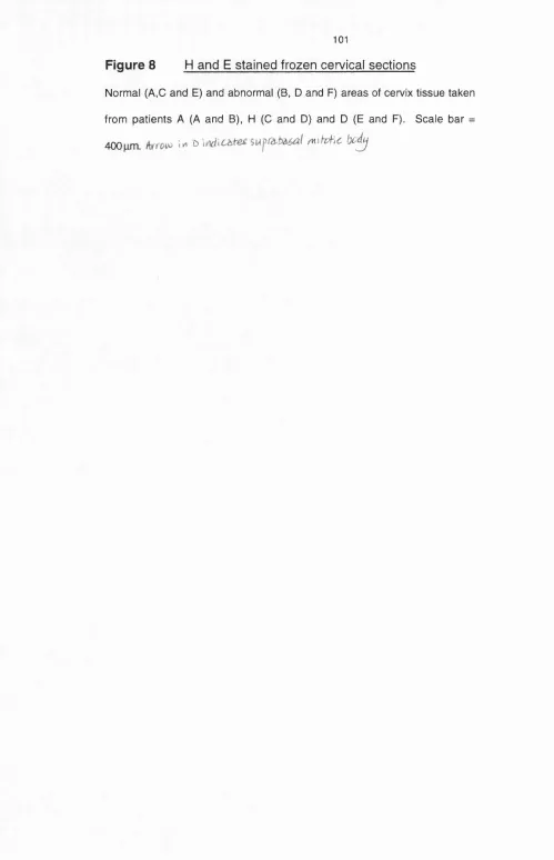
Figure 8 H and E stained frozen cervical sections
Figure 9 Indirect immunofluorescence staining of cervical tissue for integrins
Table 1 Migration rates of u, up, upr, v and vp cells
Table 2 Summary of the levels of CIN, HPV and integrins in abnormal cervical tissue
Chapter 5
Diagram 1 Calcium-induced stratification
Diagram 2 Cross-section of in sliu hybridisation pattem of integrin mRNA in standard calcium cultures
Figure 1 Integrin and involucrin distribution during calcium induced stratification
Figure 2 Double label immunofluorescence staining of permeabilised low calcium keratinocyte cultures
Figure 3 Adhesion assays
Figure 4 Integrin expression, synthesis and half-life
Figure 5 Integrin expression by terminally differentiating kératinocytes
Figure 6 Effect of anti-cadherin antibodies on calcium-induced stratification Figure 7 Effect of anti-cadherin antibodies on integrin distribution
Figure 1 The effect of cycloheximlde on integrin ioss during caicium induced stratification
Figure 2 Effects of cytochaiasin D or coichicine during caicium induced stratification
Figure 3 Effect of cytochaiasin D and colchicine on integrin and E-cadherin redistribution
Figure 4 Motiiity assay
Figure 5 Substrate adherent adhesion assay
Figure 6 Effect of microinjecting anti-catenin antibodies on E-cadherin redistribution
Figure 7 Effect of microinjecting anti-catenin antibodies on integrin bi-subunit redistribution
G
e n e r a l
I
n tr o d u c tio n
1)
STRUCTURE OF THE SKIN
The skin is one of the largest organs of the body (Goldsmith, 1990). Covering an
average area of 1.5 - 2.0 it acts as a flexible, protective layer keeping your 'insides' in and
the 'outside' out. Other functions of the skin include regulating the body temperature and
providing a sense of touch, pain, heat and cold. The thickness also varies: thus the skin of
the eyelids is very thin (less than 1mm), whilst that of the callus areas (palms and soles) is much thicker (3-4mm).
Some of the earliest observations of skin structure were made by
Auffhammer (1869). In those days of wax candles and horse-drawn carriages the existence
of the three major layers of the skin was already evident: a layer of subcutaneous fat that
forms the innermost layer with an overlaying dermis and outermost epidermis (reviewed in
Goldsmith, 1983; Wood and Bladon, 1985; Watt, 1990). The epithelium of the skin is
continuous with those of the digestive, respiratory and genito-urinary systems at their external
orifices (Wood and Bladon, 1985). The dermis and epidermis are separated by the basement
membrane . Hair, sweat glands and sebaceous glands are other structures that are part of
Diagrammatic representation of the structures identifiable in the skin
cpid.^rrv'.i< y A :
w m
i}QÎ0V0t''
Pml[?pr\/ blO<xi. \jeSSH
r!€MV5 L fy ÿ ^
Miedrv'pil
v v u i s d e ■
dw V tïm s \
mecujÊ. f â t
1a) Subcutaneous fat
The subcutaneous fat provides thermal insulation, mechanical protection and
acts as an energy reserve.
1b) Dermis
The functions of the dermis are to provide physical support and nutrition to
the epidermis. The dermis is a moderately dense fibroelastic connective tissue composed of
collagen fibres, elastic fibres and an interfibrillar gel of glycosaminoglycans, salts and water.
contains loose connective tissue and the reticular dermis is denser. The papillary dermis is
the outermost portion of the dermis. Dermal papillae project upwards from the dermis and
are moulded against the folds of the epidermis known as rete ridges. This undulating junction
provides mechanical support for the epidermis (Rook eta!., 1992). The papillary dermis
contains smaller and more loosely distributed elastic and collagen type I and III fibres than
those found in the reticular dermis. It also has a higher proportion of interfibrillar gel,
connective tissue cells (e.g. macrophages, fibroblasts and lymphocytes) to enclose nerve
endings, the microcirculatory blood vessels and lymphatic plexus (Goldsmith, 1983). The
microcirculatory system provides nutrition to the basal cells in the epidermis and acts in
thermoregulation.
The reticular dermis constitutes the large bulk of the dermis and is
distinguished from the papillary dermis by having relatively fewer cells, and blood vessels,
containing dense interwoven strands of collagen and elastin fibres.
1c)
Basement membrane
In the skin the basement membrane (also known as the basal lamina) is a
specialised layer of extracellular matrix proteins that lies between the papillary dermis and the
epidermis Its main functions are to provide an attachment substrate for epithelial cells
determine cell polarity and control epithelial cell differentiation (Adams and Watt, 1989,1990;
Streuli et al., 1991; Staiano-Coico and Higgins, 1992; Adams and Watt, 1993). Components
of the basement membrane include type IV collagen, laminin (TimpI, 1989), heparan sulfate,
proteoglycans (eg. perlecan), entactin (Moscher et al., 1993) also known as nidogen, and
epiligrin (Carter et al., 1991) also known as kalinin (Rousselle et ai., 1991) and nicein
(Marchisio eta!., 1993; Marinkovich et a!., 1993). The basement membrane is a meshwork of
the components listed above and is largely synthesized by the cells that rest on it. Electron
microscope studies have identified what appears to be three layers to the basement
membrane: the lamina lucida (uppermost), the lamina densa and the lamina reticularis that
Hairs grow out of tubular Invaginations of the epidermal surface called
follicles and each follicle with its associated sebaceous gland is called a pilosebaceous unit
(reviewed in Goldsmith, 1983). The epidermis is continuous with the epithelium lining the
pilosebaceous unit. A bundle of smooth muscle fibres (arrector pill) joins the wall of the
follicle to the dermis. The most basal region of the hair follicle expands to form the bulb
which resides in the dermis. It is here that the dead anucleated epidermal cells are pushed
up by continuous cell proliferation to form the hair shaft. The hair shaft consists of keratinised
fusiform cells.
There are two types of sweat glands, the eccrine and apocrine glands.
Eccrine glands are simple tubular glands arising embryologicaily from the epidermis and are
distributed all around the body. The apocrine glands are branching sweat giands that are
localised to the armpit, groin, face, scalp and abdomen regions of the body. Eccrine glands
secrete sweat to the skin surface via the eccrine sweat duct, but apocrine glands secrete into
the hair follicle. The production of sweat aids excretion and thermoregulation
1e)
Epidermis
The epidermis is a stratified squamous epitheiium that consists
predominantly of multiple layers of cells known as kératinocytes. In normal skin keratinocyte
proliferation occurs in the basal layer. Basal kératinocytes are undifferentiated and cells
undergo terminal differentiation as they migrate upwards through the suprabasai layers.
Apart from the stem cells, a population of transit amplifying cells that have a lower capacity
for self-renewal and higher probability of terminally differentiating also exist in the basal layer
(Barrandon and Green, 1987; Watt, 1988). Evidence for this includes the observation that
even though 60% of the cells in the basal layer are cycling (reviewed by Rotten and Morris,
1988), 10% of them are able to form foci after severe radiation damage to the skin (Whithers,
1967; Rotten and Hendry, 1973). It is within that 10% population that a subset of stem ceils is
foreskin tissue are located at the apical regions of the rete ridges,but that in palm and sole
they are concentrated in the trough region of the ridges (Jones, Harper and Watt, in
preparation).
One of the earliest events in the pathway of keratinocyte terminal
differentiation is withdrawal from the cell cycle. This is associated with a down regulation of
adhesion to the extracellular matrix (Adams and Watt, 1989,1990,1993; also see Part 5b of
this Chapter), the loss of gap junctional communication (Kam et al., 1987) and the onset of
expres.9Ci:of differentiation markers. The suprabasai layers of the epidermis include the spinous layer, the granular layer and cornified layer (Goldsmith, 1983; Wood and Bladon,
1985; Watt, 1987, 1989; Rook et a!., 1992) (see Diagram 2). As kératinocytes terminally
differentiate their morphology changes and in normal epidermis this coincides with the
precise suprabasai layer that the cells are located in. In spinous layers kératinocytes become
flattened and take on a 'spikey' appearance. These ‘spikes’ have been identified as areas of
desmosomal cell-cell contact. In the granular layer cells become larger and flatter and cells in
this layerare characterised by containing basophilic granules called keratohyaline granules.
This layer is most highly developed in the palm and sole regions of the skin and marks the
transition to the overlaying, anucleate cornified layer. Cornified cells are rich in keratin,
possess a tough comified envelope and represent terminally differentiated kératinocytes.
Comified layer is thickest in the palm and sole. The inert nature of keratin renders the skin
Diagram 2
Diagrammatic representation of a cross-section through human epidermis
Dermis
The different layers of kératinocytes can be distinguished by their morphology. Note the correlation between terminal differentiation and migration upwards from the basal layer. Red represents integrin expression and green represents cells at various stages of terminal differentiation. CL = cornified layer; GL = granular layer; SL = spinous layer; BL = basal layer; bm = basement membrane.
Cornified cells are continuously sloughed off and replaced by cells from the deeper layers of
the skin. In normal skin the rate of mitosis in the basal layer normally approximates the rate
of surface loss and therfore the epidermis remains a constant thickness.
Non-keratinocyte cell types found in the epidermis include melanocytes,
Langerhan’s cells and Merkel cells. Melanocytes are dendritic cells that produce melanin and
transfer it in melanosomes to neighbouring basal kératinocytes. Recent work has shown that
the m elanocytes adhere to kératinocytes via cadherins and that transformed melanocytes
display reduced levels of cadherins (Tang et al., 1994; see also Part 6 of this chapter).
Melanin protects the skin from ultraviolet (UVB) radiation present in sunlight and provides
mid-suprabasal layers of the epidermis where they act in association with the body's immune
system. Merkel cells are found bound to the basal kératinocytes and they act as a junction
between nerve endings that penetrate the basement membrane (reviewed in Rook et al.,
1992).
2)
MARKERS FOR KERATINOCYTE TERMINAL DIFFERENTIATION
Keratins, filaggrin and the precursors of the comified envelope are the most widely
studied markers of keratinocyte terminal differentiation. In addition, there are important
changes in lipid metaboiism during terminal differentiation, resulting in the deposition of
interceilular lipid in the cornified layers.
2a)
Keratins
Keratins are subdivided into two families, acidic and basic, and one member
of each subfamily must be present for keratin filament formation to occur. Basal cells of all
stratified squamous epithelia express K5 and K14 (Purkis et a!., 1990). During terminal
differentiation in the epidermis, K1 and KID are expressed. In vitro K6 and K16 are usually
expressed and in vivo these keratins are also expressed in areas of hyperproliferation in
preference to K1 and KID (Weiss eta!., 1984; Kopan and Fuchs, 1989).
2b)
Protein precursors of the comified envelope
As kératinocytes enter the comified layer each cell produces a toughened
and inert structure that encapsulates it known as the cornified envelope. The comified
envelope is derived from thickened plasma membrane of differentiating cells. Protein
precursors of the comified envelope include involucrin, loricrin and comifin. The genes that
encode for these proteins all contain common tandem repeats within the coding regions.
Involucrin and loricrin are the best characterised protein
precursors of the cornified envelope and are good markers for terminal differentiation.
Involucrin, a 92kD soluble protein precursor of the comified envelope, is synthesised in the
directed against human involucrin have shown that involucrin decorates the cytoplasmic face
of the envelope (Haftek et al., 1991 ). Thus, large, terminally differentiating cells contain
involucrin and smaller, undifferentiated basal cells do not (Watt and Green, 1981). Involucrin
becomes cross-linked in the cornified envelope in the presence of a keratinocyte specific
transglutaminase through the formation of e(-y-glutamyl)-lysine isopeptide bonds (Thacher
and Rice, 1985).
Loricrin is a 315 amino acid protein in humans and a 481 amino acid protein
in mice that accumulates in the granular layer of both species (Mehrel et al., 1990). Like
involucrin, loricrin also decorates the cytoplasmic face of the envelope. However, in contrast
to involucrin , loricrin expression is regulated by retinoids (Nagae et al., 1987)
2c)
Filaggrin
As kératinocytes move from the granular to the cornified layers the keratin
filaments become organised into a densely packed aggregates, a process that involves
filaggrin (Dale et al., 1985). Filaggrin is a histidine-rich basic protein that is synthesized in
granular cells as a high-molecular-weight precursor, profilaggrin. Profilaggrin is found in
keratohyalin granules (see part 1e of this Chapter). During the conversion of a granular cell
to a cornified cell, profilaggrin is dephosphorylated and proteolytically cleaved to form
filaggrin.
2d)
Lipids
Lamellar bodies that contain lipids are synthesized in the spinous cells and
move to the apex and periphery of the granular cells where they fuse with the plasma
membrane, releasing their contents into the intercellular spaces. Here, the lipids covalently
bind to form part of the comified envelope (Wertz et ah, 1989). During keratinocyte terminal
differentiation there is an increase in neutral lipids and sphingolipids and a decrease of
2e)
Peanut lectin-binding glycoproteins
Peanut agglutinin (RNA) is a lectin that binds readily to suprabasai
kératinocytes in vivo and in vitro (Reano et al., 1982; Watt, 1983). RNA binds the terminai
gaiactose residue of a 250kD glycoprotein on the surface of suprabasai cells. Although the
250kD glycoprotein is present in basai ceiis the gaiactose residue is masked by terminai sialic
acid residues, thus preventing RNA binding (Keebie and Watt, 1990). Recent work has
shown that the RNA binding glycoprotein is CD44 (Hudson and Watt, in preparation).
3)
PROUFERAT!VE DISORDERS OF THE EPIDERMIS
In normal skin the rate of loss of cornified celis from the surface of the skin is
balanced with the rate of proliferation in the basal layer. However, in wound healing (Grinnell,
1992) and benign hyperproliferative disorders such as psoriasis (Wood and Bladon, 1985) the
rate of proliferation in the basai layer is greater than the ioss of cells from the cornified iayer.
Psoriasis Vulgaris is characterised by scaly brown/red patches and thickening of the
epidermis, both in living and cornified layers (Lever, 1983). Hyperproliferation in both
psoriasis and wound healing has been demonstrated by the premature appearance of
involucrin in the iower spinous layers of the epidermis (Hertle et al., 1992). Psoriasis patients
are usually individuals with a genetic predisposition to the disease and certain physicai,
chemicai or emotional traumas can provoke the appearance of the disorder.
Squamous celi carcinoma is an invasive carcinoma of the epidermis. The tumour
consists of irregular masses of normal and abnormal kératinocytes that proliferate downward
into the dermis (MacKie, 1989) and display reduced levels of differentiation. Many squamous
ceil carcinomas arise as a resuit of exposure to environmental carcinogens, radiation, chronic
UVB irradiation from excessive sunbum (Wood and Bladon, 1985).
The possible role of viruses is the cause of squamous cell carcinoma is currently
under intensive investigation. Human papillomaviruses (HPV) are a group of small DNA
viruses which predominantly infect squamous epitheiia where they can cause benign
epithelial proliferations, e.g. cutaneous warts and genital condyloma and are thought to
over 60 different HPV types and the different types display tropism for specific epithelia. HPV
infection has been most notably associated with squamous cell carcinomas, in particular
cervical cancer. HPVs are believed to primarily infect basal epithelial cells which are exposed
to the virus through trauma. HPV-associated skin carcinomas usually develop in areas
exposed to high levels of UV light. HPV infection is most commonly associated wi/h
immunosuppressed patients. HPV types 16, 18 and 33 have been found to be more
commonly associated with cervical carcinomas than with normal cervix. Cervical
intraepithélial neoplasia (CIN) is a classification of the abnormalities of the cervix (see
Chapter 4). With increasing severity of CIN i.e. with progression from CIN I to III the
presence of HPV types 16 and 18 become more frequent (Niedobitek and Herbst, 1991).
4)
IN VITRO KERATINOCYTE CULTURE
Human kératinocytes can be grown in culture using the Rheinwald and Green
method (1975) and under such conditions the cells will stratify and show many of the
characteristics of human epidermis in vivo. The details of the method are given in Chapter 2.
Briefly, single cell suspensions of foreskin kératinocytes are cultured on an irradiated feeder
layer of mouse 313 fibroblasts in a culture medium that is composed of 1 part Ham's FI 2
medium plus 3 parts Dulbecco's modified Eagle's medium supplemented with adenine, foetal
calf serum, hydrocortisone, cholera toxin, epidermal growth factor and insulin.
Hydrocortisone stimulates proliferation and gives the growing colonies a more orderly
appearance (Rheinwald and Green, 1975). Cholera toxin also stimulates keratinocyte
proliferation (Green, 1977). Epidermal growth factor increases the number of cell generations
prior to senescence (Rheinwald and Green, 1977) and stimulates proliferation by stimulating
the outward migration of the rapidly proliferating cells at the edges of the growing colonies
(Barrandon and Green, 1987). The 3T3 feeder layer conditions the culture medium and
substratum to encourage attachment and proliferation of the kératinocytes. Kératinocytes
from newborn foreskin epidermis can be routinely passaged more than 15 times prior to
The Rheinwald and Green culture method can be adapted not only to
grow normal keratlnocytes, but abnormal keratlnocytes from squamous cell carcinomas too
(for example, Pel et al., 1991; Suglyama et a!., 1993). Culturing keratlnocytes In vlW
provides us with a valuable model to Investigate the regulation of terminal differentiation In
normal and abnormal situations (Watt, 1988;Watt 1991). Recent studies have highlighted the
Importance of two families of cell adhesion molecules namely the Integrins (Adams and Watt,
1990; Peltonen eta!., 1989; Marchisio ef a/.,1990; Nicholson and Watt, 1991; Hertle et a!.,
1991; Hotchin and Watt, 1992; Hotchin eta!., 1993; Jones and Watt, 1993; Adams and Watt,
1993; Watt eta!., 1993) and the cadherins (reviewed In TakelchI, 1990,1991; Fleming, 1991;
Wheelock and Jensen, 1992; Fujlta et al., 1992; Geiger and Ayalon, 1992; Burdsal et al.,
1993) In the regulation of epidermal cell behaviour.
5)
INTEGRINS
5a)
The intearin family, structure and function
The term 'Integrin' was proposed by Richard Hynes to describe a family of Integral
membrane receptors thought to link or 'Integrate' the cytoskeieton of cells with the
extracellular matrix. In substrate adhesive cultured cells areas of cell to substrate adhesion
are known as focal contacts. Focal contacts are sites on the cell surface where a high
density of Integrins, Integrin-assoclated cytosolic proteins and filamentous actin concentrate
together to bind the cell to the substrate. Integrins comprise a superfamily of
transmembrane, heterodlmeric glycoproteins that consist of one a- and one p-subunit that are
non-covalently linked (see Diagram 3). The Integrin superfamily was originally subdivided
Into three main groups based on the sharing of a common p-subunit (Pi, P2 or P3) by various a-subunlts. These three major groups are named VLA proteins (pi type), leukocyte Integrins
(CD11/CD18 family; P2 type) and the cytoadheslons (pa-type) consisting of platelet
glycoprotein llbllla and the vitronectin receptor (reviewed In Hogg, 1991; Hynes, 1992). To
form complexes with one or two p-subunlts, e.g., a i, «2, « 3 and eg can be found associated with p i only.
Ligand specificity depends on integrin heterodimer composition and also the cell type
in which the integrin is expressed (review in Tuckwell and Humphries, 1993). The ligand
binding region involves the extracellular domain of both subunits (Hynes, 1992). Some
integrins (eg. p i a 4 and p2aL) of the immune system recognise counter receptors on
endothelial cells that are integral membrane proteins of the immunoglobulin superfamily,
VCAM-1, ICAM-1, ICAM-2, andlCAM-3 . Such integrin counter receptor interactions mediate
direct cell-cell contact. Different integrin heterodimers recognise specific peptide sequences
(or binding sites) in various ligands. The first binding site to be defined was the Arg-Gly-Asp
(RGD) sequence present in fibronectin, vitronectin and a variety of other adhesive proteins.
o5p l, a l lb p 3 and ocvp integrins all recognise the RGD sequence, however allbp3 also recognises an additional sequence Lys-GIn-Ala-Gly-Asp-Val (KQAGDV) in fibrinogen. ot2p l binds Asp-Gly-Glu-Ala (DGEA) in collagen type I, and a 4 p i binds Glu-lle-Leu-Asp-Val
(EILDV) in an altematively spliced form of fibronectin (Hynes, 1992).
Both subunits of integrins are transmembrane glycoproteins, each
with a single hydrophobic transmembrane segment. In contrast to most of the a and p
subunits the cytoplasmic domain of the P4 subunit is notably long (1000 amino acids). The
extracellular domain of a-subunits are characterised by having multiple cation binding sites
and vary in size between 120 and 180kD. The extracellular domain of p-subunits are
characterised by having four cysteine rich repeats (90-1 lOkD). The association between the
a and p subunits to form a heterodimer is divalent cation dependent and appears to be
mediated predominantly by the integrin extracellular regions (Hayashi etal., 1990; Solowska
et al., 1991). Both subunits contain extensive internal disuphide bonding that contribute to
Diagram 3
A schematic representation of an intearin complex
The integrin integrates the extracellular matrix (ECM) with the actin (An) cytoskeieton.
a = a subunit; p = (3 -subunit; IVI++ = cation binding sites; cys = four cysteine rich repeats; cm = cell membrane; I n = talin; Vn = vinculin; Px = paxillin; a-Act = a actinin.
5a(i) p-subunits
Mutations in the P3 integrin cation binding domain has been shown to abolish ligand
binding (Loftus et al., 1990). It has also been shown that cation-binding is also associated with conformational change. For example, the binding of magnesium to integrin «|_P2 causes
a conform ational change in this integrin revealing the epitope for monoclonal antibody,
Mab24. Expression of the Mab24 epitope coincides with the ability of a \_^2 to bind ligand
(Dransfield and Hogg, 1989; Dransfield et al . , 1992; also see Hynes, 1992; Tuckwell and
Humphries, 1993). Sim ilar states are presumed to exist for other integrins. Several
antibodies have been reported to activate Pi integrin function, probably by converting or
Reichardt, 1991; Kovach et al., 1992; Fault eta!., 1993; Hotchin eta!., 1993). Point mutations
in the pi ligand binding site (Asp^^O Ala) have blocked the binding of aspi to fibronectin,
but not blocked recruitment to focal contacts (Takada eta!., 1992).
The cytoplasmic tails of the integrin subunits are believed to interact with actin
cytoskeleton via cytosolic proteins such as talin, vinculin, a-actinin and paxillin (see Diagram
3). The evidence for cytoskeletal connections comes from a variety of sources including light
and electron microscopic colocalization studies (Burridge et a!., 1988 ) and biochemical
evidence for interactions of integrins or cytoplasmic domain peptides with the cytoskeletal
proteins, talin (Horwitz eta!., 1986) and a-actinin (Otey eta!., 1990). Thus, the pi-subunit
cytoplasmic tail acts as a link between the extracellular domain with associated ligand and
the cytoskeleton (Reszka et a!., 1992). It has been shown that the cytoplasmic domain of the
p subunit is necessary for Pi integrin-ligand binding (Hayashi et a/., 1990). Deletion of all or
most of the p i cytoplasmic domain interferes with localisation to focal contacts (Solowska et
a!., 1989, Marcantonio eta!., 1990; Geiger et a!., 1992). These results suggest that the
distribution of integrins is determined by the p-subunit cytoplasmic domain. The hypothesis
is further emphisised in a study by LaFlamme and co-workers (1992) where extracellular and
transmembrane domains of the interleukin-2 (IL-2) receptor was coupled to the cytoplasmic
tails of either p i or a5 subunit. The results showed that neither chimeras coupled to
partnering integrin subunits, but the p i chimera was able to localise to focal contacts. anbPa
is the fibrinogen receptor on the surface of platelets. Studies have shown that trunca^i^D: of
the p3-subunit cytoplasmic domain abolishes cell spreading, recruitment of the anbp3 integrin to focal adhesions, and also prevents the transmission of intracellular contractile forces to
fibrin gels but does not reduce the affinity of anbp3(Ylânne eta!., 1993).
5a(li)
a-subunit
In experiments described by LaFlamme and co-workers (1992) where o5-subunit
cytoplasmic domain was coupled with the transmembrane and extracellular domain of the IL-
2 receptor (see part 5a(i) of this chapter) the chimera was not localised to focal contacts.
focal contacts. Truncation of the a-subunit cytopiasmic domain has no effect on cell
spreading, gel contraction or recruitment to focal contacts but allowed for indiscriminate
recruitment of anbPa to focal adhesions formed by other integrins (Ylanne et a/.,1993). Thus,
the aiib subunit cytoplasmic tail maintains the fidelity of recruitment of the anbPa integrins to
bind specifically to fibrinogen in focal adhesions. O'Toole and co-workers (1991) have
evidence to imply that the a-subunit of GPIIb-llla controls ligand binding affinity.
Chimeric a-subunit studies have shown that the extracellular domain of ag and the
intracellular domain of either a2, a * or as-produced different cell phenotypes. Cells containing all the different constructs bound to laminin and collagen. The cells that contained
the a2- and as-cytoplasmic domain constructs supported collagen gel contraction, but migrated poorly on collagen or laminin, whereas ceils with the a^-cytoplasmic construct
behaved in the opposite manner (Chan etal., 1992). More recent work has shown that the
region of the as-cytoplasmic domain adjacent to the membrane seems to play a role in
cytoskeletal organisation and cell motility (Bauer etal., 1993).
5a(lll)
Integrins and signalling
It
has become clear over the last few years that integrins are not only adhesion molecules, but also play a role in signal transduction both into and out of the cell(see Hynes, 1992 for several examples of 'outside-in' and 'inside-out' signalling). A
particularly important feature of integrins is that they undergo activation. During 'outside-
inside' signalling activation involves coupling of integrin to soluble mediators (eg. hormones,
cytokines etc.) and/or solid phase reactants (ECM or other cells) to cause a cascade of signal
triggered events . One of the first events identified was the activation of the Na/H antiporter
that causes a rapid and transient rise in intracellular pH as a consequence of adhesion of
clustered integrins to fibronectin. Other examples of such events include a transient rise in
intracellular calcium levels and the phosphorylation of tyrosines on associated proteins eg.
tyrosine kinases of the src family and FAK (Hynes, 1992; Schwartz, 1993). The focal
binding. The phosphorylation of FAK modulates FAK activity and thus affects other signalling
molecules.
How an integrin molecule conducts information out of the cell is of considerable
importance (Williams etal., 1994). Recent work by O'Toole and co-workers (O'Toole etal.,
1994) has identified the highly conserved GFFKR region in the cytoplasmic domain adjacent
to the transmembrane region of the allb integrin subunit in the control of integrin activation.
They showed that mutations in the GFFKR region result in high affinity binding of the allbp3
molecule to ligand. In their model they suggest that activation of an unidentified 'integrin
activator complex' interacts with the integrin cytoplasmic domains to provoke changes in the
spatial relationships or conformation of the a and p subunit cytoplasmic tails. Such changes
then traverse the membrane proximal GFFKR sequence to alter the extracellular domain
conformation of the integrin heterodimer.
In contrast to these activation events the phosphorylation of serine and tyrosine
residues on integrin p i subunits has been associated with the inactivation of this receptor
(reviewed in Hynes,1992). The correct spatio-temporal control of activation and inactivation of
integrins is vital for the precise regulation of cell behaviour and any deviation from this would
result in disaster (Schwarz, 1993).
5b)
Keratinocvte integrins
Basal kératinocytes adhere to extracellular matrix protein via integrins.
Kératinocytes express a range of integrins, including ogpi, «2^1, «sPi, «vPs and aep4. The receptor for fibronectin is ctSpl (Adams and Watt, 1990,1991; Carter etal., 1990a), «2pi, is the receptor for collagen and laminin (Adams and Watt, 1991; Carter etal., 1990 a,b), CC3P1 is a receptor for laminin and epiligrin (Adams and Watt, 1991; Carter et al., 1990 a,b, 1991),
OvPs the vitronectin receptor (Adams and Watt, 1991) and a6p4is a laminin receptor (Lee et a!., 1992) and acomponent of hemidesmosomes (Sonnenberg etal., 1991).
reason for the expression of integrins on the lateral surfaces of these cells is unclear. It is
suggested that integrins on the lateral faces of kératinocytes play a role in cell-cell
adhesion (Larjava etal., 1990; Carter et a!., 1990b; Symington et a!., 1993; but also see
Tenchini etal., 1993). Another hypothesis for the expression of integrins on the lateral faces
of kératinocytes is that the integrins on the cell-cell interfaces have in fact undergone
functional-downregulation, and have been redistributed away from the basal surface before
being internalised. However, this idea remains to be tested.
The precise spatial and temporal expression of various keratinocyte integrins
and during animal development have been studied in detail (for example Hertle et al., 1991;
Muschler and Horwitz, 1991; Sutherland e t a l, 1993). Recently, « 5 subunit knockout
experiments in mice have shown that the homozygous null mutation is an embryonic lethal
and although these mutant mice grow to around day 10-11 of gestation they show severe defects (Yang etal., 1993; Watt and Hodivala, 1994).
Keratinocyte integrins have also been shown to play a role during
stratification (Adams and Watt, 1990; Hotchin and Watt, 1992) and in the regulation of the
onset of terminal differentiation (Adams and Watt, 1989). The upward migration of cells out
of the basal layer occurs because the kératinocytes lose adhesiveness to extracellular matirx
proteins (Watt and Green, 1982; Watt, 1984). This is due to a down regulation of integrin
function in terminally differentiating cells. Keratinocyte differentiation can be induced in vitro
by placing the cells in suspension in methylcellulose (Green, 1977). Over the first 10 hr of
suspension-induced terminal differentiation, no change in the amount of cell surface agpi,
02pi or «3pi integrin is detected, yet by 5 hr the ability of the receptor to bind to fibronectin is markedly decreased (Adams and Watt, 1990). This decrease in receptor-ligand binding is
probably due to a change in integrin conformation (Hotchin e ta l., 1993). By 24 hr in
suspension the amount of 05^1 is greatly decreased and approximately 80% of cells express involucrin (Adams and Watt, 1989). Thus, there is a two stage down regulation of integrin
expression during keratinocyte terminal differentiation: firstly, there is a reversible down-
regulation of integrin function followed by an irreversible loss of integrin expression upon
When ke^ùjtmcUjp^ are placed in suspension for 24 hr in the presence of
0.19pM fibronectin the percentage of cells expressing involucrin is reduced to 50% (Adams
and Watt, 1989). Thus, the presence of fibronectin inhibits terminal differentiation of
kératinocytes. Some adhesion blocking anti-integrin antibodies or a combination of, type IV
collagen (Coll IV), laminin (Lm) and antibodies can also block the differentiation of
kératinocytes under similar conditions (Watt etal., 1993).
Further evidence for the role of integrins during keratinocyte terminal
differentiation comes from recent work carried out on locating human epidermal stem cells.
Given that stem cells are defined as having the capacity for unlimited division both in vivo and
in vitro (i.e. do not undergo terminal differentiation) it would be expected that these cells
would never downregulate their integrins and would express the highest density of functional
integrins per cell in the basal layer. This has indeed been shown to be true (Jones and Watt,
1993). 15% of cells from the basal layer will adhere to type IV collagen in 20 min and the rest
will adhere at later times. It has been shown that these 'early-stickers' have increased
surface expression of a2|3l, a3pl, o5pl but not a6p l . Moreover, these cells, when plated onto feeders, display a greater proliferative capacity than other cells isolated from the basal
layer. These results are in acordance with epidermal stem cells possessing the highest
density of functional integrins.
5c)
Aberrant integrin expression in abnormal epidermis
Changes in integrin expression occur during wound healing (reviewed by
Clark, 1990; Grinnell, 1992). Increased levels of 0% have been observed during the migration of kératinocytes to heal excision wounds in pig skin (Clark, 1990) and during migration of
human kératinocytes from explants in vitro (Guo etal., 1990).
At the time of closure of suction blister wounds, when the epidermis is
hyperproliferative the «2, cxg, oe and subunits are found in all living suprabasal layers, and
these cells coexpress markers for terminal differentiation (Hertle et al., 1992). Ov, 0 5 and P4
remain predominantly confined to the basal layer. Although 'embryonic' isoforms of
during wound closure (Brown etal., 1993) fibronectin, type IV collagen and laminin remain
confined to the basement membrane zone (Hertle et a!., 1992). One week after wound
closure the expression of all integrins is once more restricted to the basal cells. Strong
suprabasal staining of ag, 0C3, olq and is also observed in healing mouth ulcers (Jones et a!., 1992). It should also be noted that in certain regions of the oral cavity rapid squamous
cell proliferation and upward migration occurs and it is in such regions that suprabasal
integrin expression occurs.
Integrin expression in a number of benign hyperproliferative diseases has
also been examined. Suprabasal expression of oq and integrins have been observed in
psoriatic lesions (Ralfkiaer etal., 1991; Horrocks etal., 1991; Hertle etal., 1991; Kettner,
1991). Integrin staining patterns in oral squamous cell carcinomas show considerable
variation, both within and between individual tumours. Expression of «2,
«
3,
og, pi and P4subunits has been shown to be highest in the basal layer of normal epithelium, but extensive
staining in suprabasal layers is especially observed in hyperplastic epithelium adjacent to
ulcers. All poorly differentiated tumours show focal loss of a6, |34, o l2 and a3 integrin expression (Jones et al., 1993). An investigation of integrin expression in oral squamous cell
carcinomas cell lines also shows a variation in integrin expression. In particular there is an
extensive loss of a6p4 integrins in cell lines derived from poorly differentiated tumours
(Sugiyama etal., 1993). The significance of changes in integrin expression associated with
epidermal wound healing and hyperproliferative skin disorders is at yet unknown. However,
one might speculate that since changes in integrins function and expression not only regulate
keratinocyte adhesion, but also the initiation of terminal differentiation any abnormal
alterations in integrin expression may contribute to the behaviour of individual tumours.
In two benign inflammatory disorders, eczema and lichen planus, extensive
suprabasal staining of has been reported (Ralfkiaer etal., 1991). Basal cell carcinomas
are locally invasive, non-metastising keratinocyte tumours usually arising from hair bearing
skin In some basal cell carcinomas fibronectin expression appears to be increased with a
6)
CADHERINS
6a)
The cadherin family, structure and function
The cadherins are cell surface transmembrane proteins that mediate
homophillic calcium-dependent cell-ceil adhesion in a wide variety of tissues and species.
The classical cadherins are E-cadherin (epithelial cadherin or uvomurulin), P-cadherin
(piacental cadherin) and N-cadherin (neural cadherin or N-CAM). They are concentrated at
the adherens or intermediate type of cell junctions which link to the actin filament network.
Each cadherin subclass displays a unique pattern of tissue distribution. Many ceii types can
express a combination of these subciasses (see Part 6b of this Chapter). The cadherin superfamily also includes glycoproteins of the desmosome type of ceii junction, which link to
intermediate filaments within epithelial cells (for reviews see Takeichi, 1990; Takeichi, 1991;
Buxton and Magee, 1992; Geiger and Ayaion, 1992; Grunwald, 1993). Cadherins were first
described as expressing their function only in the presence of caicium. Cadherin-calcium
binding invoives resistance to proteoiytic cleavage (Takeichi, 1977).
The classicai cadherins are composed of a large extracellular domain, that
contains four internai repeats with a cysteine rich C-terminal region and six putitive calcium
binding sites; a transmembrane domain and a short cytoplasmic domain (see Diagram 2).
Homology between subclasses within a singie species is particulariy striking in the
cytoplasmic domain. In the extracellular domain the most N-terminal domain is the
hemophilic binding region. Cadherins have been suggested to be central to cell sorting
mechanisms and seiective ceil adhesion, allowing groups of cells to associate and form
organs (Takeichi, 1990,1991; Steinberg and Takeichi, 1994). In vitro aggregation assays of
a mixed population of L-ceiis transfected with the cDNAs encoding E-, P- or N-cadherin have
shown that cells expressing identical cadherin types bind to each other (Nose etal., 1989).
This preference for adherence is evidence for the homophiiic binding of the cadherins. In
aggregation studies where celis express different densities of P-cadherin the celis with the
capsule that excludes those which display lower densities of cadherin. This phenomenon is
thought to be mechanistic during morphogenesis (Steinberg and Takeichi, 1994)
The N-terminus of the extracellular domains of the classical cadherins display
a common histidine-alanine-valine (HAV) motif that acts as the recognition sequence in the
homophiiic binding of two cadherin molecules. HAV motifs have been identified in both
influenza virus hemagglutinin and in fibroblast growth factor receptors and play a general role
in protein-protein interactions (Byers etal., 1992). The penultimate N-terminal domain of the
extracellular region is the calcium binding region.
There is increasing evidence that cadherin function is modulated by cytosolic
proteins that bind to the C-terminus of cadherins (Nagafuchi and Takeichi, 1988). The
cytoplasmic domain of the classical cadherins is highly conserved and the C-terminus
associates with a distinct group of cytosolic proteins termed catenins (Nagafuchi and
Takeichi, 1988). Deletion of the catenin-binding region generates cadherins that cannot
mediate cell-cell adhesion (Nagafuchi and Takeichi, 1988).
Three distinct catenins have been identified by co-immuno-precipitation with
E-cadherin (Ozawa e ta l., 1989): a-catenin (102-105kD) that has been shown to have
homology to vinculin (Herrenknecht et al., 1991), p-catenin (92-97kD) that has partial
homology with plakoglobin (a peripheral cytoplasmic protein that also localizes to the
cytoplasmic face of desmosomes) (McCrea etal., 1991), and y-catenin (82-86kD). y-catenin
comigrates with plakoglobin on SDS-PAGE and it is suggested that they are the same protein
(Knudson and Wheelock, 1992) although other studies have suggested that on 2D SDS-
PAGE y-catenin and plakoglobin can be resolved separately (Piepenhagen and Nelson,
1993). Recent personal communication with James Nelson indicates that y-catenin and
plakoglobin are infact identical and that the distinct band previously seen on the 2D gels is an
Diagram 2
Schematic representation of the classical cadherin complex
%
OUT
Cadherin cytoplasmic tails complex with the catenins which in
turn interact with the actin cytoskeleton (An). HAV = HAV region in the homophillic binding
domain; Ca++ = cation binding site; IR = internal repeats; cys = cysteine rich domain; cm =
cell membrane; a = a-catenin; (3 = (3-catenin; pik = plakoglobin.
The im portance of cadherin-cytoskeletal interactions in
normal cadherin function is illustrated in a study where a truncated mutant of N-cadherin
lacking the extracellular domain was expressed in Xenopus embryos. Expression of this
mutant in Xenoous embryos caused a dramatic inhibition of cell adhesion. Moreover, two
point mutations in the N-cadherin cytoplasmic tail were sufficient to inhibit adhesion and effect
catenin binding to E-cadherin (Kinter, 1992). Another study that illustrates the importance of
cadherin-catenin interactions has shown that transfection of a subtype of a-catenin known as
a N-catenin into non-adherent cells (that normally lack a - and a N-catenin, but possess
The desmosomal cadherins include the desmosomal glycoproteins DGII,
DGIII (desmocollins) and DG-1 (desmoglein). To date other human desmosomal cadherins
include the desmocollins DGIV and DGV and the desmogleins HDGC and PVA (reviewed in
Buxton etal,, 1993).DGI has 29% identity to N-CAM, and DGII and DGIII have 35% identity
(Wheeler et a!., 1991). Like the cadherins the desmosomal cadherins have four internal
repeats which contain six putative calcium binding sites. Just before the transmembrane
domain the cadherins and DGII and DGIII have four srongly conserved cysteine residues.
The cytoplasmic tail of the desmosomal cadherins shows less than 25% Identity with the
cadherin cytoplasmic tail and this probably reflects the selective association of these
molecules with the intermediate filaments. DGII and DGIII differ only in that DG-III has a
smaller cytoplasmic domain (Holton etal., 1990; Parrish et a!., 1990; Collins efa/., 1991;
Parker etal., 1991). In contrast to DGII and DGIII DG-I has a much longer cytoplasmic tail
than the classical cadherins (Niles et al., 1991). Another major difference between the
classical cadherin and the desmosomal cadherin cytoplasmic tails is that the former have no
cysteine residues, but the latter can possess up6>ten.
6b)
Keratinocyte cadherins
The classical cadherins expressed in normal epidermis are P-cadherin and
E-cadherin. P-cadherin is expressed in the basal layer of cells whereas E-cadherin is
expressed in all layers of the epidermis (Hirai etal., 1989; Nicholson etal., 1991; Fujita etal.,
1992). It has been shown that E-cadherin is concentrated in adherens junctions both in vivo
(Kaiser etal., 1993) and in vitro (Green etal., 1987; O'Keefe etal., 1987).
The roles of E- and P-cadherin during skin development (Fujita et al., 1992)
and in epidermal morphogenesis have been reported (Wheelock and Jensen, 1992). In fetal
skin, while the patterns of E- and P-cadherin expression are similar to that in the adult, P-
cadherin is temporarily concentrated in the developing sweat ducts. After the sweat ducts
have formed P-cadherin is evenly expressed in the basal layer (Fujita etal., 1992).
Recent evidence for the role of cadherins during morphorgene)^ has been
blocking antl-E-cadherin antibodes to expiants of epiblast tissue from the prim itive streak
stage of mouse embryos. The results showed that perturbation of E-cadherin function
induced epiblast cells to take on mesenchymal cell morphology, ie. treatment with anti-E-
cadherin antibodies triggered steps of differentiation characteristic of the differentiation
pathway of primative streak mesoderm (Burdsal ef a/., 1993). In addition to this work, studies
in Xenopus development have shown that the absence o f E- and P- cadherin causes a
significant reduction in the adhesion between blastomeres (Heasman et al., 1994), and that
truncation of the cytoplasmic tail causes lesions in the developing ectoderm during
gastrulation (Levine etal., 1994).
Studies on kératinocytes grown in low calcium concentrations and then
switched to standard calcium concentrations have shown an apparent redistribution of
cadherin expression to the cell-cell borders and the induction of stratification (see Part 1 of
Chapter 5 for more details) Addition of an anti-E-cadherin antibody to the standard calcium
medium has inhibited the redistribution of E-cadherin in these celis. In particular, addition of
such an antibody also prevents calcium induced stratification. These studies suggest a role
for E-cadherin in the regulation of epidermal morphogenesis (Wheelock and Jensen, 1992).
The distribution of desmosomal cadherins has been described by Arneman
and co-workers (Ameman etal., 1993). Desmocollins DGIV and V are expressed only in the
upper spinous granular layers of the epidermis, whereas DGII and III expression is enriched
in the basal layers. Amongst the desmogleins expression of DGI appears to be similar to
desmoplakin and plakoglobin, i.e. expressed throughout the epidermis, gradually
accumulating during differentiation; PVA is more prevalent in the lower spinous layers,
whereas HDGC expression is detected only in the basal! layer. The precise location of
specific desmosomal cadherins in the epidermis probably reflects the slightly varied functions
of these molecules and this may be related to the need for changes in keratinocyte adhesion
6c)
Cadherin expression and cancer
It is known that cadherins are cell-cell adhesion molec as in tumours as
well as in normal tissues. The perturbation of cadherin function causes a temporary or
permanent disaggregation of tumour cells and may thus promote the Invasion and metastasis
of such cells. E-cadherin expression has been studied in various tumours jn vivo and ig vitro.
Investigations of the expression of E-cadherin in vivo include work on
squamous cell carcinoma of the hypopharynx (Schipper et al., 1991), the esophagus,
stomach and breast (Shiozaki et a!., 1991; Oka et a!., 1993). In general the E-cadherin
expression was reduced or lost in the most poorly differentiated tumours and primary tumours
when nur^r^diM to the normal tissue. However, Kinsella and co-workers (Kinselia et al., 1993)
found no correlation between the changes in E-cadherin expression and tumour spread in
carcinomas of the bowel.
E-cadherin but not P-cadherin mRNA levels have been shown to be lower
in sqamous cell carcinomas than in normal kératinocytes in vitro (Nicholson et al., 1991).
Genetic manipulation of E-cadherin in in vitro studies has shed light on the role of E-cadherin
in tumour metastisis. E-cadherin cDNA was transfected into highly invasive epithelial tumour
cell lines. The resulting transfectants showed a reproducible loss of invasion activity.
Alternatively, a plasmid encoding E-cadherin specific antisense RNA was introduced into
non-invasive ras transformed cells. This resulted in a downregulation of endogenous E-
cadherin expression the cells invasive (VIeminckx etal., 1991). The reductionin
E-cadherin expression and increased invasive potential has also been observed in esophageal
cancer cell lines (Doki etal., 1993), mouse epithelial squamous cell carcinoma lines (Stoler et
al., 1993) and in epithelial cells (Behrens etal., 1989). Interestingly, in some tumour cell lines
that have normal E-cadherin and p-catenin levels a reduced capcity for adhesion correlates
7)
aim é
The aims of my thesis have been to investigate two in vitro models in which integrin
expression is abnormal. The plan of my aims are as follows:
1 Prodiuce anti-integrin monoclonal antibodies that block adhesion to extracellular
matrix proteins in vitro and also block keratinocyte terminal differentiation.
2 Examine the levels of a large variety of integrin subunits on keratinocyte cell lines
transfected with HPV16 and or v-Harvey ras and compare them by immunofluorescence,
surface iodination and immunoprécipitation, FACS analysis, adhesion and migratory abilities
with their normal parental cell lines. In addition to this work I intend to examine the expression
of integrins on a panel of HPV infected cervical biopsies and compare their patterns of
expression with the in vitro models.
3 Investigate the expression of integins on terminally differentiating kératinocytes
grown in low calcium medium and examine the functional ability if the integrins on terminally
differentiating cells. Observe the changes in cell adhesion that occur during calcium induced
stratification and the role of cadherins in the possible control of integrin funcetion and
expression during keratinocyte terminal differentiation.
4 The final part of my project was an investigation of the possible mechanism of integrin-
cadherin cross-talk during calcium induced stratification via the cytoskeleton and cytosolic