C A S E R E P O R T
Open Access
Desmoplastic small round cell tumor of the
parotid gland-report of a rare case and a
review of the literature
Kanako C. Hatanaka
1*, Emi Takakuwa
1, Yutaka Hatanaka
1, Akira Suzuki
2, Satoshi IIzuka
3, Nayuta Tsushima
3,
Tomoko Mitsuhashi
1, Shintaro Sugita
4, Akihiro Homma
5, Shojiroh Morinaga
6, Tadashi Hashegawa
5and Yoshihiro Matsuno
1Abstract
Background:Desmoplastic small round cell tumor (DSRCT) is a rare soft tissue tumor that generally involves the retroperitoneum, pelvis, omentum and mesentery in younger patients. However, extra-abdominal DSRCT is very rare. Case presentation:A 49-year-old Japanese man noticed a mass in the right parotid gland. Ultrasound examination revealed a solid tumor about 2 cm in diameter. Computed tomography (CT) of the whole body revealed no other tumors or lymph node swelling. Superficial parotidectomy was performed. Histologically, the tumor was composed of various-sized tumor cell nests in an abundant fibromyxoid and collagenous background. The tumor cells were small to medium-sized. Immunohistochemistry showed that the tumor cells were immunoreactive for epithelial markers and desmin. They also showed strong nuclear staining with a Wilms tumor 1 (WT1) antibody detecting the C-terminal region (C-WT1), but not the N-terminal region (N-WT1). We also performed 3′/5′expression imbalance assay based on reverse transcription polymerase chain reaction (RT-PCR) to determine whether aberrant WT1 gene expression was present. This tumor was found to lack 5′-regional expression of the WT1 gene, as well as immunoreactivity with the N-WT1 antibody. Finally, fluorescence in situ hybridization (FISH) and RT-PCR analyses revealed the presence of a gene showing fusion between exon 7 of EWSR1 and exon 8 of WT1. The tumor was diagnosed as a DSRCT of the right parotid gland. The patient has been followed for 3 years without recurrence or metastasis.
Conclusions:Although DSRCT in the salivary gland is extremely rare, it should be included in the differential diagnosis of poorly differentiated salivary gland neoplasms, especially with a fibromyxoid background. Pathologists should bear in mind that DSRCT may occur in major salivary glands and should perform immunohistochemistry with appropriate antibodies, not only those against keratin and desmin, but also one detecting the C-terminal region of WT-1. Furthermore, molecular detection ofEWSR1-WT1fusion gene conclusively confirmed the diagnosis of DSRCT in this uncommon location.
Keywords:Desmoplastic small round cell tumor, Salivary gland, WT1, C-terminal region, 3′/5′expression imbalance assay
© The Author(s). 2019Open AccessThis article is distributed under the terms of the Creative Commons Attribution 4.0 International License (http://creativecommons.org/licenses/by/4.0/), which permits unrestricted use, distribution, and reproduction in any medium, provided you give appropriate credit to the original author(s) and the source, provide a link to the Creative Commons license, and indicate if changes were made. The Creative Commons Public Domain Dedication waiver (http://creativecommons.org/publicdomain/zero/1.0/) applies to the data made available in this article, unless otherwise stated.
* Correspondence:kyanack@huhp.hokudai.ac.jp
1Department of Surgical Pathology, Hokkaido University Hospital, N14W4,
Kita-ku, Sapporo, Japan
Background
Desmoplastic small round cell tumor (DSRCT) is rare and a highly aggressive neoplasm that typically involves the soft tissues of the abdomen or pelvis in children or young adults, showing a male predilection. Although it occurs over a wide age range, the peak incidence is in the third decade of life. DSRCT usually shows widespread abdominal serosal involvement, and overall patient survival is poor. On the other hand, extra-abdominal DSRCT is very rare. Previous cases have been reported to arise in the lung [1], pleura [2], paranasal sinuses [3], central nervous system [4], and scalp soft tissue [5] . DSRCT in major salivary glands has been reported, but it is extremely rare. To our know-ledge, only 5 cases occurring in the salivary gland have been reported in the English literature [6–10]. Here, we report a case of a primary parotid gland DSRCT in a 49-year-old man, who is the oldest patient known to have been affected by this tumor, and who has survived for 3 years without re-currence. We also summarize the clinicopathological fea-tures of DSRCT in the salivary gland.
Case presentation
Clinical features
A 49-year-old Japanese man noticed a mass in the right parotid gland without pain. There was no history of weight loss, fever, or night sweats. Ultrasound examin-ation demonstrated that the tumor was a solid mass about 2 cm in diameter. T1-weighted magnetic resonance im-aging showed a low-intensity, well-defined mass in the right parotid gland unaccompanied by lymph node swell-ing (Fig. 1). Abdominal computer tomography (CT) and
whole-body positron emission tomography (PET) scan re-vealed no other tumor elsewhere. Although fine-needle aspiration was performed several times, it was difficult to obtain tumor cells for diagnosis, except for cells from nor-mal salivary glands. Superficial parotidectomy was there-fore performed and the tumor was successfully resected without facial nerve paralysis. After parotidectomy, the pa-tient received radiotherapy and is currently alive and well with no evidence of recurrence after 3 years.
Pathological findings
Grossly, the tumor occupied the superficial lobe of the right parotid gland, and was solid and firm, measuring 2.7 × 2.7 × 2.3 cm. It was well circumscribed without a fibrous capsule, and the cut surface was grayish tan in color show-ing some lobulation at the tumor borders (Fig.2a). Macro-scopic necrosis or intratumoral hemorrhage was not evident. Histologically, the tumor predominantly showed a border that was well-defined from the surrounding tissue, although it was focally infiltrative in some areas. There was no fibrous capsule around the tumor. The tumor was com-posed of sharply demarcated cellular nests of various-sizes, growing in a paucicellular fibromyxoid or collagenous stroma (Fig.2b). The tumor cells were round to polygonal and small to medium-sized, with scant cytoplasm and hyperchromatic irregular round nuclei with granular chro-matin (Fig.2c). Tumor cells with clear cytoplasm were also found in tumor nests, but rhabdoid cells were not identified. Apoptotic bodies were occasionally found, but necrotic foci were not evident. Venous invasion was detected (Fig. 2d). Some non-neoplastic salivary ducts were found between tumor nests, demonstrating that the tumor infiltration had extended within the parotid parenchyma. There was no evi-dence of regional lymph node metastasis.
Immunohistochemical findings
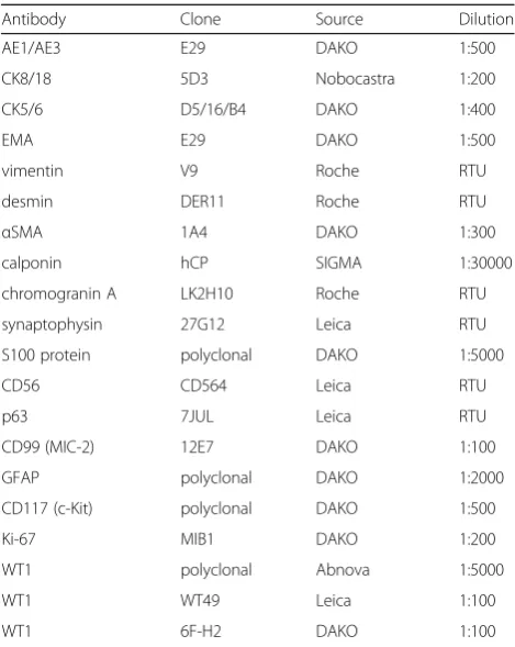
An immunohistochemical study was performed using formalin-fixed paraffin embedded (FFPE) sections of rep-resentative tumor blocks, using the antibodies summa-rized in Table 1. The tumor cells were positive for cytokeratin (AE1/AE3, CK8/18), EMA, vimentin, desmin, and focally positive for CD56 (Fig.2e). Desmin immuno-reactivity showed a diffuse cytoplasmic pattern for the most part, as well as a paranuclear dot-like pattern in a smaller proportion of the tumor (Fig.2f ). The cells were negative for chromogranin A, synaptophysin, S100 pro-tein, CK5/6, p63, CD99 (MIC2), GFAP, and CD117 (KIT). They were also negative for calponin and α-smooth muscle actin (αSMA), in contrast to the positivity shown by myofibroblast-like spindle cells in the stroma. The Ki-67 labeling index was almost 50%. For WT1, the tumor cells showed strong nuclear staining with an antibody rec-ognizing the C-terminal region of WT1 (C-WT1) (poly-clonal, Abnova). However, neither of two N-terminal Fig. 1T1-weighted magnetic resonance imaging (MRI) showing a
antibodies (N-WT1), WT49 (Leica) nor 6F-H2 (Dako), elicited positive nuclear staining, although the latter showed nonspecific cytoplasmic staining, (Fig.3a-c).
Molecular analyses
Dual-colored fluorescence in situ hybridization (FISH) ana-lysis using EWSR1 break-apart probes (Abbott Molecular, Abbott Park, IL) on FFPE tissue detectedEWSR1split sig-nals in 94% of the tumor cells (Fig.3d). We then performed 3′/5′ expression imbalance assay based on reverse tran-scription polymerase chain reaction (RT-PCR) according to the methods described by Suehara et al [11], in order to
determine whether aberrant WT1 gene expression was present. The result clearly showed that 5′-regional expres-sion of theWT1 gene was lacking in the tumor (Fig. 3e), being consistent with absence of immunoreactivity with the N-WT1 antibody at the protein level revealed by immuno-histochemistry (Fig. 3b-c). To confirm these gene alter-ations, RT-PCR for the EWSR1-WT1 fusion gene was performed using a forward primer (5′ -TCCTACAGC-CAAGCTCCAAGT-3′,EWSR1exon 7) and reverse primer (5′-ACCTTCGGTTCACAGTCCTTG-3′, WT1 exon 8) [12]. This revealed the characteristic EWSR1-WT1 fusion gene (Fig.3f).
a
b
c
d
e
f
Fig. 2Macroscopic and histological findings of the salivary gland tumor.aThe resected right parotid gland mass, appearing as a firm tan-colored tumor.bWell-defined nests of tumor cells varying in size are separated by a fibromyxoid stroma. (Hematoxylin-eosin, scale bar: 200μm).cThe nests are composed of small blue cells with scant cytoplasm and medium-sized cells with clear cytoplasm (insert). The normal salivary glands can be seen adjacent to the tumor nests. (scale bar: 100μm).dVenous invasion (arrows) is evident (Elastica van Gieson staining).e
Discussion
DSRCT is an uncommon malignant neoplasm that first described in two boys in 1989 [13]. DSRCT occurs mainly in the abdominal cavity, retroperitoneum, and pelvis, but Gerald et al. reported that 6% of DSRCTs can occur in an extra-abdominal location [14]. Histologically, DSRCT is characterized by various-sized nests composed of small neoplastic cells with a prominent desmoplastic, fibromyxoid, or collagenous stroma. Immunohistochemi-cally, DSRCT shows a distinctive and characteristic pat-tern of multi-phenotypic differentiation. Tumor cells express proteins associated with epithelial, muscular and neural differentiation. The distinctive dot-like staining pat-tern of desmin is typical, but a diffuse cytoplasmic patpat-tern has also been described [7], as seen in the present case. Hill et al. reported that nuclear immunoreactivity with C-WT1 antibody, which was observed in this case and is considered attributable to EWSR1-WT1 gene fusion, is useful for differentiating DSRCT from other small blue cell tumors, such as Ewing sarcoma [9]. The variable im-munohistochemical reactivity of WT1 protein according to the anti-WT1 antibody employed may be a diagnostic pitfall. Obviously, it is critically important to use C-WT1 antibody for diagnosis of DSRCT, because N-WT1 anti-body would give negative results, as was confirmed by the
lack of the 5′-regional expression of theWT1gene in the present case.
Although the present case showed typical histological features and immunohistochemical profiles, successful detection of the EWSR1-WT1 gene rearrangement in-volving t(11,22)(q13;q12) by FISH and RT-PCR assays using FFPE tissues conclusively confirmed the diagnosis of DSRCT in this uncommon location.
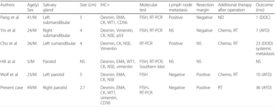
The clinicopathological features of salivary gland DSRCT reported previously in the English literatures are summarized in Table 2. All 6 cases, including the present one, occurred in males, and the age at the diagnosis ranged from 5 to 49 years, with a median age of 28 years. Two of these cases occurred in the fifth decade, although DSRCT is generally considered as a differential diagnosis in children or young adults. The tumors ranged in size from 2.7 to 5 cm, with an average of 4.1 cm. Among the six patients, three were still undergoing follow-up, and one had died due to systemic metastasis. Salivary gland DSRCT showed histological features similar to those of abdominal DSRCT. The overall survival in abdominal DSRCT patients is generally poor, despite multimodality ther-apy [15], According to the present case as well as previous cases, only one among 6 patients with saliv-ary gland DSRCT died due to metastasis. The prog-nosis of salivary gland DSRCT is unclear because of its rarity, but early detection or complete resection of salivary gland DSRCT might result in better prognosis.
The origin of DSRCT remains unclear. It has been speculated that DSRCTs are derived from mesothelial or submesothelial cells because a vast majority of pa-tients develop DSRCTs in cavities that are lined with mesothelial cells or because tumor cells show immu-nohistochemical positivity for epithelial and mesen-chymal antigens including desmin, and WT-1 [12].
DSRCT is not usually included in the differential diagnosis in primary salivary gland tumors in view of its rarity. Because DSRCT is composed of small nests with cohesive small to medium-sized cells and shows immunoreactivity for epithelial markers, it might be diagnosed as carcinomas, such as small cell carcin-oma, poorly differentiated carcincarcin-oma, undifferentiated carcinoma, without staining for desmin. Primary small cell carcinoma of the salivary glands is also rare, ac-counting for approximately 2% of all salivary gland tumors, and most arise in patients 50 years old or more [16]. Positivity for synaptophysin or chromogra-nin is useful for identification of small cell carcinoma. It is also difficult to rule out poorly differentiated car-cinoma or undifferentiated carcar-cinoma, although an abundant desmoplastic or fibromyxoid stroma is an unusual feature in these carcinomas. Therefore, this
Antibody Clone Source Dilution AE1/AE3 E29 DAKO 1:500 CK8/18 5D3 Nobocastra 1:200 CK5/6 D5/16/B4 DAKO 1:400 EMA E29 DAKO 1:500 vimentin V9 Roche RTU desmin DER11 Roche RTU
αSMA 1A4 DAKO 1:300 calponin hCP SIGMA 1:30000 chromogranin A LK2H10 Roche RTU synaptophysin 27G12 Leica RTU S100 protein polyclonal DAKO 1:5000 CD56 CD564 Leica RTU p63 7JUL Leica RTU CD99 (MIC-2) 12E7 DAKO 1:100 GFAP polyclonal DAKO 1:2000 CD117 (c-Kit) polyclonal DAKO 1:500 Ki-67 MIB1 DAKO 1:200 WT1 polyclonal Abnova 1:5000 WT1 WT49 Leica 1:100 WT1 6F-H2 DAKO 1:100
αSMAα-smooth muscle actin,EMAepithelial membrane antigen,WT1Wilms
b
a
d
c
e
f
might be a diagnostic clue for DSRCT. Other differ-ential diagnosis, such as malignant melanoma, meta-static neuroblastoma, or lymphoma, are excluded with positivity for epithelial markers. Merkel cell carcin-oma is also the differential diagnosis, but it is ex-cluded with staining for neuroendocrine markers and its characteristic dot-like staining pattern for cytoker-atin 20.
Primary non-lymphoid mesenchymal neoplasms of major salivary glands are also rare and account for only 1.9–5% of cases [8]. Most mesenchymal tumors are be-nign. The most common mesenchymal tumor is lipoma, [8] [17]. Malignant cases are extremely rare [8]. Because of the histological features of small round cell tumors, Ewing sarcoma and rhabdomyosarcoma are also in-cluded as differential diagnoses. Positivity for desmin and WT1 nuclear staining in addition to positive epithe-lial markers strongly favors a diagnosis of DSRCT over that of Ewing sarcoma and rhabdomyosarcoma. Further-more, molecular biology studies are useful and import-ant in differentiating DSRCT from Ewing sarcoma and rhabdomyosarcoma. In the present case, the tumor nests
were composed of not only small-sized cells with a high nuclear/cytoplasmic ratio, but also medium-sized cells with clear cytoplasm. These cells with clear cytoplasm were also described in a previous report [6]. Because both two cell types share the same immunohistochemi-cal profiles in the present case, cells with clear cytoplasm should also be recognized as neoplastic, and not as non-neoplastic bystander cells such as myoepithelial cells.
Conclusions
In summary, we have reported a rare primary DSRCT with venous invasion arising from the parotid glands of a middle-aged man. It is very important to be aware of the fact that DSRCT may occur in major salivary glands. To ensure accurate diagnosis with immunohistochemis-try, desmin and the C-terminal region of WT1 are very useful markers in addition to epithelial markers. Further-more, molecular detection of EWSR1-WT1 fusion gene conclusively confirmed the diagnosis of DSRCT in this uncommon location.
(See figure on previous page.)
Fig. 3Immunohistochemical findings and molecular analyses of the salivary gland tumor.aC-WT1 shows nuclear positivity (scale bar: 50μm).b N-WT1 (WT49) shows nuclear and cytoplasmic negativity.cN-WT1(6F-H2) shows cytoplasmic positivity.dFISH analysis using a break-apart probe for the EWSR1 gene region demonstrates the rearrangement in most of the cells.eThe 3′/5′expression imbalance assay based on RT-PCR reveals that the tumor lacked 5′-regional expression of the WT1 gene. The primers were designed to measure the expressions at two regions for each gene transcript: a 5′probe pair located far upstream of the exons and a second pair located within the exons located further 3′in the WT1 gene. PCR analysis was performed using these 5′and 3′primers, respectively. The Ct data were normalized to wild-type control tissue, and the normalized data was expressed as the relative gene expression level. WT cont; wild-type control (ovarian serous carcinoma), MT cont; mutant type control (typical DSRCT).fRT-PCR analysis showed that the EWS-WT1 fusion gene was present in the sample. M; marker, WT cont and MT cont; same as above
Table 2Clinicopathological summary of Major Salivary Gland DSRCT
Authors Age(y) Sex
Salivary gland
Size (cm) IHC+ Molecular test
Lymph node metastasis
Resection margin
Additional therapy after operation
Outcome (mo) Pang et al 41/M Left
submandibular
5 Desmin, EMA, CK, WT1, CD56
FISH, RT-PCR Positive Negative ND 1 (DOC) Yin et al 24/M Right
submandibular
4 Desmin, Vimentin, CK, NSE, p53
FISH, RT-PCR NS Negative Chemo, RT 7 (AFD) Cho et al 26/M Left sumandibular 4 Desmin, CK, NSE,
Vimentin
RT-PCR Positive NS Chemo, RT 23 (DOD) systemic metastasis Hill at al 5/M Parotid NS Desmin, EMA, WT1,
CK, NSE, vimentin
FISH, RT-PCR, Southern blot
NS NS NS
Wolf et al 23/M Left parotid 5 Desmin, EMA, CK, NSE
FISH Negative Positive Chemo, RT 10 (AFD) Present case 49/M Right parotid 2.7 Desmin, EMA,
CK, WT1, vimentin, CD56
FISH、 RT-PCR
Negative Positive RT 36 (AFD)
IHCimmunohistochemistry,momonth,EMAepithelial membrane antigen,CKcytokeratin,WT1Wilms tumor 1,NSEneuro-specific enolase,PRprogesterone
Abbreviations
CK:cytokeratin; CT: Computed tomography; C-WT1: WT1 antibody detecting the C-terminal region; DSRCT: Desmoplastic small round cell tumor; FFPE: formalin-fixed paraffin embedded; FISH: fluorescence in situ hybridization; GFAP: glial fibrillary acid protein; N-WT1: WT1 antibody detecting the C-terminal region; PET: positron emission tomography; RT-PCR: reverse transcription polymerase chain reaction; RTU: ready-to-use; SMA: smooth muscle actin; WT1: Wilms tumor 1
Acknowledgments
The authors thank Ms. Sayaka Honjo and Ms. Asami Okumura for technical supports.
Funding Not applicable.
Availability of data and materials
Data sharing is not applicable to this article as no datasets were generated or analysed during the current study.
Authors’contributions
KCH, ET, AS, TM, and SM performed pathologic diagnosis and writing of manuscript. SI, NT, and AH collected clinical data and follow-up of the patient. YH performed immunohistochemical and molecular analyses. SS and TH performed pathologic diagnosis and FISH analysis. YM redacted the manuscript. All authors read and approved the final manuscript prior to submission.
Ethics approval and consent to participate
Approval for this case report was obtained from the Ethical Committee in Hokkaido university hospital. Consent to participate was obtained from the participants.
Consent for publication
Informed consent was obtained from the patient for the publication of this case report.
Competing interests
The authors declare that they have no competing interests.
Publisher’s Note
Springer Nature remains neutral with regard to jurisdictional claims in published maps and institutional affiliations.
Author details
1Department of Surgical Pathology, Hokkaido University Hospital, N14W4,
Kita-ku, Sapporo, Japan.2Department of Pathology, KKR, Sapporo Medical
Center, 1-6, hiragishi, Toyohira-ku, Sapporo, Japan.3Department of
otorhinolaryngology, Hakodate Central General Hospital, 33-2, Honcho, Hakodate, Japan.4Department of Surgical Pathology, Sapporo Medical
University School of Medicine, S1W16, chou-ku, Sapporo, Japan.5Department
of Otolaryngology-Head & Neck Surgery, Faculty of Medicine and Graduate School of Medicine, Hokkaido University, N15W7, Kita-ku, Sapporo, Japan.
6Department of Diagnostic Pathology, Hino Municipal Hospital, 4-3-1,
Tamadaira, Hino, Tokyo, Japan.
Received: 9 November 2018 Accepted: 13 May 2019
References
1. Syed S, Haque AK, Hawkins HK, Sorensen PH, Cowan DF. Desmoplastic small round cell tumor of the lung. Archives of pathology & laboratory medicine. 2002;126(10):1226–8.
2. Karavitakis EM, Moschovi M, Stefanaki K, Karamolegou K, Dimitriadis E, Pandis N, et al. Desmoplastic small round cell tumor of the pleura. Pediatr Blood Cancer. 2007;49(3):335–8.
3. Finke NM, Lae ME, Lloyd RV, Gehani SK, Nascimento AG. Sinonasal desmoplastic small round cell tumor: a case report. Am J Surg Pathol. 2002;26(6):799–803. 4. Neder L, Scheithauer BW, Turel KE, Arnesen MA, Ketterling RP, Jin L, et al.
Desmoplastic small round cell tumor of the central nervous system: report of two cases and review of the literature. Virchows Archiv : an international journal of pathology. 2009;454(4):431–9.
5. Lae ME, Roche PC, Jin L, Lloyd RV, Nascimento AG. Desmoplastic small round cell tumor: a clinicopathologic, immunohistochemical, and molecular study of 32 tumors. Am J Surg Pathol. 2002;26(7):823–35.
6. Pang B, Leong CC, Salto-Tellez M, Petersson F. Desmoplastic small round cell tumor of major salivary glands: report of 1 case and a review of the literature. Applied immunohistochemistry & molecular morphology : AIMM / official publication of the Society for Applied Immunohistochemistry. 2011; 19(1):70–5.
7. Yin WH, Guo SP, Yang HY, Chan JK. Desmoplastic small round cell tumor of the submandibular gland--a rare but distinctive primary salivary gland neoplasm. Hum Pathol. 2010;41(3):438–42.
8. Cho KJ, Ro JY, Choi J, Choi SH, Nam SY, Kim SY. Mesenchymal neoplasms of the major salivary glands: clinicopathological features of 18 cases. European archives of oto-rhino-laryngology : official journal of the European Federation of Oto-Rhino-Laryngological Societies (EUFOS) : affiliated with the German Society for Oto-Rhino-Laryngology - Head and Neck Surgery. 2008;265(Suppl 1):S47–56.
9. Hill DA, Pfeifer JD, Marley EF, Dehner LP, Humphrey PA, Zhu X, et al. WT1 staining reliably differentiates desmoplastic small round cell tumor from Ewing sarcoma/primitive neuroectodermal tumor. An immunohistochemical and molecular diagnostic study. Am J Clin Pathol. 2000;114(3):345–53. 10. Wolf AN, Ladanyi M, Paull G, Blaugrund JE, Westra WH. The expanding
clinical spectrum of desmoplastic small round-cell tumor: a report of two cases with molecular confirmation. Hum Pathol. 1999;30(4):430–5. 11. Suehara Y, Arcila M, Wang L, Hasanovic A, Ang D, Ito T, et al. Identification
of KIF5B-RET and GOPC-ROS1 fusions in lung adenocarcinomas through a comprehensive mRNA-based screen for tyrosine kinase fusions. Clinical cancer research : an official journal of the American Association for Cancer Research. 2012;18(24):6599–608.
12. Lee YS, Hsiao CH. Desmoplastic small round cell tumor: a clinicopathologic, immunohistochemical and molecular study of four patients. Journal of the Formosan Medical Association = Taiwan yi zhi. 2007;106(10):854–60. 13. Gerald WL, Rosai J. Case 2. Desmoplastic small cell tumor with divergent
differentiation. Pediatric pathology / affiliated with the International Paediatric Pathology Association. 1989;9(2):177–83.
14. Gerald WL, Ladanyi M, de Alava E, Cuatrecasas M, Kushner BH, LaQuaglia MP, et al. Clinical, pathologic, and molecular spectrum of tumors associated with t(11;22)(p13;q12): desmoplastic small round-cell tumor and its variants. Journal of clinical oncology : official journal of the American Society of Clinical Oncology. 1998;16(9):3028–36.
15. Philippe-Chomette P, Kabbara N, Andre N, Pierron G, Coulomb A, Laurence V, et al. Desmoplastic small round cell tumors with EWS-WT1 fusion transcript in children and young adults. Pediatr Blood Cancer. 2012;58(6):891–7. 16. Gnepp DR, Corio RL, Brannon RB. Small cell carcinoma of the major salivary
glands. Cancer. 1986;58(3):705–14.