R E S E A R C H A R T I C L E
Open Access
Is there a correlation between
inflammatory markers and coagulation
parameters in women with advanced
ovarian endometriosis?
Shaojie Ding
†, Qiao Lin
†, Tianhong Zhu, Tiantian Li, Libo Zhu, Jianzhang Wang and Xinmei Zhang
*Abstract
Background:Endometriosis is defined as a chronic inflammatory disease. Recent studies have shown that increased coagulation parameters including fibrinogen and platelets are associated with endometriosis. The objective of this study was to determine the levels of inflammatory markers and coagulation parameters and their correlations in women with endometriomas compared to those with benign ovarian cysts or normal pelvic anatomy.
Methods:Between June 2015 and June 2017, a total of 548 women who underwent laparoscopic/laparotomic surgery for ovarian endometriomas (OMA group,n= 226), non-endometriosis benign ovarian cysts (Cyst group,n= 210) and tubal reanastomosis (Control group,n= 112) were recruited in this study. Inflammatory markers including c-reactive protein (CRP), neutrophil-to-lymphocyte ratio (NLR), platelet-to-lymphocyte ratio (PLR) and coagulation parameters including platelet count, thrombin time (TT), prothrombin time (PT), activated partial thromboplastin time, and plasma fibrinogen as well as CA-125 were determined.
Results:Compared with Cyst group and Control group, TT and PT in OMA group were significantly shorter and plasma fibrinogen levels were significantly higher (P< 0.05). Moreover, the levels of plasma fibrinogen were positively correlated with CRP, NLR and PLR (P< 0.05). In addition, the confidence intervals for the area under the curve (AUC) for CA-125 × fibrinogen were significantly higher than those for CA-125 (0.904–0.952 vs. 0.899–0.949) in the diagnosis of endometrioma.
Conclusions:These results indicate that women with endometriomas demonstrate a hypercoagulable status due to the inflammatory nature of endometriosis. The combined determination for CA-125 and fibrinogen demonstrate a higher area under the curve than the single detection of CA-125 in those with endometriomas compared to these with benign ovarian cysts.
Trial registration:This study was approved by the Human Ethics Committee of the Women’s Hospital, School of Medicine, Zhejiang University (No.20170174) and all women provided written informed consent.
Keywords:Endometriosis, Fibrinogen, Inflammation, Coagulation, Ovarian cyst
© The Author(s). 2019Open AccessThis article is distributed under the terms of the Creative Commons Attribution 4.0 International License (http://creativecommons.org/licenses/by/4.0/), which permits unrestricted use, distribution, and reproduction in any medium, provided you give appropriate credit to the original author(s) and the source, provide a link to the Creative Commons license, and indicate if changes were made. The Creative Commons Public Domain Dedication waiver (http://creativecommons.org/publicdomain/zero/1.0/) applies to the data made available in this article, unless otherwise stated.
* Correspondence:zhangxinm@zju.edu.cn
†Shaojie Ding and Qiao Lin contributed equally to this work.
Background
Endometriosis, defined as the presence of functional endo-metrial tissue outside the uterine cavity, is considered to be a chronic inflammatory disease, which is related to the increase of pro-inflammatory cytokines and chemokines [1]. The inflammatory process of endometriosis is associ-ated with increased levels of activassoci-ated macrophages se-creted cytokines in the endometriotic lesions and peritoneal fluid (PF), which provides a local microenviron-ment suitable for the growth and maintenance of endo-metriosis. The expression levels of interleukin (IL)-1β, IL-6 and tumor necrosis factor (TNF)-α in lesion, endome-triotic cyst fluid, and PF have been demonstrated to be in-creased in women with endometriosis [2–5]. The ratio of neutrophil-to-lymphocyte ratio (NLR) and platelet-to-lymphocyte ratio (PLR) in the peripheral blood of women with endometriosis also increased [6–8]. Moreover, the levels of cytokines [5], free iron, reactive oxygen species [9], matrix metalloproteinases (MMPs) [10], activins [11], and even plasminogen activated system components [12] have also been shown to be elevated in women with ovar-ian endometriomas compared with other benign ovarovar-ian cysts. These different inflammatory and coagulation medi-ators may identify pathways activated or associated with the development and progression of endometriosis [13].
It has been shown that women with endometriosis ap-pear to be in a hypercoagulable and hyperfibrinolytic status because platelets aggregate in endometriotic lesions [14]. In women with endometriosis, platelet counts (PLT) and plasma fibrinogen levels increase [15, 16], thrombin time (TT) [15] and activated partial thromboplastin time (APTT) decrease [15,17], while prothrombin time (PT) re-mains at normal level. Moreover, tissue factor (TF, coagula-tion factor III) is also increased in the endometriotic lesions and PF in women with endometriosis [14,18]. TF binds to coagulation factor VIIa, activates coagulation factors IX and X, and induces thrombin formation. Fibrinolysis is a re-sponse to elevated levels of coagulation factors and pro-motes the levels of urokinase-plasminogen activator (u-PA) and plasminogen activator inhibitors (PAIs) in the eutopic endometrium and PF [19,20].
Inflammatory processes can initiate and promote co-agulation, increasing the risk of bleeding, microvascular thrombosis and organ dysfunction [21]. In the coagula-tion cascade reaccoagula-tion, activated platelets and TF bind to coagulation factors and thrombin to induce inflamma-tion [22, 23]. Activated fibrinogen can also induce thrombin generation, which further activates chemokine production and macrophage adhesion [24].
In this study, we aimed to determine the correlations between coagulation parameters (TT, PT, APTT, and PLT), inflammatory biomarkers [C-reactive protein (CRP), NLR and PLR], compare the differences between patients with endometriomas and patients with
non-ovarian endometriosis cysts and without non-ovarian cysts, and to explore how these parameters can be used as auxiliary biomarkers for diagnosis.
Methods Patients
This study was approved by the Ethics Committee of the Women’s Hospital, Zhejiang University School of Medi-cine (No.20170174) in accordance with the Declaration of Helsiniki, and we obtained the written informed con-sent of each participant in this study.
Before the implementation of this study, we used formu-las for mismatched case-control studies (α= 0.05, β= 0.1) to determine the size of samples. According to the cut-off of fibrinogen at 2.8 g/L for the diagnosis of endometrio-mas reported by Chmaj-Wierzchowska et al. [16], P1 and P2 were 0.83 and 0.64 in our small sample test, and the size of samples was at least 111. As a results, a total of 548 women who underwent laparoscopic/laparotomic surgery for ovarian endometriomas (OMA group, n= 226), non-endometriosis benign ovarian cysts (Cyst group,n= 210) and tubal anastomosis (Control group, n= 112) at the Women’s Hospital affiliated to Zhejiang University School of Medicine from June 2015 and June 2017 were recruited in this study. In OMA group, endometriosis was graded according to the Revised American Fertility Society Scor-ing (rAFS) system (III = 91, IV = 135) [25]. Of the 226 women with endometriosis, 128 (56.6%) women had dys-menorrhea. In Cyst group, 117 (55.7%) women had ovar-ian mature teratomas, 21 (10.0%) benign ovarovar-ian serous tumors, 24 (11.4%) benign ovarian mucinous tumors, 33 (15.7%) simple cysts, 8 (3.8%) follicular cysts, 5 (2.4%) par-oophoritic cysts and 2 (1.0%) corpus luteum cysts. In Con-trol group, all women underwent tubal anastomosis because of the requirement of fertility after tubal ligation.
The patients’demographic and clinical data such as age, gravidity, parity, body mass index (BMI), dysmenorrhea, pelvic pain, and cyst size were extracted and recorded from patients’ original electronic medical record. Due to the lack of women in secretory phase at admission, only women in proliferative phase were included in the study. All patients had no history of hypertension, diabetes, liver and kidney dysfunction, leiomyoma, adenomyosis, and co-agulation disorder. No steroid hormone or anticoco-agulation therapy was given 6 months before surgery.
Blood assays
C16000). Levels of APTT, PT, TT and fibrinogen were determined with an automatic blood coagulation analyzer (STAGO, Evolution ISTA-R-IV, Germany). The levels of serum cancer antigen (CA) 125 were mea-sured by chemiluminescence with an endocrine de-tector (Roche, COBAS800, France).
Statistical analysis
Statistical Package for the Social Sciences Statistics v22.0 were used for data analysis. Continuous data were pre-sented as mean ± standard error of the mean (SEM). The comparison of the distribution of continuous data among the three groups was performed with Kruskal–Wallis H test. Mann–Whitney U andχ2 tests were used to compare the medians and frequencies among the groups. Spearman analysis was conducted to determine the correlation among the measured parameters. The optimal cut-off values of the parameters differentiating endometriomas were evaluated with the receiver operating characteristic (ROC) curve analysis. Then, the area under the curve (AUC) was calculated and the sensitivity and specificity of each parameter were determined. P values of less than 0.05 (P< 0.05) is considered to have statistical significance.
Results
Patients’characteristics
There were no statistical differences with regard to age, body weight, height or BMI between OMA, Cyst and Con-trol groups (P> 0.05). Compared with the Cyst and Control groups, the gravidity and parity were significantly lower, and the incidence of dysmenorrhea was significantly higher in the OMA group (P < 0.05). No statistical difference in the size of ovarian cysts between the OMA and Cyst groups was observed (P > 0.05, Table1).
Comparisons of levels of coagulation parameters and inflammatory markers between groups
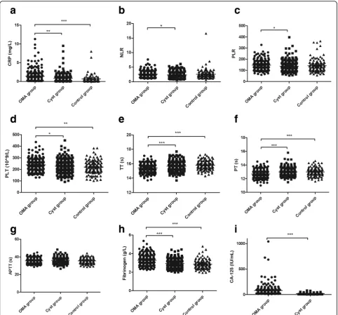
The mean (± SEM) levels of CRP (mg/L) were signifi-cantly higher in the OMA group (1.38 ± 0.11) than those in the Cyst group (0.86 ± 0.08, P< 0.01) and Control group (0.72 ± 0.15, P< 0.0001, Fig.1a). More-over, the levels of NLR and PLR were significantly higher in the OMA group (2.56 ± 0.07 and 146.4 ± 2.8) than those in the Cyst group (2.34 ± 0.07 and 137.7 ± 3.4, P< 0.05; Fig. 1b, c). Furthermore, the levels of PLT (10^9/L) and plasma fibrinogen (g/L) were also significantly higher in the OMA group (239.8 ± 3.6 and 3.29 ± 0.04) than those in the Cyst group (228.4 ± 4.0 and 2.93 ± 0.03, P< 0.05) and the Control group (220.0 ± 5.4 and 2.88 ± 0.05, P < 0.05; Fig. 1d, h). How-ever, the time of TT (s) and PT (s) were significantly shorter in the OMA group (15.42 ± 0.04 and 12.69 ± 0.04) than those in the Cyst group (15.68 ± 0.05 and 13.00 ± 0.04, P< 0.05) and the Control group(15.78 ±
0.06 and 12.99 ± 0.06, P < 0.05; Fig.1E–1F). No signifi-cant differences with regard to CRP, PLT, fibrinogen, TT or PT between the Cyst and Control groups were found (P> 0.05). There were no statistical differences in the APTT between groups (P > 0.05, Fig.1g).
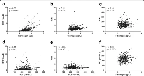
Spearman analysis showed that fibrinogen was positively correlated with CRP (r = 0.56,P< 0.0001; Fig.2a), NLR (r = 0.11,P< 0.01; Fig.2b), and PLR (r = 0.10, P< 0.05; Fig.2c). PLT was positively correlated with CRP (r = 0.16, P < 0.01; Fig.2d) but not significantly correlated with NLR (P= 0.45; Fig. 2e). Fibrinogen and PLT demonstrated a significantly positive correlation (r = 0.20, P < 0.0001; Fig.2f). Moreover, fibrinogen (r =−0.41, P < 0.0001; r =−0.37, P < 0.0001 re-spectively) and PLT (r =−0.10, P< 0.05; r =−0.19, P < 0.0001, respectively) were negatively correlated with TT and PT but not significantly correlated with APTT (P> 0.05).
Coagulation parameters and inflammatory biomarkers in women with ovarian endometriosis
In the OMA group, no significant correlation between co-agulation parameters or inflammatory markers and dys-menorrhea, cyst size or endometriosis stage was found (Table 2). However, the levels of PLT in women with endometriosis who had severe pelvic adhesions were sig-nificant higher when compared with women with endo-metriosis who had mild or no pelvic adhesions (P < 0.05; Table2). In addition, the time of TT, PT and APTT were shorted, and the levels of fibrinogen were higher in women with endometriosis who had severe pelvic adhe-sions as compared with women with endometriosis who had mild or no pelvic adhesions, but the differences did not reach statistical significance (P > 0.05; Table2).
Diagnostic value of CA-125, coagulation and inflammatory parameters in ovarian endometrioma The levels of CA-125 in OMA group were significantly higher than those in Cyst group (80.0 ± 7.1 vs. 19.2 ± Table 1Patients’characteristics
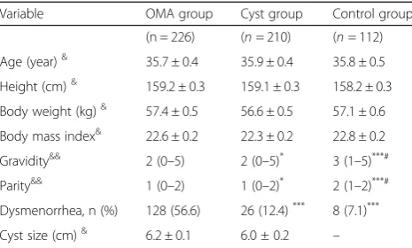
Variable OMA group Cyst group Control group (n = 226) (n= 210) (n= 112) Age (year)& 35.7 ± 0.4 35.9 ± 0.4 35.8 ± 0.5 Height (cm)& 159.2 ± 0.3 159.1 ± 0.3 158.2 ± 0.3 Body weight (kg)& 57.4 ± 0.5 56.6 ± 0.5 57.1 ± 0.6 Body mass index& 22.6 ± 0.2 22.3 ± 0.2 22.8 ± 0.2 Gravidity&& 2 (0–5) 2 (0–5)* 3 (1–5)***# Parity&& 1 (0–2) 1 (0–2)* 2 (1–2)***# Dysmenorrhea, n (%) 128 (56.6) 26 (12.4)*** 8 (7.1)*** Cyst size (cm)& 6.2 ± 0.1 6.0 ± 0.2 –
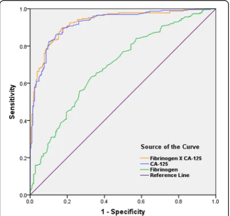
0.8;P< 0.0001; Fig.1i), with a cut-off value at 30.75 IU/ mL. The AUC of CA-125 was 0.924 (95% confidence interval: 0.899–0.949) with sensitivity and specificity reaching 82.3 and 90.0%, respectively (Table 3). The optimal cut-off points for inflammatory markers, co-agulation parameters, and the combined marker (plus CA-125) for determining endometriosis were evaluated by ROC analysis (Table3). The results also showed that the sensitivity and specificity of using any of these
factors alone for diagnosis of endometrioma were lower than those of CA-125. For fibrinogen, the cut-off value was 3.09 g/L with a validity power at 0.9998 (P1 = 0.63, P2 = 0.28, β= 0.0002) in present study. However, the combination of CA-125 and fibrinogen showed the highest AUC of 0.928 (0.904–0.952) with sensitivity of 0.898 and specificity of 0.829 (Fig.3). The diagnostic ef-fects of TT or PT combined with CA-125 were also better than that of CA-125 alone. (Table3).
Fig. 1Levels of inflammatory biomarkers and coagulation parameters in groups.a-c, Inflammatory biomarkers of CRP (a), NLR (b) and PLR (c)
were measured among groups.d-i, Coagulation parameters of PLT (d), TT (e), PT (f), APTT (g) and fibrinogen (h), as well as cancer antigen CA-125
(i) were measured among groups. OMA group, women with ovarian endometriomas; Cyst group, women with non-endometriosis benign ovarian
cysts; Control group, women undergoing tubal anastomosis; CRP, C-reactive protein; NLR, neutrophil-to-lymphocyte ratio; PLR;
platelet-to-lymphocyte ratio; PLT, platelet count; TT, thrombin time; PT, prothrombin time; APTT, activated partial thromboplastin time; OMA group; Cyst
Discussion
Women with ovarian endometriomas demonstrated a hy-percoagulable and inflammatory status, based on increased levels of CRP, PLT, and fibrinogen as well as shortened TT and PT.
Evidence shows that systemic inflammation activates the coagulation system in response to TF-mediated thrombin generation [26]. TF can be secreted by activated monocytes
and endothelial and polymorphonuclear cells, which are regulated by TNF-α, IL-1β, and lipopolysaccharide [27–29]. Ding et al. have [14] reported that TF concentrations are significantly elevated in primary endometriotic stromal cells. TF binds to circulating factor VIIa to mediate the acti-vation of factors IX and X and generates thrombin [30]. It has been reported in endometriotic stromal cells, thrombin and proteinase-activated receptor (PAR)-1 agonist induce Fig. 2Correlations of coagulation parameters and inflammatory biomarkers.a-c, The correlations of fibrinogen with CRP (a), NLR (b) and PLR (c)
were tested using Spearmen analysis.d-f, The correlations of PLT with CRP(d), NLR (e) and fibrinogen (f) were tested using Spearmen analysis.
CRP, C-reactive protein; NLR, neutrophil-to-lymphocyte ratio; PLR; platelet-to-lymphocyte ratio; PLT, platelet count
Table 2Coagulation parameters and inflammatory biomarkers in women with ovarian endometriosis
Variables CRP NLR PLR PLT TT PT APTT Fb Dysmenorrhea
Yes (n= 128) 1.46 ± 0.01 2.60 ± 0.10 148.7 ± 3.9 242.3 ± 4.9 15.45 ± 0.05 12.72 ± 0.05 35.86 ± 0.27 3.32 ± 0.05 No (n= 98) 1.27 ± 0.02 2.50 ± 0.10 143.3 ± 3.8 236.5 ± 5.2 15.37 ± 0.06 12.65 ± 0.06 35.11 ± 0.28 3.25 ± 0.05 Cyst size, cm
< = 5 cm (n= 66) 1.32 ± 0.02 2.66 ± 0.14 153.2 ± 5.8 237.3 ± 7.3 15.48 ± 0.08 12.71 ± 0.07 35.06 ± 0.34 3.29 ± 0.07 > 5 cm (n= 160) 1.40 ± 0.01 2.52 ± 0.08 143.5 ± 3.1 240.8 ± 4.0 15.39 ± 0.04 12.68 ± 0.05 35.73 ± 0.23 3.29 ± 0.04 Stage
III (n= 91) 1.22 ± 0.02 2.58 ± 0.10 145.6 ± 4.5 243.8 ± 5.4 15.38 ± 0.06 12.64 ± 0.06 35.68 ± 0.30 3.24 ± 0.05 IV (n= 135) 1.48 ± 0.01 2.54 ± 0.09 146.9 ± 3.5 237.1 ± 4.7 15.44 ± 0.05 12.72 ± 0.05 35.44 ± 0.26 3.32 ± 0.06 Pelvic adhesions#
Yes (n= 204) 1.38 ± 0.01 2.52 ± 0.07 147.7 ± 3.0 242.6 ± 3.8* 15.41 ± 0.04 12.68 ± 0.04 35.51 ± 0.20 3.29 ± 0.04 N0 (n= 22) 1.37 ± 0.10 2.90 ± 0.32 134.2 ± 6.8 214.0 ± 10.0 15.51 ± 0.13 12.76 ± 0.14 35.77 ± 0.71 3.25 ± 0.11
IL-6 and IL-8 secretion and cell proliferation [23]. Throm-bin also can Throm-binds to another type of PAR expressed in endometriotic stromal cells, PAR-2, mediating the produc-tion of chemokines and cytokines such as IL-8, monocyte chemotactic protein-1, MMP-2, and cyclooxygenase-2 [22].
Studies have reported that inflammatory also in-duces fibrinolysis activation in endometriosis. Plasmin,
an active enzyme, can degrade various extracellular matrix proteins and activate MMPs [31]. The eutopic endometrium of women with ovarian endometriosis has been shown to express high levels of MMP-3, which can hydrolyze and inactivate PAI-1, regulating cell-associated plasmin activities [32]. Higher levels of PAI-1 and tissue inhibitor of metalloproteinase-1 in ovarian endometriomas prevent endometriotic cysts from invading surrounding ovarian tissues [33, 34]. Meanwhile, activated plasmin may induce expression of proinflammatory cytokines such as IL-1α, IL-1β, TNF-α, and TF [28]. Inflammatory changes and acti-vated fibrinolytic systems in women with endometrio-mas may play an important role in the development and progression of endometriosis.
Fibrinogen influences thrombin formation, platelet ag-gregation, blood rheology and blood viscosity. Fibrino-gen levels are elevated in a variety of diseases such as diabetes and nephrotic diseases, and are associated with an increased risk of cardiovascular disease [35, 36]. Fi-brinogen is closely associated with hypercoagulation. Kurata et al. [37] reported that TT, APTT, and PT were all significantly shortened in canines injected with brinogen. In the present study, the levels of plasma fi-brinogen were significantly higher in women with ovarian endometriomas than those in women with non-endometriosis benign ovarian cysts and those in women without ovarian cysts. These results are in agreement with those of previous reports [15, 16]. We also found Table 3The diagnostic value of CA-125, coagulation and inflammatory parameters in ovarian endometrioma
Parameters AUC (95% CI) Sensitivity (%) Specificity (%) Cutoff value CA-125 (IU/mL) 0.924 (0.899–0.949) 82.3 90.0 30.75 CRP (mg/L) 0.630 (0.578–0.682) 78.8 40.1 0.35 NLR 0.575 (0.522–0.629) 77 39.5 1.88 PLR 0.584 (0.530–0.638) 65.9 51.4 128.3 PLT (10^9/L) 0.557 (0.503–0.611) 41.2 69.5 253.5 TT (s) 0.613 (0.560–0.665) 69.5 49.1 15.35 PT (s) 0.643 (0.592–0.695) 56.7 65 12.85 APTT (s) 0.546 (0.492–0.600) 33.3 76.1 37.45 Fibrinogen (g/L) 0.692 (0.643–0.741) 63.3 67.1 3.09 CA-125 * CRP (IU*mg/mL*L) 0.832 (0.794–0.869) 69.5 82.6 24.17 CA-125 * NLR (IU/mL) 0.899 (0.870–0.927) 84.1 81.0 60.06 CA-125 * PLR (IU/mL) 0.907 (0.879–0.935) 88.1 80.0 3383.16 CA-125 * PLT (IU*10^9/mL*L) 0.909 (0.882–0.937) 80.1 87.6 6695.85 CA-125 / TT (IU*s/mL) 0.926 (0.902–0.951) 85.0 88.1 1.84 CA-125 / PT (IU*s/mL) 0.927 (0.903–0.952) 87.2 85.7 2.06 CA-125 / APTT (IU*s/mL) 0.924 (0.899–0.949) 88.1 84.3 0.75 CA-125*Fibrinogen(IU*g/mL*L) 0.928 (0.904–0.952) 89.8 82.9 73.77
AUCarea under the curve,CRPC-reactive protein,NLRneutrophil-to-lymphocyte ratio,PLRplatelet-to-lymphocyte ratio,PLTplatelet count,TTthrombin time,PT prothrombin time,APTTactivated partial thromboplastin time
that TT and PT were significantly shortened in patients with endometrioma, but there was no difference in APTT. In coagulation cascade, the procoagulation factor, PT, measures the extrinsic coagulation pathway. PT is most sensitive to factor VII (FVII) levels as the latter ex-hibits a short half-life [38]. Given PT is initiated by TF, our findings correspond to previous studies that have re-ported the elevation of TF in endometriotic lesions and PF in women with endometriosis [14, 18]. However, Paola et al. [17] demonstrated shortened APTT and con-stant TT, whereas Guo et al. [15] reported shortened TT and APTT and constant PT in women with endometri-osis. These different results may be attributed to the dif-ferent sample sizes, conditions and techniques applied between the studies, considering that blood assays are highly dependent on the combination of reagents and in-struments. For coagulation parameters, specific reagents and different manufacturers usually lead to variable re-sults [38]. Obviously, the coagulation function of women with endometriosis needs further study.
CA-125 is a marker and often used in the diagnosis of endometriomas. Some studies reported that NLR as an adjunct to CA-125 is a useful diagnostic marker [6, 7]. However, some studies refute this claim because NLR has not yet been fully investigated and is not suitable as a diagnostic tool for advanced endometriosis [39,40]. In our study, we demonstrated that the coagulation factors TT, PT, and fibrinogen were more reliable as comple-mentary auxiliary markers of CA-125 for identifying ovarian endometrioma from non-endometriosis benign ovarian cysts. Obviously, the discrepancies among the studies can be attributed to the differences in sample size, experimental measurement methods, and instru-ments used.
The primary limitation of our study is its retrospective design. All the recruited women were in proliferative phase of menstrual cycle. The coagulation stability in women with endometriosis remains unknown while co-agulation status is usually unaffected by menstrual cycle in healthy women [41]. Moreover, only women with ad-vanced endometriosis (stages III–IV) were included in the study. Therefore, the coagulation function and its in-fluence on inflammation in women with early-stage endometriosis are still uncertain.
A combined influence of inflammatory response and hypercoagulation status may exist in advanced stages of endometriosis. At present, the treatment of endometriosis often focuses on anti-inflammatory control. Modulation of the coagulation pathway in endometriosis may provide an-other potential treatment option. Studies have reported in-hibition of inflammation by blocking formation of TF-PAR-2 and the TF-VIIa signaling pathway [42]. In a mouse model of endometriosis, a chimeric immunoconjugate mol-ecule specifically targeting endothelial TF in ectopic
implants has been shown to obliterate the endometriotic implant by vascular disruption without reducing fertility [43]. Guo et al. [44] reported that targeting P-selectin-mediated platelet adhesion can reduce the size of endome-triotic lesions, improving general hyperalgesia and result-ing in reduction of macrophage infiltration and fibrotic tissue content. Thus, in order to determine whether ab-normal coagulation parameters contribute to the diagnosis and treatment of endometriosis, a more detailed study of large samples is needed.
In conclusion, our findings suggest that women with ovarian endometriomas demonstrate a hypercoagulable sta-tus potentially attributable to the inflammatory nature of endometrioma. Plasma fibrinogen is an auxiliary marker of serum CA-125 in the diagnosis of endometriosis.
Abbreviations
APTT:Activated partial thromboplastin time; AUC: Area under the curve; BMI: Body mass index; CA: Cancer antigen; CRP: C-reactive protein; IL: Interleukin; MMP: Matrix metalloproteinase; NLR: Neutrophil-to-lymphocyte ratio; PAIs: Plasminogen activator inhibitors; PAR: Proteinase-activated receptor; PF: Peritoneal fluid; PLR: Platelet-to-lymphocyte ratio; PLT: Platelet count; PT: Prothrombin time; ROC: Receiver operating characteristic; SEM: Standard error of the mean; TF: Tissue factor; TNF: Tumor necrosis factor; TT: Thrombin time; uPA: urokinase plasminogen activator
Acknowledgements
None.
Consent to publication
Not applicable.
Authors’contributions
SD: Data Collection, Manuscript writing; QL: Data collection, Manuscript writing; TZ: Data collection; TL: Data analysis; LZ: Data analysis; JW: Data analysis, Manuscript writing; XZ: Project development, Manuscript editing. All authors have read and approved the manuscript in its current state.
Funding
This work was supported by National Key R&D Program of China (2017YFC1001202), The National Natural Science Foundation of China (81471433, 81671429), and Key Medical Science (Innovation) Project of Zhejiang Province. The funders had no role in the design of the study, in the collection, analysis, and interpretation of the data, or in the writing or approval of the manuscript.
Availability of data and materials
The datasets used and/or analyzed during the current study are available from the corresponding author on reasonable request.
Ethics approval and consent to participate
This study is approved by the Human Ethics Committee of the Women’s
Hospital, School of Medicine, Zhejiang University (No.20170174) and all women provided written informed consent.
Competing interests
The authors declare that they have no competing interests.
Received: 13 March 2018 Accepted: 29 November 2019
References
1. Burney RO, Giudice LC. Pathogenesis and pathophysiology of
endometriosis. Fertil Steril. 2012;98(3):511–9.
2. Sikora J, Mielczarek-Palacz A, Kondera-Anasz Z. Imbalance in cytokines from
interleukin-1 family - role in pathogenesis of endometriosis. Am J Reprod
3. Wickiewicz D, Chrobak A, Gmyrek GB, Halbersztadt A, Gabrys MS, Goluda M, et al. Diagnostic accuracy of interleukin-6 levels in peritoneal fluid for
detection of endometriosis. Arch Gynecol Obstet. 2013;288(4):805–14.
4. Birt JA, Nabli H, Stilley JA, Windham EA, Frazier SR, Sharpe-Timms KL.
Elevated peritoneal fluid TNF-alpha incites ovarian early growth response factor 1 expression and downstream protease mediators: a correlation with
ovulatory dysfunction in endometriosis. Reprod Sci. 2013;20(5):514–23.
5. Velasco I, Acien P, Campos A, Acien MI, Ruiz-Macia E. Interleukin-6 and other
soluble factors in peritoneal fluid and endometriomas and their relation to
pain and aromatase expression. J Reprod Immunol. 2010;84(2):199–205.
6. Tokmak A, Yildirim G, Oztas E, Akar S, Erkenekli K, Gulsen P, et al. Use of
neutrophil-to-lymphocyte ratio combined with CA-125 to distinguish
Endometriomas from other benign ovarian cysts. Reprod Sci. 2016;23(6):795–802.
7. Cho S, Cho H, Nam A, Kim HY, Choi YS, Park KH, et al. Neutrophil-to-lymphocyte
ratio as an adjunct to CA-125 for the diagnosis of endometriosis. Fertil Steril.
2008;90(6):2073–9.
8. Yang H, Zhu L, Wang S, Lang J, Xu T. Noninvasive diagnosis of moderate to
severe endometriosis: the platelet-lymphocyte ratio cannot be a neoadjuvant biomarker for serum cancer antigen 125. J Minim Invasive
Gynecol. 2015;22(3):373–7.
9. Yamaguchi K, Mandai M, Toyokuni S, Hamanishi J, Higuchi T, Takakura K,
et al. Contents of endometriotic cysts, especially the high concentration of free iron, are a possible cause of carcinogenesis in the cysts through the iron-induced persistent oxidative stress. Clin Cancer Res : Official J Am Assoc
Cancer Res. 2008;14(1):32–40.
10. Mizumoto H, Saito T, Ashihara K, Nishimura M, Takehara M, Tanaka R, et al.
Expression of matrix metalloproteinases in ovarian endometriomas:
immunohistochemical study and enzyme immunoassay. Life Sci. 2002;71(3):259–73.
11. Reis FM, Di Blasio AM, Florio P, Ambrosini G, Di Loreto C, Petraglia F.
Evidence for local production of inhibin a and activin a in patients with
ovarian endometriosis. Fertil Steril. 2001;75(2):367–73.
12. Boss EA, Massuger LF, Thomas CM, Geurts-Moespot A, van Schaik JH,
Boonstra H, et al. Clinical value of components of the plasminogen
activation system in ovarian cyst fluid. Anticancer Res. 2002;22(1A):275–82.
13. Sanchez AM, Vigano P, Somigliana E, Panina-Bordignon P, Vercellini P,
Candiani M. The distinguishing cellular and molecular features of the endometriotic ovarian cyst: from pathophysiology to the potential endometrioma-mediated damage to the ovary. Hum Reprod Update. 2014;
20(2):217–30.
14. Ding D, Liu X, Duan J, Guo SW. Platelets are an unindicted culprit in the
development of endometriosis: clinical and experimental evidence. Hum
Reprod. 2015;30(4):812–32.
15. Wu Q, Ding D, Liu X, Guo SW. Evidence for a Hypercoagulable state in
women with ovarian Endometriomas. Reprod Sci. 2015;22(9):1107–14.
16. Chmaj-Wierzchowska K, Kampioni M, Wilczak M, Sajdak S, Opala T. Novel markers
in the diagnostics of endometriomas: Urocortin, ghrelin, and leptin or leukocytes,
fibrinogen, and CA-125? Taiwan J Obstet Gynecol. 2015;54(2):126–30.
17. Vigano P, Ottolina J, Sarais V, Rebonato G, Somigliana E, Candiani M.
Coagulation status in women with endometriosis. Reprod Sci. 2017; 1933719117718273.
18. Lin M, Weng H, Wang X, Zhou B, Yu P, Wang Y. The role of tissue factor and
protease-activated receptor 2 in endometriosis. Am J Reprod Immunol.
2012;68(3):251–7.
19. Bruse C, Bergqvist A, Carlstrom K, Fianu-Jonasson A, Lecander I, Astedt B.
Fibrinolytic factors in endometriotic tissue, endometrium, peritoneal fluid, and plasma from women with endometriosis and in endometrium and
peritoneal fluid from healthy women. Fertil Steril. 1998;70(5):821–6.
20. Gilabert-Estelles J, Ramon LA, Espana F, Gilabert J, Vila V, Reganon E, et al.
Expression of angiogenic factors in endometriosis: relationship to fibrinolytic
and metalloproteinase systems. Hum Reprod. 2007;22(8):2120–7.
21. Levi M, van der Poll T. Coagulation and sepsis. Thromb Res. 2017;149:38–44.
22. Hirota Y, Osuga Y, Hirata T, Harada M, Morimoto C, Yoshino O, et al.
Activation of protease-activated receptor 2 stimulates proliferation and interleukin (IL)-6 and IL-8 secretion of endometriotic stromal cells. Hum
Reprod. 2005;20(12):3547–53.
23. Hirota Y, Osuga Y, Hirata T, Yoshino O, Koga K, Harada M, et al. Possible
involvement of thrombin/protease-activated receptor 1 system in the
pathogenesis of endometriosis. J Clin Endocrinol Metab. 2005;90(6):3673–9.
24. Szaba FM, Smiley ST. Roles for thrombin and fibrin(ogen) in cytokine/
chemokine production and macrophage adhesion in vivo. Blood. 2002;
99(3):1053–9.
25. Revised American Society for Reproductive Medicine classification of
endometriosis: 1996. Fertility and sterility. 1997;67(5):817–21.
26. Levi M, Keller TT, van Gorp E, ten Cate H. Infection and inflammation and
the coagulation system. Cardiovasc Res. 2003;60(1):26–39.
27. Parry GC, Mackman N. Transcriptional regulation of tissue factor expression
in human endothelial cells. Arterioscler Thromb Vasc Biol. 1995;15(5):612–21.
28. Syrovets T, Jendrach M, Rohwedder A, Schule A, Simmet T. Plasmin-induced
expression of cytokines and tissue factor in human monocytes involves
AP-1 and IKKbeta-mediated NF-kappaB activation. Blood. 200AP-1;97(AP-12):394AP-1–50.
29. Rauch U, Bonderman D, Bohrmann B, Badimon JJ, Himber J, Riederer MA,
et al. Transfer of tissue factor from leukocytes to platelets is mediated by
CD15 and tissue factor. Blood. 2000;96(1):170–5.
30. Eilertsen KE, Osterud B. Tissue factor: (patho)physiology and cellular biology.
Blood Coagul Fibrinolysis : Int J Haemost Thromb. 2004;15(7):521–38.
31. Murphy G, Knauper V, Atkinson S, Gavrilovic J, Edwards D. Cellular
mechanisms for focal proteolysis and the regulation of the
microenvironment. Fibrinolysis Proteol. 2000;14(2–3):165–74.
32. Ugwu F, Lemmens G, Collen D, Lijnen HR. Modulation of cell-associated
plasminogen activation by stromelysin-1 (MMP-3). Thromb Haemost. 1999;
82(3):1127–31.
33. Gilabert-Estelles J, Estelles A, Gilabert J, Castello R, Espana F, Falco C, et al.
Expression of several components of the plasminogen activator and matrix
metalloproteinase systems in endometriosis. Hum Reprod. 2003;18(7):1516–22.
34. Ramon L, Gilabert-Estelles J, Castello R, Gilabert J, Espana F, Romeu A, et al.
mRNA analysis of several components of the plasminogen activator and matrix metalloproteinase systems in endometriosis using a real-time
quantitative RT-PCR assay. Hum Reprod. 2005;20(1):272–8.
35. Zhao Y, Zhang J, Zhang J, Wu J. Diabetes mellitus is associated with
shortened activated partial thromboplastin time and increased fibrinogen values. PLoS One. 2011;6(1):e16470.
36. Zwaginga JJ, Koomans HA, Sixma JJ, Rabelink TJ. Thrombus formation and
platelet-vessel wall interaction in the nephrotic syndrome under flow
conditions. J Clin Invest. 1994;93(1):204–11.
37. Kurata M, Sasayama Y, Yamasaki N, Kitazawa I, Hamada Y, Horii I. Mechanism
for shortening PT and APTT in dogs and rats--effect of fibrinogen on PT and
APTT. J Toxicol Sci. 2003;28(5):439–43.
38. Ng VL. Prothrombin time and partial thromboplastin time assay
considerations. Clin Lab Med. 2009;29(2):253–63.
39. Yavuzcan A, Caglar M, Ustun Y, Dilbaz S, Ozdemir I, Yildiz E, et al. Evaluation
of mean platelet volume, neutrophil/lymphocyte ratio and platelet/ lymphocyte ratio in advanced stage endometriosis with endometrioma. J
Turk Ger Gynecol Association. 2013;14(4):210–5.
40. Kim SK, Park JY, Jee BC, Suh CS, Kim SH. Association of the
neutrophil-to-lymphocyte ratio and CA-125 with the endometriosis score. Clin Exp Repro
Med. 2014;41(4):151–7.
41. Tchaikovski SN, Thomassen MC, Costa SD, Bremme K, Rosing J. Changes in
haemostatic parameters during the menstrual cycle and subsequent use of
drospirenone-containing oral contraceptives. Thromb Res. 2014;134(5):1032–7.
42. Ahamed J, Versteeg HH, Kerver M, Chen VM, Mueller BM, Hogg PJ, et al.
Disulfide isomerization switches tissue factor from coagulation to cell
signaling. Proc Natl Acad Sci U S A. 2006;103(38):13932–7.
43. Krikun G, Hu Z, Osteen K, Bruner-Tran KL, Schatz F, Taylor HS, et al. The
immunoconjugate "icon" targets aberrantly expressed endothelial tissue
factor causing regression of endometriosis. Am J Pathol. 2010;176(2):1050–6.
44. Guo SW, Ding D, Geng JG, Wang L, Liu X. P-selectin as a potential
therapeutic target for endometriosis. Fertil Steril. 2015;103(4):990–1000 e8.
Publisher’s Note