RESEARCH
Is a positive intracutaneous
test induced by penicillin mediated
by histamine? A cutaneous microdialysis study
in penicillin-allergic patients
Line K. Tannert
1*, Sidsel Falkencrone
2, Charlotte G. Mortz
1, Carsten Bindslev‑Jensen
1and Per Stahl Skov
1,2Abstract
Background: Diagnostic workup of penicillin allergy comprises skin testing with penicillins, and patients are deemed allergic if skin test is positive. However, the literature suggests that skin test‑positive patients may be challenge‑nega‑ tive, indicating that the skin test may be falsely positive.
Objective: To investigate real‑time histamine release from a positive intracutaneous test induced by penicillin in patients with positive and negative challenges to penicillin.
Methods: Skin microdialysis was performed in 21 penicillin‑allergic patients with positive skin test, 13 non‑allergic volunteers serving as negative controls, and 7 grass pollen‑allergic patients serving as positive controls. Histamine was measured by microdialysis after skin test with penicillin/grass/NaCl. Penicillin challenge was subsequently performed in 12 of the patients.
Results: Only 10/21 patients (47.6%) were skin test positive at microdialysis. During microdialysis 13 single intracu‑ taneous tests were positive and histamine was detected in 4/13 occurring in four challenge positive patients. Thir‑ teen/21 patients (61.9%) were deemed allergic to penicillin; eight had positive skin test. Two patients with positive skin test were challenge negative. In grass pollen allergic patients, 7/7 had a positive intracutaneous test to grass and all released histamine in the wheals. All 13 negative controls had negative intracutaneous test to penicillin and no histamine release.
Conclusion: Histamine was only detected in the minority of positive intracutaneous tests with penicillin in penicillin‑ allergic patients. Other mediators may be involved.
Keywords: Histamine release, Penicillin‑allergy, Penicillin challenge, Penicillin intracutaneous test, Skin microdialysis
© The Author(s) 2017. This article is distributed under the terms of the Creative Commons Attribution 4.0 International License (http://creativecommons.org/licenses/by/4.0/), which permits unrestricted use, distribution, and reproduction in any medium, provided you give appropriate credit to the original author(s) and the source, provide a link to the Creative Commons license, and indicate if changes were made. The Creative Commons Public Domain Dedication waiver (http://creativecommons.org/ publicdomain/zero/1.0/) applies to the data made available in this article, unless otherwise stated.
Background
Identification of penicillin allergy is important, and the diagnostic work-up of patients with suspected penicillin allergy consists of several steps according to guidelines
[1–3]. One step is skin testing, and if either the skin prick
test (SPT) or the intracutaneous test (ICT) is positive, the
patient is deemed to have a life-long allergy to penicillin. Positive skin testing is considered reliable for the diagno-sis of penicillin allergy, but we recently demonstrated that 60% of patients with a positive skin test were
challenge-negative to the culprit penicillin [4]. A few other studies
also reported negative challenges in patients with a
posi-tive skin test [5–12].
A positive SPT or ICT to an allergen is generally accepted to be due to histamine that is released in an IgE-mediated reaction from the skin mast cells, although these cells contain a large number of other mediators and are considered to be the orchestrating cells in initiation
Open Access
*Correspondence: line.tannert@rsyd.dk
1 Department of Dermatology and Allergy Center, Odense Research Center for Anaphylaxis, Odense University Hospital, Kløvervænget 15, 5000 Odense C, Denmark
and dissemination of the allergic reaction [13]. Previ-ous skin microdialysis studies have confirmed histamine release in wheals induced by grass in pollen-allergic
patients [14]. Histamine release in wheals has also been
demonstrated in non-IgE mediated reactions such as ice
cube challenge in cold urticaria patients [15].
The aim of this study was to use skin microdialysis to investigate histamine release in patients with positive intracutaneous test to penicillin, and to compare the results with challenge outcome.
Methods
Participants
The following groups of patients and controls were
included in the microdialysis study (Table 1).
• Patients (n = 21) with a case history of allergic
reaction to a penicillin and positive intracutane-ous test to at least one type of penicillin; 6 men and 15 women, mean age 51 years (range 23–70 years). Of the 21 patients, 5 also had low levels of specific IgE to one or more penicillin (s-IgE). Median time interval between initial reaction and inclusion in the study was 40 months (range 1–240 months). Two of the patients had a case history of a non-immediate reaction occurring > 1 h after last administration of
penicillin (Patient 12 and 13, Table 1). Two patients
(Patient 8 and 17, Table 1) could not remember the
exact timing from administration until reaction, and the remaining 17 had immediate reactions (occur-ring < 1 h after last penicillin administration).
• Healthy volunteers (n = 13) without any
aller-gic reactions to penicillin treatment; 2 men and 11 women, mean age 45 years (range 27–62 years), served as negative controls.
• Grass pollen-allergic patients (n = 7); 3 men and 4
women, mean age 35 years (range 27–51 years), with a positive skin prick test and s-IgE to grass, suffering from rhinoconjunctivitis during the grass pollen sea-son, and serving as positive “classical allergic” controls.
Skin testing
An intracutaneous test with penicillin was performed during microdialysis with the penicillin(s) previously shown to induce a positive test. The following concentra-tions were used: benzylpenicillin 20 mg/mL, amoxicillin 20 mg/mL, ampicillin 20 mg/mL, dicloxacillin 1 mg/mL, and mecillinam 4 mg/mL both in patients and controls. Except for benzylpenicillin, concentrations were accord-ing to European Network for Drug Allergy (ENDA)
guidelines [16].
During microdialysis, ICT was performed with the penicillin(s) that was positive at first ICT. None of the
patients were tested with the major or minor determi-nants, PPL and MDM, during microdialysis. Skin testing with these reagents was not part of the routine testing at the Allergy Center, because Hjortlund et al. demonstrated that all patients with a positive PPL or MDM were also
positive to benzylpenicillin [17]. However, 5/21 patients
had a previous PPL and MDM skin test; one patient was positive to both but concomitantly positive to benzylpen-icillin. Controls were tested with the same types of peni-cillin as the patients.
Intracutaneous test with grass was performed with Phleum pratense extract in a dilution of 1000 SQ-U/mL (ALK-Abello, Hørsholm, Denmark).
The non-IgE dependent histamine releaser, codeine
(codeine phosphate 1 mg/mL RefLab®, Copenhagen,
Denmark) was used as positive control to demon-strate releasability of histamine from skin mast cells and to release residual histamine after a positive ICT induced by penicillin or grass. Intracutaneous test with isotonic saline 0.9% was the negative control in all participants.
ICT was always performed by injecting 50 µL, and reactions were considered positive if the wheal size diam-eter was 3 mm larger than the initial bleb. ICT was read
after 20 min, according to ENDA guidelines [2].
Regarding interpretation of the ICTs, all tests with pen-icillin in the group of 21 patients were assessed blinded with photographs of the reactions by three independent consultants with experience in skin testing. Evaluations were in full compliance with the investigators’ primary evaluation.
Measurements of IgE to penicillins (s‑IgE)
S-IgE against penicillin V, penicillin G, amoxicillin (AX), and ampicillin (AMP) were measured using ImmunoCap
(Thermo Fischer, Uppsala, Sweden). Results ≥ 0.35 kU/L
were considered positive.
Penicillin challenge
Patients with a present or previous positive ICT elicited by penicillin were challenged with the culprit penicillin with increasing doses: starting at 1/100 of a therapeutic dose, followed by 1/10 and finally 1/1 with 30 min inter-vals. Therapeutic doses were as follows: Pen V 800 mg, DX 1000 mg, MC 400 mg, AMP 500 mg, AX 750 mg, and
AX + Cla 500/125 mg. For safety reasons, patients who
had a delayed positive reaction to ICT (n = 3) or a recent
anaphylactic reaction to penicillin (n = 2) were not
chal-lenged. Three patients refused challenge.
Skin microdialysis
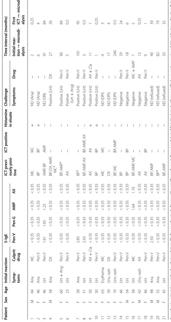
Table 1 Demo gr aphic da ta and r esult of sk in t est , s-IgE , micr odialy
sis and challenge in pa
tien
ts with a pr
evious p ositiv e IC T t o p enicillin Age r ef ers t o pa tien ts
’ age a
t the da
y of micr
odialy
sis
. Challenge r
ef
ers t
o challenge with penicillin per
for
med af
ter micr
odialy
sis
. * Not sk
in t
est
ed with amo
xicillin because it w
as not a
vailable a
t the time of t
esting
. P
atien
t
16 w
as challenged a
t t w o separ at e oc casions Pen V penicillin V , Pen G penicillin G, AMP ampicillin, AX amo xicillin, MC mecillinam, Ana rec en t anaph ylaxis , DPI dela yed positiv e IC T, SRI sy st emic r eac tion dur ing IC T, Ur t ur ticar ia, Ang ang ioedema, ND not done Pa tien t Sex Ag e Initial r eac tion S‑ IgE IC T pr evi ‑ ously posi ‑ tiv e IC T positiv e H istamine in elua te Challenge Time in ter val (mon ths) Symp ‑ toms
Culprit drug
Pen V Pen G AM P AX Sympt oms D rug Initial r eac ‑ tion → micr odi ‑ aly sis
First ICT
substances. In this study, we performed microdialysis
as described by Petersen et al. [18]. The microdialysis
probes (EP Medical Aps®, Copenhagen, Denmark) were
semipermeable, linear, and equipped with a guide wire. The membrane had a molecular cut-off weight of 2 kDa
(outer diameter = 216 µm, wall thickness = 8 µm)
allow-ing passive diffusion of small molecular substances. The probes were inserted intradermally and as superficially as possible into the volar forearm at a length of 2 cm of the skin using a 23G cannula. They were perfused with isotonic NaCl 0.9% at a rate of 3 µL/min. Prior to probe insertion, a local anesthetic and vasoconstrictor cream
containing prilocaine and lidocaine (Emla®,
AstraZen-eca, Södertälje, Sweden) was applied to the skin for 1 h to diminish pain and bleeding from the injection sites.
Emla® does not affect histamine degranulation from mast
cells [19]. The dialysate was sampled in glass fiber-coated
microtiter wells at 2-min intervals for 72 min (Fig. 1).
Depending on the number of previous positive ICTs to penicillin, the participants had 2–4 probes inserted at least two cm apart. One probe in each participant was always a control probe where first ICT was done with NaCl and second ICT with codeine.
Baseline histamine values were collected for the first 24 min after which two ICTs were performed above each probe. The first ICT was with penicillin(s) in the penicil-lin-allergic (culprit penicillin(s)) patients and controls, grass in grass-pollen allergics, codeine in controls (posi-tive control), or isotonic NaCl (nega(posi-tive control) that were injected at a distance of 1 mm from each probe. The second ICT after 48 min was always performed by injec-tion of codeine to collect residual histamine from the skin around the probe. When a wheal developed, it always extended across the probe. Histamine collected from the microdialysate from the 2-min sampling period was
ana-lyzed spectrofluorometrically [20]. Detection level was 5
nanogram of histamine.
Histamine release was determined in:
• 12 baseline samples
• 12 samples after first ICT
• 12 samples after second ICT
Histamine area under curve (AUC)
As a supplement to peak histamine in wheals, the hista-mine under the curve, histahista-mine AUC, was calculated by
summing up the trapezoids; area = Σ((Y1 + Y2)/2*(X2
− X1)) + ((Yn + Yn − 1)/2*(Xn − Xn − 1)).The AUC
comprises histaminerelease added for all 36 samples (baseline, first and second ICT).
Statistical analysis
We analyzed data using Kruskal–Wallis test and Mann– Whitney test for ordinal data. Wilcoxon Signed Rank Test was used to determine whether ICT with penicillin
induced significant release of histamine. P values < 0.05
were considered significant. The statistical analyses were performed with SigmaPlot 13.0, Alfasoft, Sweden.
Results
Microdialysis experiments on grass pollen‑allergic patients and controls
As expected, ICT with grass pollen extract induced a wheal reaction in all seven grass pollen-allergic patients (median size 13 mm, range 7.5–16.5 mm) and a significant
hista-mine release from 2 min after first ICT (Fig. 2i). When
codeine was injected at second ICT at the same site,
non-significant increases in wheal size (p = 0.46) and peak
his-tamine (p = 0.07) were observed, indicating that the grass
pollen allergen released almost all the histamine from the skin mast cells. None of the controls developed wheals or
released histamine after skin testing with penicillin (Fig. 2g).
When injecting codeine twice, the first codeine ICT induced, as expected, a wheal reaction (median size 11 mm, range 10.0–12.5 mm) in all six controls and a significant histamine release from 2 min after the ICT
(Fig. 2j). The second codeine ICT induced a
non-signifi-cant increase in wheal size in 3 of 7 controls (p = 0.12),
and no peak histamine was observed (p = 0.39). We
therefore use codeine 1 mg/mL as a surrogate marker for total histamine content in the skin.
Control probe–codeine and histamine release
All penicillin patients (n = 21), grass pollen patients
(n = 7) and healthy controls (n = 13) reacted to ICT with
First ICT
Second ICT
Baseline sampling Sampling Sampling
0-24 min 26-48 min 50-72 min
Control probe in patients with a positive ICT penicillin, no histamine release, n=6
Time [min]
0 20 40 60
0 100 200 300
Positive ICT penicillin with histamine release, n=4
Time [min]
0 20 40 60
Histamine [ng/mL]
0 100 200
300 Control probe in patients with a positive ICT penicillin
with histamine release, n=4
Time [min]
0 20 40 60
0 100 200 300
Negative but previously positive ICT penicillin, n=11
Time [min]
0 20 40 60
Histamine [ng/mL]
0 100 200 300
Control probe in patients with a previous positive ICT penicillin, n=11
Time [min]
0 20 40 60
0 100 200 300
Controls, ICT penicillin negative, n=13
Time [min]
0 20 40 60
Histamine [ng/mL
]
0 100 200
300 Control probe, controls, n=13
Time [min]
0 20 40 60
0 100 200 300
Controls, codeine-codeine, n=6
Time [min]
0 20 40 60
0 100 200 300
ICT penicillin ICT codeine ICT NaCl ICT codeine
ICT NaCl ICT codeine
ICT NaCl ICT codeine
ICT NaCl ICT codeine ICT penicillin ICT codeine
ICT penicillin ICT codeine
ICT penicillin ICT codeine
ICT grass ICT codeine ICT codeine ICT codeine Positive ICT penicillin, no histamine release, n=6
Time [min]
0 20 40 60
Histamine [ng/mL]
0 100 200 300
Histamine [ng/mL]
Histamine [ng/mL]
Histamine [ng/mL]
Histamine [ng/mL
]
Histamine [ng/mL
]
a b
c d
e f
g h
i Grass allergics, positive control, n=7 j
Time [min]
0 20 40 60
Histamine [ng/mL
]
0 100 200 300
codeine with a wheal response showing a median diam-eter of 12.5 mm (range 7.5–23.5 mm) and a concomitant
significant histamine release (p < 0.001), with a median
peak histamine of 75.4 ng/mL (range 12.6–350.0 ng/
mL) (Fig. 2a–h). Compared with baseline, the ICT with
physiological saline gave no significant histamine release
(p = 0.79), median peak histamine 8.6 ng/mL (range
−7.6–35.5 ng/mL), and no wheals developed (Fig. 2b, d,
f, h).
Microdialysis in penicillin‑allergic patients
During microdialysis, 13 positive ICTs with penicil-lin developed in 10 patients; one patient was positive to three different penicillins, and one patient was posi-tive to two penicillins. Histamine was detected in four of these positive ICTs, which had median size 13.8 mm (range 12.0–20.5 mm) and a significant histamine release
from 2 min after the first ICT (Fig. 2c). These positive
ICTs occurred in four patients with a case history of recent anaphylactic reaction or severe urticaria to
phe-noxymethylpenicillin (n = 3) or mecillinam (n = 1);
all had negative s-IgE (Table 1). The nine positive ICTs
without histamine had a median size of 11.3 mm (range
8.5–15.0 mm) (Fig. 2a). The size did not differ
signifi-cantly from the ICTs with positive histamine release
(p = 0.17). No histamine was released in the negative
ICTs in patients with a previous positive ICT to
penicil-lin (Fig. 2e). Histamine release after ICT with codeine in
patients with a positive ICT was similar for patients with a penicillin-induced histamine release (median 165.4, range 88.2–270.1 ng/mL) and patients without a penicil-lin-induced histamine release (median 166.2, range 5.5–
308.1 ng/mL) (p = 0.82). Mast cells in all patients were
thus able to release histamine in detectable amounts. In patients with a positive ICT to penicillin (with or without histamine release), there was a significantly higher his-tamine release after the second ICT with codeine com-pared to patients with negative ICT to penicillin, controls
or grass pollen-allergic patients (p < 0.05) (Fig. 2a, c, e, g,
i, j).
Penicillin challenge
Intracutaneous test with the culprit penicillin was posi-tive in only 10/21 (47.6%) of patients who previously had had a positive ICT. There was a tendency that the time interval from initial reaction until microdialysis was shorter for patients with positive ICT penicillin (median 7.5 months) than patients with negative ICT (median
27 months) (p = 0.084). There were no differences
regarding age or gender in the two groups. In all patients, the penicillin(s) eliciting the positive ICT was identical to that causing the initial reaction.
Among the 21 penicillin-allergic patients with a previ-ous positive ICT, 13 were deemed allergic to penicillin: 7 were challenge-positive, 2 had recent anaphylaxis, 1 had a systemic reaction to ICT, and 3 had a delayed positive
ICT (Table 1). All seven patients with a positive challenge
developed urticaria and/or angioedema during challenge, and all reactions were immediate i.e. commencing within 1 h after intake of penicillin. Four of the seven challenge-positive patients had become ICT negative since the ini-tial evaluation whereas three were still positive; two of them had histamine release in the wheals. Additionally, two patients had positive s-IgE: one had negative reac-tions to penicillin ICT and challenge, and the other had positive reactions to penicillin ICT and challenge (but no histamine release).
One patient with a systemic reaction to ICT (Patient 3,
Table 1) had participated in microdialysis six months
pre-viously, where she had been s-IgE positive and had devel-oped a positive ICT with no histamine release (and no systemic reaction). In the planning of challenge, micro-dialysis was performed again and during this procedure the patient developed a systemic reaction 8 min after ICT with penicillin. Microdialysis was discontinued immedi-ately, but the eluate already collected was analyzed and showed an increase in histamine significantly above base-line from 4 min after the first ICT (data not shown and
not included in Fig. 2).
Both of the patients with recent anaphylaxis (one also IgE positive) had positive ICT with histamine release in the wheal.
A positive ICT was present in 8 of 13 patients who were deemed penicillin-allergic in this setting (Patient
1–13, Table 1), providing a sensitivity of 62%. Two of five
patients with a negative challenge had positive ICT, giv-ing a specificity of 60%. In contrast, the sensitivity of ICT with grass was 100%.
For positive ICTs with histamine release, a sensitivity of 30% and specificity of 100% were found.
Total histamine released in the skin
There was no significant difference in total histamine release in patients with a positive ICT to penicillin with
or without histamine release (p = 0.777), data not shown,
and there was no difference in total histamine in patients with a previous positive ICT compared with controls
(p = 0.729). Figure 3 compares all patients with a
posi-tive ICT to penicillin (with or without histamine release) and all with a negative ICT to penicillin (previously tive ICT to penicillin and controls). Patients with a posi-tive ICT to penicillin had significantly higher histamine levels (median 1877.9 ng/mL, range 84.0–3368.2 ng/mL) than patients with a negative ICT to penicillin (median
Similar results were obtained from the negative control probe: a significantly higher total histamine release in the group with positive ICT (median 1606.1 ng/mL, range 91.2–2934.8 ng/mL) than in the group with negative ICT to penicillin (median 605.8 ng/mL, range 79.2–4448.1 ng/
mL) (Fig. 3b).
Discussion
Previously, the microdialysis technique has been applied on grass pollen allergic patients demonstrating histamine
release in grass pollen induced wheals [21]. Further, it has
been demonstrated that there is a correlation between
size of wheal and histamine concentration in wheal [22].
To our knowledge, skin microdialysis has never been used to investigate histamine release after skin testing with an antibiotic.
All penicillin-allergic patients included in this study had a previous positive ICT to penicillin, but only 10 of 21 patients had a positive ICT to penicillin when entering the study 3–30 months after the initial positive ICT. This is in line with other studies describing declining rates of
positive ICTs over time [4, 5, 23, 24]. In contrast, three
patients (Patient 11–13, Table 1) showed a dual ICT
response with an immediate reaction followed hours later by a delayed reaction that was persistently positive in our study, which is in agreement with data from Hjortlund
et al. [17]. The fluctuating ICT response in the group of
penicillin-allergic patients differs from the reproducible
positive skin test to grass in the group of pollen allergic-patients with a “classical” s-IgE-mediated reaction. The difference between the two groups was further empha-sized by the fact that only a few of the penicillin-allergic patients were s-IgE positive to penicillin.
A positive ICT to penicillin was only partially corre-lated to a positive challenge: eight ICT positive patients were deemed allergic whereas two were challenge nega-tive. Four of the challenge positive patients had negative ICT to penicillin, but all had had positive ICT at the ini-tial evaluation.
The four ICTs induced by histamine release all occurred in patients who were challenge-positive or had recent anaphylaxis to penicillin. Strikingly, none of these four patients had positive s-IgE, although one of the reac-tions was caused by mecillinam where no s-IgE is avail-able. Interestingly, there was no detectable histamine in the ICT of any of the patients with both immediate and delayed ICT reactions nor in challenge-negative patients.
We included a control group of non-allergic healthy individuals and a control group of verified grass pollen-allergic patients. In accordance with previously published results, we found that all those with grass pollen allergy had a positive ICT to grass pollen and codeine, as well
as histamine release in the wheal areas [14]. In the group
of healthy individuals, only codeine caused a positive ICT and histamine release in the ICT area. These control experiments showed that the positive ICT was mediated
by histamine release from the mast cells. As expected, codeine induced only a little histamine release in an ICT area previously challenged with grass pollen, indicating that the allergen caused release of almost all of the his-tamine from the mast cells of patients with grass pollen allergy.
When codeine was injected at the same skin site as the previously injected penicillin, all penicillin-allergic patients showed identical codeine-induced histamine release independent of a positive or negative ICT to penicillin. It might therefore be questioned whether his-tamine always is a key mediator in a positive penicillin-induced ICT. This is further emphasized by our finding that only 4 of 13 positive ICTs showed histamine release.
We consider codeine-induced histamine release as a surrogate marker of total mast cell histamine content in the ICT area and codeine has previously been used to evaluate total histamine in skin from patients with cold
urticaria [15]. This is based on two observations. First,
codeine-induced histamine release was usually higher than allergen-induced histamine release, demonstrating that codeine is a very potent histamine-releasing agent. Secondly, we found that a second injection of codeine at the same skin site induced only a marginal, and not significant, increased histamine release, indicating that most histamine was already released from mast cells by the first codeine injection.
The lack of histamine release in most of the penicillin-allergic patients during penicillin ICT points to other mechanisms than mast cell histamine release speaking against the general concept that histamine plays a pivotal role in these reactions. Other mediators such as
leukot-rienes [25], prostaglandins [26], platelet-activating
fac-tor (PAF) [27], bradykinin [26], or cytokines [28] might
thus be upregulated in the patients’ skin. It is also possi-ble that other cell types than mast cells are involved, and/ or that non-IgE mediated mechanisms are involved such
as nerve-mast cell interactions [29]. Patients with a
posi-tive ICT to penicillin released more total histamine than patients with a previous positive ICT and controls. This may indicate that patients with a positive ICT to penicil-lin have more mast cells in the skin, or that the histamine content in each mast cell is higher.
Conclusion
This study demonstrates that the majority of cutaneous reactions to penicillin in penicillin-allergic patients may be caused by other mast cells mediators than histamine and may be non-IgE mediated. This contrasts with the results in patients with classic IgE mediated allergic reac-tions (to grass pollen), who all had near-maximum hista-mine release in wheals after grass pollen ICT.
In order to elucidate the complexity of penicillin-aller-gic reactions, future studies should be aimed at detecting other mediators in penicillin ICT wheals and search for possible non-IgE mediated allergy-like reactions.
Abbreviations
Ana: Recent anaphylaxis; Ang: Angioedema; AX: Amoxicillin; AMP: Ampicillin; DPI: Delayed positive intracutaneous test; DX: Dicloxacillin; ENDA: European Network for Drug Allergy; ICT: Intracutaneous test; MC: Mecillinam; MDM: Minor determinant mixture; Pen G: Penicillin G; Pen V: Penicillin V; PPL: Penicilloyl poly‑l‑lysine; S‑IgE: Specific IgE to penicillin; SPT: Skin prick test; SRI: Systemic reaction during ICT; Uns: Unspecific; Urt: Urticaria.
Authors’ contributions
LT performed the experiments, analyzed the data and drafted the manu‑ script. All coauthors have contributed to conception and design of the study; interpretation and discussion of the results; critically review of the manuscript and all approved the final version. All authors read and approved the final manuscript.
Author details
1 Department of Dermatology and Allergy Center, Odense Research Center for Anaphylaxis, Odense University Hospital, Kløvervænget 15, 5000 Odense C, Denmark. 2 Reflab®, Copenhagen, Denmark.
Acknowledgements
We would like to thank Professor Knut Brockow (Department of Dermatology and Allergy Biederstein, Technical University Munich, Germany), Dr Lene Heise Garvey (Department of Dermatology and Allergy, Danish Anaesthesia Allergy Centre, Copenhagen, Denmark) and Dr Flemming Andersen (Department of Dermatology and Allergy Center, Odense, Denmark) for evaluating the photographs of skin tests.
Competing interests
The authors declare that they have no competing interests.
Availability of data and materials
The datasets used during the current study are available from the correspond‑ ing author on reasonable request.
Consent for publication
All participants gave written informed consent to publish data.
Ethical approval and consent to participate
The project was approved by The Regional Committees on Health Research Ethics for Southern Denmark (Project‑ID: S‑20120221), and all participants gave informed consent.
Funding
The project was funded by the department’s internal ORCA Research Fund.
Publisher’s Note
Springer Nature remains neutral with regard to jurisdictional claims in pub‑ lished maps and institutional affiliations.
Received: 25 August 2017 Accepted: 6 November 2017
References
1. Torres MJ, Blanca M, Fernandez J, Romano A, Weck A, Aberer W, et al. Diagnosis of immediate allergic reactions to beta‑lactam antibiotics. Allergy. 2003;58(10):961–72.
• We accept pre-submission inquiries
• Our selector tool helps you to find the most relevant journal
• We provide round the clock customer support
• Convenient online submission
• Thorough peer review
• Inclusion in PubMed and all major indexing services
• Maximum visibility for your research
Submit your manuscript at www.biomedcentral.com/submit
Submit your next manuscript to BioMed Central
and we will help you at every step:
3. Romano A, Blanca M, Torres MJ, Bircher A, Aberer W, Brockow K, et al. Diagnosis of nonimmediate reactions to beta‑lactam antibiotics. Allergy. 2004;59(11):1153–60.
4. Tannert LK, Mortz CG, Skov PS, Bindslev‑Jensen C. Positive skin test or specific IgE to penicillin does not reliably predict penicillin allergy. J Allergy Clin Immunol Pract. 2017;5(3):676–83.
5. Bourke J, Pavlos R, James I, Phillips E. Improving the effectiveness of peni‑ cillin allergy de‑labeling. J Allergy Clin Immunol Pract. 2015;3(3):365‑34e1. 6. Sogn DD, Evans R 3rd, Shepherd GM, Casale TB, Condemi J, Greenberger
PA, et al. Results of the National Institute of Allergy and Infectious Dis‑ eases Collaborative Clinical Trial to test the predictive value of skin testing with major and minor penicillin derivatives in hospitalized adults. Arch Intern Med. 1992;152(5):1025–32.
7. Goldberg A, Confino‑Cohen R. Skin testing and oral penicillin challenge in patients with a history of remote penicillin allergy. Ann Allergy Asthma Immunol. 2008;100(1):37–43.
8. Solley GO, Gleich GJ, Van Dellen RG. Penicillin allergy: clinical experi‑ ence with a battery of skin‑test reagents. J Allergy Clin Immunol. 1982;69(2):238–44.
9. Green GR, Rosenblum AH, Sweet LC. Evaluation of penicillin hypersensi‑ tivity: value of clinical history and skin testing with penicilloyl‑polylysine and penicillin G. A cooperative prospective study of the penicillin study group of the American Academy of Allergy. J Allergy Clin Immunol. 1977;60(6):339–45.
10. Caubet JC, Kaiser L, Lemaitre B, Fellay B, Gervaix A, Eigenmann PA. The role of penicillin in benign skin rashes in childhood: a prospective study based on drug rechallenge. J Allergy Clin Immunol. 2011;127(1):218–22. 11. Padial A, Antunez C, Blanca‑Lopez N, Fernandez TD, Cornejo‑Garcia JA,
Mayorga C, et al. Non‑immediate reactions to beta‑lactams: diagnos‑ tic value of skin testing and drug provocation test. Clin Exp Allergy. 2008;38(5):822–8.
12. Confino‑Cohen R, Rosman Y, Meir‑Shafrir K, Stauber T, Lachover‑Roth I, Hershko A, et al. Oral challenge without skin testing safely excludes clini‑ cally significant delayed‑onset penicillin hypersensitivity. J Allergy Clin Immunol Pract. 2017;5(3):669–75.
13. da Silva EZ, Jamur MC, Oliver C. Mast cell function: a new vision of an old cell. J Histochem Cytochem. 2014;62(10):698–738.
14. Petersen LJ, Church MK, Skov PS. Histamine is released in the wheal but not the flare following challenge of human skin in vivo: a microdialysis study. Clin Exp Allergy. 1997;27(3):284–95.
15. KringTannert L, Stahl‑Skov P, Bjerremann‑Jensen L, Maurer M, Bindslev‑ Jensen C. Cold urticaria patients exhibit normal skin levels of functional mast cells and histamine after tolerance induction. Dermatology. 2012;224(2):101–5.
16. Brockow K, Garvey LH, Aberer W, Atanaskovic‑Markovic M, Barbaud A, Bilo MB, et al. Skin test concentrations for systemically administered
drugs—an ENDA/EAACI Drug Allergy Interest Group position paper. Allergy. 2013;68(6):702–12.
17. Hjortlund J, Mortz CG, Skov PS, Bindslev‑Jensen C. Diagnosis of penicillin allergy revisited: the value of case history, skin testing, specific IgE and prolonged challenge. Allergy. 2013;68(8):1057–64.
18. Petersen LJ. Measurement of histamine release in intact human skin by microdialysis technique. Clinical and experimental findings. Dan Med Bull. 1998;45(4):383–401.
19. Pipkorn U, Andersson M. Topical dermal anaesthesia inhibits the flare but not the weal response to allergen and histamine in the skin‑prick test. Clin Allergy. 1987;17(4):307–11.
20. Skov PS, Mosbech H, Norn S, Weeke B. Sensitive glass microfibre‑based histamine analysis for allergy testing in washed blood cells. Results compared with conventional leukocyte histamine release assay. Allergy. 1985;40(3):213–8.
21. Petersen LJ, Mosbech H, Skov PS. Allergen‑induced histamine release in intact human skin in vivo assessed by skin microdialysis technique: characterization of factors influencing histamine releasability. J Allergy Clin Immunol. 1996;97(2):672–9.
22. Petersen LJ. Quantitative measurement of extracellular histamine con‑ centrations in intact human skin in vivo by the microdialysis technique: methodological aspects. Allergy. 1997;52(5):547–55.
23. Blanca M, Torres MJ, Garcia JJ, Romano A, Mayorga C, de Ramon E, et al. Natural evolution of skin test sensitivity in patients allergic to beta‑lactam antibiotics. J Allergy Clin Immunol. 1999;103(5 Pt 1):918–24.
24. Macy E, Schatz M, Lin C, Poon KY. The falling rate of positive penicillin skin tests from 1995 to 2007. Perm J. 2009;13(2):12–8.
25. Soter NA, Lewis RA, Corey EJ, Austen KF. Local effects of synthetic leu‑ kotrienes (LTC4, LTD4, LTE4, and LTB4) in human skin. J Invest Dermatol. 1983;80(2):115–9.
26. Wallengren J, Hakanson R. Effects of capsaicin, bradykinin and prosta‑ glandin E2 in the human skin. Br J Dermatol. 1992;126(2):111–7. 27. Basran GS, Page CP, Paul W, Morley J. Cromoglycate (DSCG) inhib‑
its responses to platelet‑activating factor (PAF‑acether) in man: an alternative mode of action for DSCG in asthma? Eur J Pharmacol. 1982;86(1):143–4.
28. Petersen LJ, Brasso K, Pryds M, Skov PS. Histamine release in intact human skin by monocyte chemoattractant factor‑1, RANTES, macrophage inflammatory protein‑1 alpha, stem cell factor, anti‑IgE, and codeine as determined by an ex vivo skin microdialysis technique. J Allergy Clin Immunol. 1996;98(4):790–6.