C A S E R E P O R T
Open Access
Anesthetic management in an adult
moyamoya disease patient undergoing
mitral valve plasty for severe mitral
regurgitation
Kazutomo Saito
1*, Hiroaki Toyama
1, Yutaka Ejima
2and Masanori Yamauchi
3Abstract
Background:Despite several previous reports, there are no established procedures for intraoperative management in moyamoya disease patients requiring cardiac surgery.
Case presentation:Herein, we report the case of a 42-year-old man who was scheduled to undergo mitral valve plasty for severe mitral regurgitation. He had been diagnosed with moyamoya disease on the onset of cerebral ischemia at 41 years of age. During the cardiac surgical procedure, the patient was maintained on inhalation anesthesia with 1 to 1.5 % sevoflurane. Sevoflurane causes cerebral vasodilation followed by increased cerebral blood flow, and moreover we expected a sevoflurane preconditioning-induced neuroprotective effect. In addition, we used pulsatile perfusion support to maintain cerebral circulation with intra-aortic balloon pumping during the cardiopulmonary bypass. We aimed to keep the mean arterial pressure constantly above 70 mmHg. We were able to maintain regional cerebral oxygen saturation at 80 % of the baseline value, and could not detect the progression of neurological deficits using follow-up brain single photon emission computed tomography. The patient was discharged 16 days after admission.
Conclusions:The details of the clinical course of his case will add to our knowledge regarding intraoperative management options in moyamoya disease patients requiring cardiac surgery. We suggest that pulsatile blood flow supported by intra-aortic balloon pumping and sevoflurane anesthesia for increasing cerebral blood flow and for possible neuroprotection may be efficacious for anesthetic management of moyamoya disease patients.
Keywords:Moyamoya disease, Cardiopulmonary bypass, Intra-aortic balloon pumping, Sevoflurane, Preconditioning
Background
Moyamoya disease (MMD) is a chronic cerebrovascular disorder characterized by steno-occlusive changes of the terminal portion of the internal carotid arteries and the development of a network of abnormal collateral vessels [1]. In MMD patients with severe cerebrovascular disorders, prevention of cerebral ischemia or cerebral hemorrhage dur-ing cardiac surgery involvdur-ing cardiopulmonary bypass (CPB) is extremely difficult because autoregulation of cerebral
blood flow is impaired. Several previous reports have described intraoperative management in MMD patients requiring cardiac surgery [2–4], but there is no established evidence regarding anesthetic agents for maintenance of general anesthesia, effects of pulsatile perfusion, appropri-ate cerebral perfusion pressure and arterial carbon dioxide partial pressure (PaCO2) during CPB.
Herein, we report the successful management of a patient who had right hemiplegia due to MMD, and who under-went mitral valve plasty for severe mitral regurgitation (MR) without deterioration of neurological function. * Correspondence:kazutomo0815@gmail.com
1Department of Anesthesiology, Tohoku University Hospital, 1-1 Seiryomachi,
Aoba-ku, Sendai 980-8574, Japan
Full list of author information is available at the end of the article
Case presentation
A 41-year-old man (height 171.5 cm, body weight 67 kg) was transferred to the regional medical center due to right hemiplegia and aphagia. Cerebral magnetic reson-ance imaging revealed cerebral infarction caused by oc-clusion of the left middle cerebral artery, while cerebral magnetic resonance angiography showed the develop-ment of a network of abnormal collateral vessels. Hence, the patient was diagnosed with MMD.
Before cerebral revascularization surgery, severe MR (III/IV) due to the prolapse of the P2 leaflet in the mitral valve was indicated by transthoracic echocardiography. Cardiac catheterization indicated elevated pulmonary ar-terial pressure (PAP) (systolic/diastolic/mean: 86/33/ 60 mmHg) and pulmonary capillary wedge pressure (32 mmHg) at the systemic arterial pressure of 120/83/ 102 mmHg. Hence, the patient was admitted to our uni-versity center for the surgical treatment of MR.
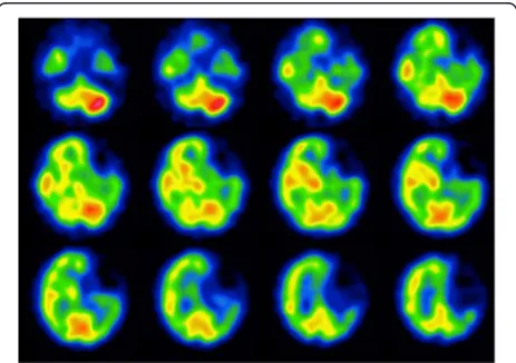
Preoperative single-photon emission computed tomog-raphy revealed reduced cerebral blood flow in the left cerebral hemisphere (especially in the external left frontal cortex; Fig. 1). Neurosurgeons at our center judged that the patient did not have an indication for cerebral revas-cularization surgery, which is used to prevent ischemic complications during the perioperative period of mitral valve plasty. This was because his left frontal lobe showed extensive cerebral infarction and no cerebral infarction symptoms were observed in the right cerebral hemisphere. Therefore, mitral valve plasty without cerebral revasculari-zation was chosen.
In the operation room, the patient’s monitoring of electrocardiogram, oxygen saturation, systemic arterial pressure via right radial artery catheter, bispectral index, and regional cerebral oxygen saturation (rSO2) at the
right and left forehead (INVOS™ 5100C, Somanetics, USA) was initiated before the administration of general anesthesia. The rSO2values for the left and right
fore-head were 72 and 81 %, respectively.
General anesthesia was induced by intravenous admin-istration of 3 mg of midazolam, 0.4 mg of fentanyl, and 50 mg of rocuronium. After tracheal intubation, a trans-esophageal echocardiography (TEE) probe was inserted. Then, a central venous catheter and right heart catheter were inserted via the right internal jugular vein, and central venous pressure, PAP, cardiac output, and mixed venous oxygen saturation were measured. The nasopha-ryngeal temperature and urinary bladder temperature were also measured.
Before CPB, general anesthesia was maintained by inhalation of sevoflurane (1–1.5 % of end-tidal concen-tration). The patient’s PaCO2 was maintained between
38 and 42 mmHg. Intra-aortic balloon pumping (IABP) was placed at the start of surgery and the augmented pressure was maintained. The mean arterial pressure was constantly above 70 mmHg. Before CPB, rSO2values were
almost above 80 % on both sides (Fig. 2).
During CPB, administration of sevoflurane via the oxy-genator was also continued because of its cerebrovascular dilatation activity and potential preconditioning effect against focal cerebral ischemia. PaCO2was maintained
between 45 and 50 mmHg, and alpha-stat management of pH was performed. Hypothermia was induced; the temperature at the bottom of the nasopharyngeal tem-prature was 28 °C. We used pulsatile perfusion assist to maintain cerebral circulation during CPB with IABP. A decrease in rSO2was observed 162 min after the
initi-ation of CPB. Our perfusionist increased the CPB pump flow from 2.2 L/min/m2to 2.8 L/min/m2in order to in-crease cerebral blood flow. Moreover, the concentration of sevoflurane was increased to 2 %. Yet, rSO2
desatur-ation (15 % reduction from baseline) was not improved. We decided to increase the depth-of-anesthesia with an-other dose of midazolam. After administration of 3 mg of midazolam, the rSO2values increased from 67 to 73 % on
the right side and from 71 to 74 % on the left side. During CPB, the lowest values (and variation) of rSO2in the left
and right forehead were 71 % (−2 %) and 67 % (−17 %), re-spectively. Mitral valve plasty was performed as planned.
At the weaning from the CPB, the disappearance of MR was confirmed by TEE; the weaning was not diffi-cult. Pulmonary hypertension also improved (PAP was 26/12 mmHg, while systemic arterial pressure was 105/56 mmHg). After CPB, inhalation of sevoflurane (1–1.5 % of end-tidal concentration) was also continued. The rSO2 values were almost above 75 % on both sides
and not below the awake rSO2 values (Fig. 2). CPB and
aortic cross-clamping lasted 352 min and 289 min, re-spectively. On the completion of the surgery, the IABP Fig. 1Preoperative single-photon emission computed tomography
was discontinued and sevoflurane administration was stopped. The patient was transferred to the intensive care unit with ventilator support under propofol sedation.
On the 1st postoperative day (POD), the patient was weaned from the ventilator, and the patient did not complain about any new neurological deficits. We monitored the rSO2 of his forehead until the
2nd POD and no significant decrease (−20 %) of the rSO2 values was confirmed. The postoperative course
was uneventful. On the 15th POD, single-photon emission computed tomography revealed that the low cerebral blood flow lesions had not changed (Fig. 3), and the patient was discharged from our hospital on the 16th POD.
Discussion
We performed anesthesia for mitral valve plasty in a pa-tient with cerebral infarction due to MMD using pulsa-tile perfusion of cardiopulmonary bypass with an assistance of IABP, without exacerbation of neurological complications.
Anesthetic management of a patient with MMD is ra-ther challenging, and we have to keep several key-points in mind. Among them, maintaining normocapnea is most critical. Hypocapnia would induce brain ischemia and brain infarction, while hypercapnia would induce vasodila-tion and hyper-perfusion of the fragile vessels in the brain, which might cause brain hemorrhage. In patients with MMD, both vasoconstriction and vasodilation would not be preferable. That is why keeping normocapnea is quite important.
During CPB, PaCO2was maintained between 45 and
50 mmHg with alpha-stat management of pH. This patient developed MMD not by the onset of cerebral hemorrhage but by the onset of cerebral infarction. We regarded that we should avoid hypocapnia or nor-mocapnea, which would induce cerebral infarction, rather than hypercapnia, which would induce cerebral hemorrhage. But, we cannot ignore that hypercapnia would induce vasodilation and hyper-perfusion of the fragile vessels in the MMD brain, which might cause brain hemorrhage.
Usually, extracorporeal circulation is maintained by non-pulsatile perfusion, and non-pulsatile perfusion can Fig. 2Intraoperative regional cerebral oxygen saturation measured by INVOS® and bispectral index. rSO2, regional cerebral oxygen saturation;
BIS, bispectral index; CPB, cardiopulmonary bypass; IABP, intra-aortic balloon pumping
induce ischemic injury, especially in organs under inad-equate perfusion [5]. In patients with restricted cerebral arterial blood supply such as MMD, decreased cerebral perfusion pressure and non-pulsatile perfusion during CPB are risk factors for cerebral ischemia.
Kashima et al. [6] reported that high-pressure pulsatile perfusion assisted by IABP was effective for brain protec-tion during coronary artery bypass grafting in a MMD pa-tient. De Buysscher et al. [4] also reported that conversion from a non-pulsatile flow to a pulsatile flow resulted in a gradual increase in rSO2 values as opposed to a sudden
decrease in rSO2 in an adult MMD patient undergoing
CPB. In addition, Cheul-Hong et al. [3] reported up to 15 % fluctuations in rSO2values during cardiac surgery in
an MMD patient who was later discharged from the hospital without any complications. In pediatric patients undergoing CPB, pulsatile flow has advantages over non-pulsatile flow as measured by near-infrared spectroscopy and transcranial Doppler ultrasound, which may improve postoperative neurodevelopmental outcomes [7]. Owing to these previous reports, we selected the pulsatile flow method supported by IABP in our case and maintained the mean perfusion pressure above 70 mmHg.
The optimum anesthetic agent for the maintenance of anesthesia in MMD patients requiring cardiac surgery is a topic of much debate. Neither inhalational anesthetics (sevoflurane or isoflurane) nor intravenous anesthetic (propofol) presents strong clinical evidence for the efficacy of the outcome. Propofol suppresses cerebral metabolism and reduces cerebral blood flow [8]. In contrast, sevo-flurane strongly dilates cerebral vessels and increases cerebral blood flow [9]. Moreover, several reports de-scribe sevoflurane preconditioning against myocardial ischemia-reperfusion injury. Recently, it has been re-ported that sevoflurane has the potential preconditioning effect against cerebral ischemia [10], although obvious preconditioning effect of sevoflurane has not been proved clinically. In rats with transient cerebral ischemia, sevo-flurane preconditioning protects mitochondria from cerebral ischemia-reperfusion injury and ameliorates long-term neurological deficits [11]. Thus, these prop-erties may support sevoflurane use over propofol for intracranial steno-occlusive arterial disease, but choice of anesthetic in patients with MMD remains an open question.
A decrease in rSO2was observed during the CPB.
Cur-rently, rSO2is considered as the average of arterial oxygen
saturation (SaO2) and internal jugular venous oxygen
sat-uration (SjO2) in the measured region. Several reasons
should be considered for changes of rSO2. It is also known
to be influenced by the skin blood flow [12]. Cutaneous vasoconstriction induced by vasoconstrictor, such as phenylephrine and norepinephrine, or hypothermia dur-ing CPB possibly affects changes of rSO2.
In this case, we increased the pump flow and concen-tration of sevoflurane to increase cerebral blood flow, but these interventions were not effective. Secondly, we deepened the anesthesia level with a supplemental intra-venous anesthetic. After another administration of mid-azolam, an improvement in bilateral rSO2 values was
observed. We didn’t know why bilateral rSO2 was
im-proved after administration of an intravenous anesthetic, but we cannot ignore the possibility that an intravenous anesthetic decreases the cerebral metabolic rate of oxygen consumption [7]. When regional cerebral oxygen desatur-ation continues during sevoflurane general anesthesia in patients with MMD, it might be efficacious to administer intravenous anesthetics concurrently. These management procedures limited the fluctuation of rSO2 values to
within 17 % in our patient, which was nearly equal to that in a previous study [3], and prevented the development of cerebral deficits.
Conclusion
Pulsatile flow supported by IABP during CPB had a pos-sible beneficial effect for brain protection in patients with restricted cerebral arterial blood supply. Sevoflurane inhal-ation was continued during CPB with an expectinhal-ation of its vasodilatory activity and its potential neuroprotective effect. We could maintain rSO2at 80 % of baseline value
during the surgical procedure, and we could not detect other neurological deficits. But, the further discussion is warranted whether inhalational anesthetic or intra-venous anesthetic might be superior for intracranial steno-occlusive arterial disease. Thus, our case provides further evidence for the efficacious use of this anesthetic in MMD patients requiring cardiac surgery.
Abbreviations
CPB, cardiopulmonary bypass; IABP, intra-aortic balloon pumping; MMD, moyamoya disease; MR, mitral regurgitation; PaCO2, arterial carbon
dioxide partial pressure; PAP, pulmonary arterial pressure; POD, postoperative day; rSO2, regional cerebral oxygen saturation; TEE, transesophageal
echocardiography
Acknowledgments
We would like to thank Editage (www.editage.jp) for English language editing.
Authors’contributions
KS was primary anesthetist and drafted the manuscript. HT supervised anesthetic management. YE and MY helped to draft the manuscript. All authors read and approved the final manuscript.
Authors’information
KS is M.D., and Staff Anesthesiologists of Department of Anesthesiology, Tohoku University Hospital; HT is M.D., PhD, Lecturer of Department of Anesthesiology, Tohoku University Hospital; YE is M.D., PhD, and Associate Director Division of Surgical Center and Supply, Sterillization, Tohoku University Hospital; MY is M.D., PhD, and Professor of Anesthesiology and Perioperative Medicine, Tohoku University School of Medicine.
Competing interests
Consent for publication
Written informed consent was obtained from the patient for publication of this Case report and any accompanying images. A copy of the written consent is available for review by the Editor-in-Chief of this journal.
Author details
1Department of Anesthesiology, Tohoku University Hospital, 1-1 Seiryomachi,
Aoba-ku, Sendai 980-8574, Japan.2Division of Surgical Center and Supply, Sterillization, Tohoku University Hospital, 1-1 Seiryomachi, Aoba-ku, Sendai 980-8574, Japan.3Anesthesiology and Perioperative Medicine, Tohoku
University School of Medicine, 2-1 Seiryomachi, Aoba-ku, Sendai 980-8575, Japan.
Received: 17 March 2016 Accepted: 29 June 2016
References
1. Suzuki J, Takaku A. Cerebrovascular“moyamoya”disease. Disease showing abnormal net-like vessels in base of brain. Arch Neurol. 1969;20:288–99. 2. Wang N, Kuluz J, Barron N, et al. Cardiopulmonary bypass in a patient with
moyamoya disease. Anesth Analg. 1997;84:1160–3.
3. Kim C-H, Yoon J-U, Lee H-J, et al. Hypothermic cardiopulmonary bypass for minimally invasive mitral valve plasty in adult moyamoya disease. J Anesth. 2012;26:259–61.
4. De Buysscher P, Moerman A, Bové T, et al. Value of cerebral oxygen saturation monitoring during cardiopulmonary bypass in an adult patient with moyamoya disease. J Cardiothorac Vasc Anesth. 2013;27:740–3. 5. Onorati F, Santarpino G, Presta P, et al. Pulsatile perfusion with intra-aortic
balloon pumping ameliorates whole body response to cardiopulmonary bypass in the elderly. Crit Care Med. 2009;37:902–11.
6. Kashima I, Inoue Y, Takahashi R. The use of intra-aortic balloon pumping as cerebral protection in a patient with moyamoya disease undergoing coronary artery bypass grafting. Interact Cardio Vasc Thorac Surg. 2008;7:522–3. 7. Su XW, Guan Y, Barnes M, et al. Improved cerebral oxygen saturation and
blood flow pulsatility with pulsatile perfusion during pediatric cardiopulmonary bypass. Pediatr Res. 2011;70:181–5.
8. Kaisti KK, Langsjo JW, Aslto S, et al. Effects of sevoflurane, propofol, and adjunct nitrous oxide on regional cerebral blood flow, oxygen consumption, and blood volume in humans. Anesthesiology. 2003;99:603–13.
9. Bundgaard H, von Oettingen G, Larsen KM, et al. Effects of sevoflurane on intracranial pressure, cerebral blood flow and cerebral metabolism. A dose-response study in patients subjected to craniotomy for cerebral tumors. Acta Anaesthesiol Scand. 1998;42:621–7.
10. Codaccioni JL, Velly LJ, Moubarik C, et al. Sevoflurane preconditioning against focal cerebral ischemia: Inhibition of apoptosis in the face of transient improvement of neurological outcome. Anesthesiology. 2009;110:1271–8. 11. Ye R, Yang Q, Kong X, et al. Sevoflurane preconditioning improves
mitochondrial function and long-term neurologic sequelae after transient cerebral ischemia: role of mitochondrial permeability transition. Crit Care Med. 2012;40:2685–93.
12. Sørensen H, Secher NH, Siebenmann C, et al. Cutaneous vasoconstriction affects near-infrared spectroscopy determined cerebral oxygen saturation during administration of norepinephrine. Anesthesiology. 2012;117:263–70.
Submit your manuscript to a
journal and benefi t from:
7Convenient online submission
7Rigorous peer review
7Immediate publication on acceptance
7Open access: articles freely available online
7High visibility within the fi eld
7Retaining the copyright to your article