RESEARCH ARTICLE
HEAD AND NECK POSTURE IN YOUNG ADULTS WITH CHRONIC NECK PAIN
*
Mohankumar, P. and Leong Wai Yie
School of Engineering, Taylor's University, Malaysia
ARTICLE INFO ABSTRACT
Neck pain is very common in the general population. Every individual would have experienced neck pain at some point in their life. Some might have had acute and some with chronic. Chronic neck pain affects range of motion and limits the functional capacity of neck. Because of chronic neck pain, neck muscles undergo spasm and gets fatigue soon. This leads to altered head and neck posture. Hence there is a need to find what trait or feature in the chronic neck pain lead to altered posture. Study group consisted of 30 subjects with chronic neck pain and control group with 30 pain free persons in the age group between 20 to 30 years. Spearman correlation revealed that there was a high level of significance between ROM & pain with significance level of 0.05 and between posture & ROM with significance level of 0.01Findings from this study showed that there was a significant difference between chronic neck pain patients and pain free subjects in frontal plane alignment, upper and lower cervical angle. There was a correlation between ROM, posture and pain. Postural changes in the neck need to be considered during therapeutic intervention of patients with chronic neck pain.
INTRODUCTION
Chronic neck pain is a common problem faced by the general population (Fejer et al., 2006), specifically the working population and it is a frequent source of disability causing
important health and economic costs (Bone et al.,
2004).Chronic neck pain is often considered to be associated with the development and persistence of abnormal neck
posture. Clinicians emphasize its importance during
examination and intervention (Borenstein et al., 2004; Kendall
et al., 2005; Magee, 2013: McKenzie, 2011). This abnormal
posture of the neck is the cause or result of cervicogenic headache, thoracic outlet syndrome, fibromyalgia and degenerative joint diseases. Although not as common as low back pain, neck pain appears to be very frequent all over the world and particularly in the Western countries. In Scandinavian countries, as many as 71% of an adult population have had neck pain during their life(Makela et al., 1991)and 75% during the past year(Rauhala K, 2000). However, lower prevalence estimates have also been reported. For example, in Sweden a lifetime prevalence of 26% was reported, and in Finland, only 17% had the neck pain for the past year (Takala J, 1982).Three literature reviews have suggested possible explanations for the diversity in the neck pain prevalence estimates, which may partly explain these variations in its occurrence (Cote P, 1998; Nachemson and Jonsson, 2000). Typically, the definitions of neck pain varied, were too broad, or different sample populations with different
*Corresponding author: Mohankumar, P.
School of Engineering, Taylor’s University, Malaysia.
age distributions were used. Unfortunately, these literature reviews suffered from various methodological shortcomings and the true prevalence of neck pain in the population is largely unknown. The presence of neck pain does not reveal its severity and impact on daily living. For example, mild neck pain may result in little or no influence on people’s daily life, whereas people with severe chronic neck pain may be highly disabled. Thus, pain intensity grading provides additional information for use in clinical and research activities (Korff et al., 1990).However, the grading of neck pain is a combination of duration, pain, intensity and disability & despite the moderate correlation between neck pain intensity and disability (Demaille et al., 2004; Clair D, 2004). Controversy exists as to how they are interrelated (Korff et al., 2000). Pain and disability are two separate dimensions. Still, people with neck pain continue to go to work with a resulting loss in productivity. Study on neck acceleration and muscle activation in chronic neck pain patients found that, the dynamic performance level of all cervical muscles was significantly lower (Tsang et al., 2016).Between 20% and 40% of the general population seek treatment for neck pain at some time during their lives(Picavet H.S, 2003). Moreover, with an increased mobile usage, the teenagers and young adults are more commonly affected by musculoskeletal disorders and neck pain(Gustafsson et al., 2017).The impact of chronic neck pain is considerable. It results in reduced range of motion, fatigued muscles and decreased functional capability. A study on cervical flexion relaxation ratio in neck pain patients found that the neck pain patients have significant difference in their neuromuscular control (Zabihhosseinian et al., 2015).Another study on head posture and neck muscle endurance in Article History:
Received 21st August, 2017
Received in revised form
04th September, 2017
Accepted 24th October, 2017
Published online 11th November, 2017
International Journal of Recent Advances in Multidisciplinary Research
Vol. 04, Issue 11, pp.2946-2951, November, 2017
Keywords:
Upper cervical angle, Lower cervical angle, Frontal plane alignment, NRS, Range of Motion,
adolescents with neck pain found that the adolescents with neck pain have less forward head posture, less neck flexor and extensor endurance when compared with the normal (Oliveira and Silva, 2015).Therefore, it is important to study the factors associated with the pain, causing neck postural deviation for effective intervention.
The need for this study is to knowand understand the changes occurred due to chronic neck pain in young adults. Also, the postural differences ofhead and neck need to be addressed during physiotherapy management.It has been postulated that treatment should not only address the pain, but also postural changes in chronic neck pain patients, in case if it should be effective. Thus indeed, there is need of adequate knowledge about the postural changes of head and neck in patients with chronic neck pain. Hence, this study is done to provide information about the changes in head and neck posture in young adults with chronic neck pain. The aim of this study is to determine whether there is a relationship among range of motion, chronic neck pain and head &neck posture in young adults when compared with pain-free persons in frontal and sagittal planes.
MATERIALS AND METHODS
Participants
Both the genders at an age group between 20 to 30 years were selected. Subjects were divided into Group A (Pain free) and Group B (Neck pain). Pain-free participants with no current neck pain and no history of neck pain for more than past one month with an optimal posture confirmed by plumb line assessment for sagittal and frontal planes were categorized into Group A. Subjects with neck pain felt posteriorly between inferior margin of occiput and T1 with a duration of more than
two months with Numerical Rating Scale (NRS) score for pain from 3 to 7 were categorized into Group B(Bogduk and McGuirk, 2006). Subjects with history of cervical or facial trauma or surgery, diagnoses of cervical radiculopathy or cervical myelopathy, diagnosis of cervical Spondylosis, congenital or acquired anomalies involving the spine and pelvis such as scoliosis, limb length discrepancy & any systemic arthritis, recurrent middle ear infections over the last 5 years or any hearing impairment requiring the use of a hearing aid, patients with vestibular dysfunction, such as vertigo, any visual impairment, any disorder of the central nervous system and pregnancy or breast-feeding were excluded from this study (Berger et al., 2006; Ris et al., 2017).
Instruments
For base line assessment like measuring height and weight, height scale and weighing machine were used.A standardised pain reporting scale called Numerical Rating Scale (NRS) was provided to the subjects to report their intensity of neck pain. This scale consists of zero to ten in numerical. Where, zero denotes no pain at all, one to three denotes a mild pain, four to six denotes a moderate pain, seven to nine as severe pain and ten denotes the maximal or worst pain. Subjects are supposed to mark the pain they perceived in the past 24 hours.Universal Goniometer was used to measure the Range of Motion (ROM) of neck. Regarding the postural assessment, Digital camera with 16 pixels mounted on the tripod stand was used to take
postural photographs for both Group A and Group B subjects. Plumb line was used to make sure that the pain free persons did not have any postural abnormalities. Subjects were asked to stand and have a similar body weight distribution through each foot, with their feet slightly apart and arms by their sides. The spinous process of C7 vertebrae was identified by palpation &
marked with a marker made of paper roll fixed on the skin, with a double side tape.
Procedure
Initially, informed consent was obtained from each subject followed by NRS and baseline assessment was done for height and weight for all subjects.
ROM measurement
Cervical ROM includes flexion, extension, side flexion and rotation. Measurements were taken as per the guidance provided by (Norkin and White, 2009).For flexion and extension movement, the subject is made to sit erect with thorax and lumbar spine well supported. Subject’s external auditory meatus is considered as the fulcrum. The stable arm is kept perpendicular to the ground and movable arm is kept parallel to the ground. Subject is asked to do flexion and extension movements and the measurements are noted. For side flexion movement, subject’s C7 spinous process is
considered as the fulcrum. The stable arm is positioned along the spine and perpendicular to the ground. The movable arm is kept along the midline of the head using occipital protuberance. Subject is asked to do side flexion on both the sides and the measurements are noted. For rotation movement, center of cranial aspect of the head is considered as the fulcrum. The stable arm is kept parallel to the acromial process and movable arm is positioned along the tip of the nose. Subject is asked to perform rotation movement on both the sides and the measurements are noted.
Sagittal plane assessment
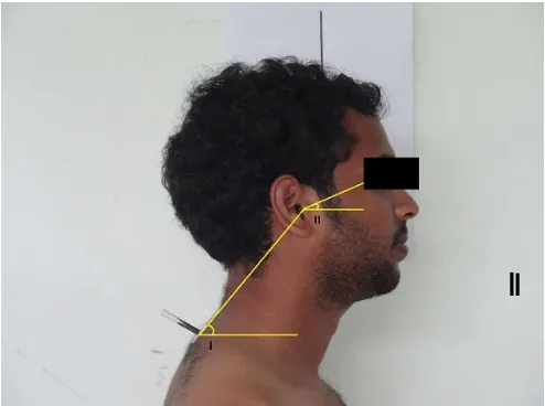
Postural assessment of neck was done by Photographic method as per the guide lines given by (Silva et al., 2009). Tripod was placed to right side of the subjects at a distance of 200 cm with right lateral malleolus as a reference point. Height of the camera was adjusted in such a manner that tragus of the right ear of the subject was at the focus point of the camera. (Refer Fig.1) Bubble level on the tripod was taken as reference for standard alignment of the camera. To facilitate their natural head posture, subjects were asked to tilt their head forward & backward in decreasing amplitude like a pendulum until they felt that a natural head posture is reached. Once settled, images were taken. For reliability purpose, this process was repeated for 3 times.
Frontal plane assessment
to both the sides in decreasing amplitude like a pendulum until they felt that a natural head posture is reached. Once settled, images were taken. For reliability purpose, this process was repeated for 3 times. From the obtained images, the below mentioned angles were measured:
1. Angle I: Line connecting C7 vertebrae to tragus of the
ear and horizontal line (Lower cervical Angle - LCA). 2. Angle II: Line connecting the tragus to canthus of the
eye and horizontal line (Upper Cervical Angle - UCA). 3. Angle III: Line connecting the inferior margins of both
ears and horizontal line (Frontal Plane Alignment - FPA).
After digitization process, the angles were measured using a software ImageJ Version 1.46f. The results of the Intra Class Correlation Coefficient to determine the agreement between measures from goniometry and the ImageJ program was 0.89 (95% CI = 0.83 – 0.93), which is considered high. Thus, the digitization process of the angles is found to be highly reliable.
Data analysis
Data analysis was done with SPSS software Version 17.0. ‘p’ value was set as 0.05 as level of significance for all comparisons. Comparison were made using Independent ‘t’ test between the groups, Group A (pain free) and Group B (neck pain) subjects for the obtained data. Results showed that there is no significant difference in the Age (t = 0.345, p = 0.768), Height (t = 0.462, p = 0.868) and Weight (t = 0.480, p
= 0.633) between the subjects of both groups. Hence the groups are homogenous in demographic characteristics. We also conducted Pearson Correlation test between the ROM, posture and pain.
RESULTS
Table 1 shows that, there is no significant difference in the Age, Height and Weightbetween the subjects of both groups. Hence the groups are homogenous in demographic characteristics. In Table 2, comparison of Lower Cervical Angle between Group A and Group B shows a statistically significant difference with t value of 2.235 and p value of 0.029.
Table 1. Demographic Analysis
Variables Group A Group B t
value p value
Mean S.D. Mean S.D.
Age 23.97 1.974 25.77 2.063 0.345 0.768
Height 167.653 8.3577 167.307 7.6776 0.167 0.868
Weight 65.617 14.3801 64.2 7.3503 0.48 0.633
Table 2. Analysis in Posture
Groups Mean S.D. t value p value
LCA
Group A 50.56437 5.474428 2.235 0.029
Group B 53.25733 3.683261
UCA
Group A 22.6704 6.89334 2.482 0.02
Group B 24.912 4.845169
FP
A Group A 2.075 1.02049 3.223 0.002
Group B 3.2785 1.77271
Note. LCA: Lower Cervical Angle; UCA: Upper Cervical Angle; FPA:
Frontal Plane Alignment.
Figure 1. Angle I: Line connecting C7 vertebrae to tragus of the
ear and horizontal line (Lower cervical Angle - LCA). Angle II: Line connecting the tragus to canthus of the eye and horizontal
line (Upper Cervical Angle - UCA)
Figure 2. Angle III: Line connecting the inferior margins of both ears and horizontal line (Frontal Plane Alignment - FPA)
Comparison of Upper Cervical Angle between Group A and Group B shows a statistically significant difference with t value of 2.482 and p value of 0.02.Comparison of Frontal Plane Alignment between Group A and Group B shows a highly significant difference with t value of 3.223 and p value of 0.002. The Spearman correlation revealed that there is a high level of significance between ROM& pain with the significance level of 0.05 (0.018) and between posture & ROM with the significance level of 0.01 (0.007). The correlation between cervical extension (ROM) and pain(NRS) is fairly positive 0.428. The correlation between UCA (Posture) and lateral flexion & rotation (ROM) is negative 0.483. The correlation between FPA (Posture) and side flexion (ROM) is positive 0.366.
DISCUSSION
participants. Our finding is supported by the study conducted by (Silva et al., 2009), who concluded that, patients with chronic neck pain have changes in neck posture, when compared to age matched controls. It may also be worth considering the clinical significance of the finding in terms of a change in head posture relating to a change in neck pain. In this study, findings show that there is a significant difference between chronic neck pain patients and pain free subjects in LCA measured in sagittal plane, with the angle measured between C7 to the tragus, and the horizontal (neck pain group:
53.257 ± 3.683, pain free group: 50.564 ± 5.474). Our findings are supported by (Shiau and Chai, 1990; Braun, 1991), and colleagues who found a statistically significant difference in the LCA between patients and control. Shiau and Chai et al
used photographs and a sample of 51 patients with neck pain aged between 19 and 66 years old and 28 pain-free participants aged 20 to 34 years old. A mean angle of 54.0° ± 4.9° for patients and 56.7° ± 3.5° for pain-free participants was reported. Braun used a personal analysis digitizing system and a sample of 9 patients with a mean age of 38 years old and 40 pain-free participants with a mean age of 28 years old and reported values of 48.2° ± 32° and 55.4° ± 4.6° for patients and controls, respectively.The results of this study also show that there is a significant difference in UCA in sagittal plane between chronic neck pain patients and pain-free subjects, for the angle been measured between the line joining the tragus to the eye and the horizontal (neck pain group: 24.912 ± 4.845; pain free group: 22.670 ± 6.893). This finding is in line with finding by Silva et al who concluded that upper cervical angle varies significantly between neck pain and control subjects (neck pain group: 21.0°± 6.4°; pain-free group: mean SD (18.8° ± 7.7°). This finding is also in line with the results reported by (Harrison et al., 1996), who concluded that a more forward head posture is associated with an increase in head extension, involving the upper cervical spine. In this study, it was found that there is a significant difference in FPA of neck between the chronic neck pain patients and pain-free subjects (neck pain group: 3.278 ± 1.7727, pain free group: 2.075 ± 1.0204). However, studies done by Shiau and Chai et al
concluded that there is no significant difference in FPA between neck pain and control subjects, where the angle is measured between the line that joins both pupils and the horizontal, (neck pain group: 1.7° ± 1.7°; pain free group: – 1.7° ± 1.6°). Studies conducted by Shiau and Chai et al., on healthy participants confirm that this angle approaches 0, indicating a symmetrical head posture with regards to this measurement.
Study on neck pain patients on the extension and flexion in upper cervical spine concluded that, upper cervical flexion and extension showed a moderate correlation with headache intensity and frequency (Ernst et al., 2015). In our study, results of Spearman correlation in the chronic neck pain subjects revealed that there is a significance between ROM & pain and between posture & ROM. The correlation between cervical extension ROM and NRS was +0.428 with a significance value of 0.018. Hence, if cervical extension increases, pain increases. The correlation between side flexion and FPA was +0.366 with a significance value of 0.047. Whereas, in contrast, instead of flexion extension, we noticed that the side flexion and rotation hadsignificant relation with UCA. Flexion, extension and upper cervical angle are noticed in the sagittal plane. But, side flexion occurs in frontal plane
and rotation occurs in transverse plane.Cervical spine consists of seven vertebrae. The first two vertebrae Atlas and Axis (C1& C2) are highly specialized and complex. Because head
movements are mainly produced by these two bones. While, the remaining five vertebrae (C3 to C7) produce the neck
movements. Flexion and extension of head mainly occurs in the atlanto-occipital joint. i.e. between the occiput of the skull and C1 vertebra. Rotation of the head occurs between the C1
and C2. In the overall flexion, extension and rotation of head
and neck, nearly 50% of the movements occur in the atlanto-occipital and atlanto-axial joints alone. Muscles like sternocleidomastoid, rectus capitis major & minor, oblique capitis superior, semispinalis capitis, splenius capitis, splenius cervicis, upper fibers of trapezius, scalene, erector spinae and levator scapulae are the prime and secondary movers of head and neck.
A study done by (Silva and Johnson, 2013)suggests that the non-optimal posture like forward head posture or tilted head could be a time dependent. Because the causes for these mal postures are primarily due to the tightness of one group of muscles or weakness in other group of muscles.This bewilderment can be avoided by finite element analysis. A 3D model of the cervical vertebrae can be created using CT scan images and using bone and ligament properties, finite element analysis can be performed to get more accurate results (P. Mohankumar, 2015).The measurement of cervicothoracic junctional structures remains as a reliable method in judging the severity of chronic neck pain (Lee et al., 2014).A five-year cohort research study on mobile usage and musculoskeletal disorders in young adults shows that there are some
associations between usage of mobile phones and
musculoskeletal disorders. The long-term impression leads to musculoskeletal disorders in the neck and upper extremities (Gustafsson et al, 2017). However, another observational study on the effects of texting on balance and gait in young adults with and without neck pain revealed that young adults with neck pain who have habit of texting do lead to discrepancy in balance and gait (R. Fraser, 2016). Another study on cervical proprioception in young adults with neck pain and prolonged usage of mobile found that these young adults show poor proprioception and have lost the ability to accurately determine their normal straight head position is (S. Reid, 2016).
A study from (Falla et al., 2007) suggests that a decrease in neck pain intensity may be independent of an improvement in forward head posture. In his study, the change in the angle between C7, the tragus, and the horizontal was measured during
head posture predisposes people to suffer neck pain. A more reliable and accurate procedure may be needed, to measure forward head posture in clinical practice, so that it can be monitored and used to inform decisions regarding treatment. A randomized controlled study on muscle energy technique, static stretching and functional disability in neck pain patients found that both the muscle energy technique and stretching are helpful in reducing pain and disability (Phadke et al, 2016).Thus the findings of our study have implications in the management of chronic neck pain in patients.Our study shows that, there is a significant difference in the neck posture between chronic neck pain patients and age matched pain free persons. Thus, postural changes in the neck needs to be considered during therapeutic intervention of patients with chronic neck pain.
Acknowledgment
We acknowledge and convey our thanks to Professor V.P.R. Sivakumar, Dean, SRM College of Physiotherapy and Dr. S. Rajesh for their constant support and guidance rendered throughout this study.
REFERENCES
Berger, M. F., Prob, R. D., Ilg, U. J., and Karnath, H.O. 2006. Deviation of eyes and head in acute cerebral stroke. BMC
Neurology, 6(23): 16-23. doi:10.1186/1471-2377-6-23
Bogduk, N., and McGuirk, B. 2006. Management of Acute and Chronic Neck Pain: An Evidence Based Approach. (1st Ed.) Elsevier, Philadelphia.
Bone, K. W., Reading, I., Coggon, D., Cooper, C., and Palmer, K. T. 2004. The anatomical pattern and determinants of pain in the neck and upper limbs: an epidemilogic study. Pain.109(1-2): 45-51.
Borenstein, D., Wiesel, S., and Boden, S. 2004. Low back and neck pain: Comprehensive diagnosis and management.(3rd Ed.) Saunders, Philadelphia.
Braun, B. L. 1991. Postural differences between asymptomatic
men and women and craniofacial pain patients. Archives of
Physical Medicine and Rehabilitation, 72(9): 653-656.
Clair, D., Edmondston. S. and Allison, G. 2004. Variability in pain intensity, physical and psychological function in non-acute, non-traumatic neck pain. Physiotherapy Research
International, 9(1): 43-54.
Cote, P., Cassidy, J. D., and Carroll, L. 1998. The Saskatchewan Health and Back Pain Survey. The prevalence of neck pain and related disability in Saskatchewan adults.Spine, 23(15): 1689-1698.
Demaille, S. W., Poiraudaeu, S., Catanzariti, J. F., Rannou, F., Fermanian, J., and Revel, M.2004. The ability to change of three questionnaires for neck pain. Joint Bone Spine,71(4): 317-326.
Ernst, M. J., Crawford, R. J., Schelldorfer, S., Rausch-Osthoff, A. K., Barbero, M., Kool, J. and Bauer, C. M. 2015. Extension and flexion in the upper cervical spine in neck
pain patients. Manual Therapy, 20(4): 1-6.
doi:10.1016/j.math.2014.12.005
Falla, D., Jull, G., Russell, T., Vicenzino, B., and Hodges, P. 2007. Effect of neck exercise on sitting posture in patients with chronic neck pain. Physical Therapy Journal, 87(4): 408-417. doi:10.2522/ptj.20060009
Fejer, R., Kyvik, K. O., and Hartvigsen, J. 2006. The prevalance of neck pain in the world population: a systematic review of the literature. European Spine Journal,
15(6): 834-848. doi:10.1007/s00586-004-0864-4
Fraser, R., and Reid, S. 2016. The effects of texting on balance and gait in young adults with and without neck pain: An
observational study. Manual Therapy, 25: 100.
doi:10.1016/j.math.2016.05.175
Gustafsson, E., Thomee, S., Grimby-Ekman, A., and Hagberg, M. 2017. Texting on mobile phones and musculoskeletal disorders in young adults: A five year cohort study. Applied
Ergonomics, 58: 208-214. doi:10.1016/
j.apergo.2016.06.012
Harrison, A. L., Barry-Greb, T., and Wojtowicz, G. 1996. Clinical Measurement of Head and Shoulder Posture Variables. Journal of Orthopaedic and Sports Physical
Therapy, 23(6): 353-361. doi:10.2519/jospt.1996.23.6.353
Kendall, F. P., McCreary, E. K., Provance, P. G., and Rodgers, M. M. 2005. Muscles: Testing and Function, with Posture and Pain.(4th Ed.). Lippincott Williams & Wilkins, Philadelphia.
Korff, M. V., Dworkin, S. F., and Resche, L. L. 1990. Graded chronic pain status: an epidemiologic evaluation. Pain, 40(3): 279-291.
Korff, M. V., Jensen, M. P., and Karoly, P. 2000. Assessing Global Pain Severity by Self-Report in Clinical and Health
Services Research. Spine, 25(24): 3140-3151.
doi:10.1097/00007632-200012150-00009
Lee, J. H., Park, Y. K. and Kim, J. H. 2014. Chronic neck pain in young adults: perspectives on anatomic differences. The
Spine Journal, 14(11): 2628-2638. doi:10.1016/
j.spinee.2014.02.039
Magee, D. J. 2013. Orthopedic Physical Assessment.(4th Ed.). Saunders, Philadelphia.
Makela, M., Heliovaara, M., Sievers, K., Impivaara, O., Knekt, P., and Aromaa, A. 1991. Prevalence, Determinants and Consequences of Chronic Neck Pain in Finland. American
Journal of Epidemiology, 134(11): 1356-1367.
McKenzie, R. 2011. Treat Your Own Neck.(4th Ed.). Orthopedic Physical Therapy Products, New Zealand. Mohankumar, P., Leong, W. Y. 2015. 3D Modelling with CT
and MRI Images of a Scoliotic Vertebrae. Journal of Engineering Science and Technology. EURECA 2015 Special issue February 2016: 188-198.
Nachemson, A. and Jonsson, E. 2000. Neck and Back Pain - The Scientific Evidence of Causes, Diagnosis, and Treatment. Lippincott Williams & Wilkins, Philadelphia. Norkin, C. C., and White, D. J. 2009. Measurement of Joint
Motion: A Guide to Goniometry (Vol. 4). F.A. Davis, Philadelphia.
Oliveira, A. C. and Silva, A. G. 2015. Neck muscle endurance and head posture: A comparison between adolescents with
and without neck pain. Manual Therapy, 22: 62-67.
doi:10.1016/j.math.2015.10.002
Phadke, A., Bedekar, N., Shyam, A., & Sancheti, P. 2016. Effect of muscle energy technique and static stretching on pain and functional disability in patients with mechanical
neck pain: A randomized controlled trial. Hong
KongPhysiotherapy Journal, 35(C): 5-11. doi:10.1016/
j.hkpj.2015.12.002
consequences and risk groups, the DMC3-study. Pain, 1(2), 167-178. doi:10.1016/s0304-3959(02)00372-x
Rauhala, K., Oikarinen, K. S., Jarvelin, M., and Raustia A. M. 2000. Facial pain and temporomandibular disorders: an epidemiological study of the Northern Finland 1966 Birth Cohort.The Journal of Craniomandibular Practice, 18(1): 40-46.
Reid, S. and Portelli, A. 2016. Cervical proprioception in young adults with and without neck pain, who spend prolonged time on mobile devices: An observational study. Manual
Therapy, 25:86-87. doi:10.1016/j.math.2016.05.146
Ris, I., Kristensen, B. J., Boyle, E., Kongsted, A., Manniche, C., and Sogaard, K. 2017. Chronic neck pain patients with
traumatic or non-traumatic onset: Differences in
characteristics. A cross-sectional study. Scandinavian
Journal of Pain, 14:1-8. doi:10.1016/j.sjpain.2016.08.008
Shiau, Y. Y. and Chai, H. M. 1990. Body Posture and Hand
Strength of Patients with Temporomandibular Disorder. The
Journal of Craniomandibular & Sleep Practice, 8(3):
244-251. doi:10.1080/08869634.1990.11678318
Silva, A. G., and Johnson, M. I. 2013. Does forward head posture affect postural control in human healthy volunteers?
Gait & Posture. 38(2):352-353. doi:10.1016/
j.gaitpost.2012.11.014
Silva, A. G., Punt, T. D., Sharples, P., Vilas-Boas, J. P., and Johnson, M. I. 2009. Head Posture and Neck Pain of Chronic Nontraumatic Origin: A Comparison Between Patients and Pain Free Persons. Archives of Physical
Medicine and Rehabilitation, 90(4): 669-674.
Takala, J., Sievers, K. and Klaukka, T. 1982. Rheumatic symptoms in the middle-aged population in southwestern Finland. Scandinavian Journal of Rheumatology, 47: 15-29. Tsang, S. M., Szeto, G. P., and Lee, R. Y. 2016. Relationship
between neck acceleration and muscle activation in people with chronic neck pain: Implications for functional
disability. Clinical Biomechanics, 35:27-36.
doi:10.1016/j.clinbiomech.2016.04.006
Zabihhosseinian, M., Holmes, M. W., Ferguson, B., and Murphy, B. 2015. Neck muscle fatigue alters the cervical flexion relaxation ratio in sub-clinical neck pain patients.
Clinical Biomechanics, 30(5): 397-404. doi:10.1016/
j.clinbiomech.2015.03.020