Original Research Article.
Comparison of Ultrasound and ERCP for Obstructive Jaundice
Ali Yasen Ali
Taif University, College of Medicine, Taif, Kingdom of Saudi Arabia.
ABSTRACT
Background: Jaundice is a term used to describe the yellowing of the skin and the whites of the eyes. It's caused by a build-up of a substance called bilirubin in the blood and body's tissues. Jaundice is considered a sign of an underlying disease process.
Objectives: Is to assess the accuracy of ultrasound U/S and Endoscope Retrograde Cholangio-Pancratography ERCP to diagnose site and cause of biliary obstruction.
Methods and Materials: Prospective, study that Compare U/S and ERCP to diagnose obstructive jaundice among Sudanese population. The study was performed at different hospitals in KSA, by performing abdominal U/S and ERCP for thirty patients with signs and symptoms of obstructive jaundice. Results: The study found that obstructive jaundice was the most common in elderly ages especially over 65 years there were 11 out of 30 cases (36.7%) their ages above 65 years. The females were forming (53.3%) v/s (46.7%) males. Most of the patients were married (90%). Abdominal U/S of the sample showed that (77.7%) of the patients had dilated common bile duct but ERCP showed (76.7%). Abdominal U/S of the sample
that (50%) of the patients had stone in CBD and (20%) carcinoma head of pancreas but ERCP showed (36.7%) of the patients with stone and (23.3%) had carcinoma head of pancreas. Accuracy of ERCP in diagnosis obstructive jaundice was (80%), and ultrasound accuracy was (70%).
Key words: U/S, ERCP, Jaundice, Sudan.
*Correspondence to:
Dr. Ali Yasen Ali Ahmed,
Taif University, College of Medicine, Taif Kingdom of Saudi Arabia. Article History:
Received: 19-07-2017, Revised: 23-08-2017, Accepted: 15-09-2017
Access this article online Website:
www.ijmrp.com
Quick Response code
DOI:
10.21276/ijmrp.2017.3.5.070
INTRODUCTION
Jaundice (icterus) is detectable clinically when the serum bilirubin
is greater than 50μmol/L (3mg/dL). The jaundice is divided into
prehepatic, hepatocellular and obstructive (cholestatic) is an over simplification as in hepatocellular jaundice there is invariably cholestasis and the clinical problem is whether the cholestasis is intrahepatic or extra hepatic.1
Jaundice will therefore be considered under the following headings: haemolytic jaundice increased bilirubin load for the liver cells, congenital hyperbilirubinaemias defects in conjugation, cholestatic jaundice, including hepatocellular (parenchymal) liver disease and large duct obstruction.1 The principal role of imaging
in the jaundiced patient is the identification and detailed assessment of major bile duct obstruction. The clinical suspicion is based on a variable combination of dark urine, pale stools, pruritus, cholangitis and cholestatic liver function tests. Ultrasound (US) is the preferred initial imaging investigation, but will usually be supplemented with a combination of computed tomography (CT), magnetic resonance cholangiopancreatography (MRCP), direct cholangiography and, in some centers, endoscopic and/or intraoperative Ultrasound US.2 US have replaced Oral
cholecystography (OCG) for the diagnosis of gallstones and in many centers the oral contrast agents are no longer available. When extracorporeal shockwave lithotripsy was popular OCG was used to prove cystic duct patency, which was necessary for the
passage of stone fragments. More recently oral contrast agents have been used for computed tomography (CT) cholangiography, although most centers use intravenous cholangiography IV biliary contrast agents.2
Endoscopic retrograde cholangiopancreatography (ERCP): ERCP provides direct opacification of bile ducts and pancreatic ducts with success rates of 92–97 per cent. It provides dynamic information during contrast medium introduction and drainage. It allows visual assessment of the duodenum and ampulla of Vater and provides an option for biopsy and brushings, as well as interventional procedures such as sphincterotomy and stone extraction, biliary stenting and biliary stricture dilatation.2
Complication rates vary depending on the indication for the procedure, the presence of co-existing disease and the experience of the endoscopist, with severe complication rates of 0.9 per cent to 2.3 per cent, and total complication rates of 8.4–
11.1 per cent, the most common significant complication being pancreatitis. The main diagnostic pitfall with ERCP is the under filling of ducts above a stricture.2 The biliary system is one of the
MATERIALS AND METHODS
The current study aimed to assess the accuracy of US and ERCP in detection of Jaundice.
This is Prospective, study for ultrasound and endoscopic retrograde cholango- pancreatography procedures in diagnosis of obstructive jaundice among Sudanese population.
The target population of this study was consecutive patients presented to the hospital with ultrasound report showed obstructive jaundice for ERCP examination.
Sample frame was comprised of thirty random cases confirmed signs and symptoms of obstructive jaundice were scanned by ultrasound and ERCP. Selection of participation was done through simple random sampling on Tuesday and Wednesday weekly. Data collection sheet which was designed to include all variables like , Gall bladder abnormality, measurement of the diameter of common bile duct , causes and site of obstructive jaundice to satisfy the study. The patient should be taking nothing by mouth for 8 hours preceding the examination, if fluid is desirable only water should be given.
Start with the patient lying supine. A coupling liberally agent was applied to the right upper abdomen. Later cover the left upper abdomen. The scanned was performed with patient holding the breath in or with the abdomen “pushed out” in full expiration.
Starting with longitudinal scans, then transverse scans and sometimes intercostals scans are performed. Then the patient was turned on to the left side for oblique scans at different angles. When excessive gas in the bowel the patient was examined standing erect. Hands/knees position was used to demonstrate gallstones more clearly, allowing the stones to move anteriorly. Shimadzu SDU-350 XL (Japan) ultrasound machine with multi-frequency curvilinear probe (3.5-5 MHz) which has variable focal
zone and frequency capability, and KIAXIN (China) with two probes curvilinear multi-frequency (2 MHz-5 MHz) and linear high frequency 6.5 MHz probe. High frequency probe 6.5 MHz was used to evaluate the gall bladder, common bile duct diameter measurements, cause of obstructive jaundice and other superficial structure.
Shimadzu SDU-350 X1 curvilinear probe was used for other abdominal organs. Proper setting of the overall gain (system) gain and time gain or depth gain compensation (TGC/ DGC) was adjusted to optimally visualize each organ.
For ERCP examination preparation was performed, asked the patients to fast8 hours before ERCP. Patients post medical history was recorded before the exam.
Patients was asked to temporarily stop taking medications that affect blood clotting or interact with sedatives, which are usually given during ERCP to help patients relax and stay comfortable. During ERCP, patients lied on their back or side on an x-ray table. An endoscope was inserted down the esophagus, through the stomach, and into the duodenum. Video is transmitted from a small camera attached to the endoscope to a computer screen
within the doctor’s view. Air is pumped through the endoscope to
inflate the stomach and duodenum, making them easier to be examined. When the doctor locates the duodenal papilla, a blunt tube called a catheter is slid through the endoscope and guided through the papillary opening. Once the catheter is inside the papilla, the doctor injected contrast media into the ducts allowing the ducts to be seen on X- rays. X- Rays are then taken to locate for narrowed areas or blockages.
Toshiba fluoroscopic unit with under table tube and T.V monitors, with following specification: Input of 150 KVP and an Output of 500 mA.
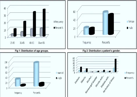
Fig 1: Distribution of age groups. Fig 2: Distribution a patient’s gender.
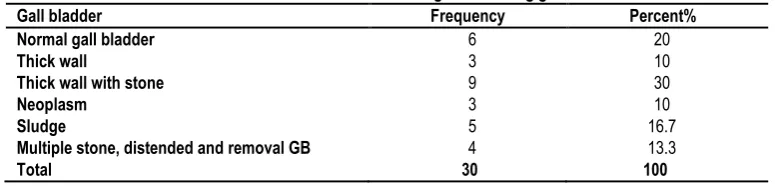
Table 1: Distribution of ultrasound findings concerning gall bladder.
Gall bladder Frequency Percent%
Normal gall bladder 6 20
Thick wall 3 10
Thick wall with stone 9 30
Neoplasm 3 10
Sludge 5 16.7
Multiple stone, distended and removal GB 4 13.3
Total 30 100
Table 2: Distribution of ultrasound findings concerning common bile duct.
Common bile duct Frequency Percent %
Normal 7 23.3
Dilated 23 77.7
Total 30 100
Table 3: Distribution of ultrasound findings concerning causes of obstructive jaundice.
Causes of obstructive observed Frequency Percent %
Common bile duct stone 15 5
Common bile duct mass 0 0
Carcinoma head of pancreas 6 20
Ultrasound failed 9 30
Total 30 100
Table 4: Distribution of ERCP findings concerning CBD diameter.
Common bile duct Frequency Percent %
Normal 1 3.3
Dilated 23 76.7
Failed 6 20
Total 30 100
Table 5: Distribution of ERCP findings concerningcauses of obstructive jaundice.
Causes of obstructive observed Frequency Percent %
Normal 1 3.3
Common bile duct stone 11 36.7
Common bile duct mass 0 0
Carcinoma head of pancreas 7 23.3
Stricture 5 16.7
ERCP failed 6 20
Total 30 100
Table 6: Distribution for diagnosis of biliary system stone by ERCP and US out of 26 patients.
Techniques Biliary stone Percentage 100%
ERCP 11 42.3
U/S 15 57.7
Total 26 100
.
Fig 5: Distribution of diagnosis carcinoma head of pancreas by ERCP and USout of 13 patients.
Fig 7: Efficiency of Ultrasound compared with ERCP to detect causes of obstructive jaundice out of 30 patients.
RESULTS AND DISCUSSION
30 jaundiced patients with biliary system disorders were investigated by ERCP and ultrasound.
It’s well known that U/S scan procedure is easy, comfortable,
noninvasive procedure and without patients or staff hazard. While ERCP procedure is invasive, performed in the painful risky, considerable conditions and with radiation hazard.
The study found that obstructive jaundice is most common in elderly ages especially over 65 years there were 11 out of 30
cases (36.7%) this might due to “elevation of the bilirubin level in
this stage of life than normal which (>50umol/L)”1, and this results
was not match the recent results reported by Zhong et al 2012 that revealed from their sample youth group of patients with Jaundice and viral hepatitis is the most common, accounting for 31.94%, much higher than the middle age group of 9.23% and 4.26% of the elderly group.3
16 cases out of thirty were females forming an incidence (53.3%) and 14 were males (46.7%). Increased incidence of female patients in this study due to excess of cholesterol in the body, multiple pregnancies, obesity and rapid weight loss, this results approach the results of Zhong et al 2012 who mentioned that In the 352 cases of jaundice, male to female ratio is 1.88:1.3 Also
these results are basically the same as Xudong et al and Qiu et al.4,5
This study found that obstructive jaundice in married patients were 27 out of 30 cases (90%) more than single patients 3 out of 30 cases (10%) because in Sudan most of the people married after 30 years, and there is relationship between obstruction jaundice and advanced age in this study. In addition to that the majority of patients were diabetic 6 out of 30 patients (20%) and this record might attributed to advanced patient age.
The ultrasound scan sensitivity in detecting the calculus cholecystitis that associated with obstructive jaundice was commonest and was 9 out of 30 patients (30%) compared with other abnormality of gall bladder, but ERCP failed to diagnosis any abnormality in the gall bladder. This results is not comparable with the study performed by Khan MA et al 1995 that detected the ultrasound accuracy in calculi, and reported that the positive predictive value for the site and etiology of obstruction by ultrasonography was 94% and 86% respectively.6
The dilated common bile duct was commonest finding that concerned with obstructive jaundice which was 23 out of 30 (77.7%), compared with ERCP which was 23 cases (76.7%), due to accumulation of bile in common bile duct. These findings to somewhat agree with the findings of Karki; et al 2013, they reported that "Ultrasound was found to have very high accuracy in detecting biliary tract dilatation with sensitivity of 94.8% and specificity of 100%. However the detection of CBD stricture by USG was not statically significant as compared to ERCP.7
Ultrasound scan sensitivity in detecting biliary stones was (57. 7%), 15 patients out of 26. Sensitivity for ERCP was (42.3%), and this record explains the increased sensitivity of U/S over ERCP because some stones were too small in size or largest to prevent entering ERCP guide wire. Also the stenosis in the site of the papilla, makes it difficult to cannulate. This study agrees with records of Rigauts et al, (1992) in their study about Comparison of ultrasound and ERCP in detection of the causes of obstructive biliary disease. They found that out of 120 patients the ultrasound correctly defined the cause of obstruction in 71% of the patients with ductal stones compared with ERCP.8 In addition to that it also
match the findings of Kiani et al in their recent study 2012, as they revealed that Choledocholithiasis was the commonest causes (twenty patients i.e., 66.6%) followed by various malignancies.9
ERCP most sensitive to detect biliary tumors, there were 7 out of 13 patients with biliary tumors (53.8%) diagnosed by ERCP, and U/S diagnosed 6 (46.2%). This record explains the sensitivity of ERCP over U/S. this findings agrees with Rigauts et al8 (1992) in
their study about Comparison of ultrasound and ERCP in the detection of the cause of obstructive biliary disease. They found that out of 120 patients the ERCP correctly defined the cause of obstructive in (90%) of patients with ductal tumors compared with U/S. also this findings comparable with the original study of Karki et al7 2013 as they concluded that ERCP was to be the much
sensitive in detecting CBD stricture as compared to USG which was similar to the study performed by Upadhyaya et.al 2006. And ERCP with advanced technology and well trained staff may be more sensitive than these findings as the findings mainly depend on the technology and experience of the staff.10
Efficiency of ultrasound to detect causes of obstructive jaundice was 21 cases out of 30 (70%) and ERCP was 24 (80%). This record explains that the ERCP more accurate than U/S to detect causes of obstructive jaundice. This study agrees with Pasanen; et al (1992), in study about ultrasonography, CT, and ERCP in the diagnosis of choledochal Stones. They found that out of 187 patients the sensitivity of U/S in detection of obstructive jaundice was (22.5%) and ERCP was (87%).11 Also this match the findings
of Satish et al 2013 as they concluded that ultrasound even with high resolution equipment and tissue harmonic imaging technique was limited by many factor such as obese patients who were poor ultrasound candidates, as well as too bowel gases which caused obscuration of distal CBD.12 Besides, smaller lesions beyond the
resolving power of ultrasound were missed.
Also U/S scanning is individual dependence, therefore it require experience and new advanced machines and it has a wide range to covering the diagnosis of the biliary system diseases and has the ability to differentiate between them, while ERCP procedure is effective in the bile ducts.
Obstructive jaundice has different etiological spectrum in males; While malignant causes predominate compared to females who have more of benign disease. Benign causes are seen at a comparatively younger age group compared to malignant causes.
RECOMMENDATIONS
U/S examinations should be done before ERCP. ERCP procedure is needed for treatment only, however good sterilization and safety considerations must be achieved to avoid the complications. Some cases missed diagnosed by U/S, so ERCP should be done to ensure the final diagnosis. More studies on this field of diagnosis were required, and the training of the staff is the principal key.
REFERENCES
1. Kumar and Clark. Clinical medicine. 6th ed. Elsevier; UK: 1993.P.)357-395(.
2. A. Adam. A. K. Dixon. Diagnostic radiology. 5th ed.an imprint of elesevier; London: 2008. volume 1 section 3 chapter 36.
3. Zhong Yu, Jun Zhan*, Chu-Qiang Li, Hui-Min Zhou. Age and gender analysis of jaundice patients; The Journal of Bioscience and Medicine 2, 2 (2012);1-3.
3. JC. Underwood. General and systematic pathology.4th ed. Elsevier; National: 2007. P. 402 407,408,425-432.
4. Xudong Xu, Yaqun Wu, Zhisu Liu, et al. The value of ERCP in the diagnosis of obstructive jaundice in elderly patients. Journal of Chinese General Surgical Basis and Clinical. 2011, (01):55. 5. Qiu YD, Bai JL, Xu FG, et al. Effect of preoperative biliary drainage on malignant obstructive jaundice: a meta-analysis. World J Gastroenterol, 2011,17 (3):391.
6. Khan Rahim M A, Khan A A, Shafqat F. Comparison of ultrasonography and cholangiography (ERCP/ PTC) in the
differential diagnosis of obstructive jaundice. J Pak Med Assoc. 1996 Sep;46(9):188-90.
7. Karki S, Joshi KS, Regmi S, Gurung RB, Malla B. Role of Ultrasound as Compared with ERCP in Patient with Obstructive Jaundice. Kathmandu Univ Med J 2013; 43(3):237-240.
8. Rigauts H, Marchal G, Van Steenbergen W, Ponette E. Comparison of ultrasound and E.R.C.P. in the detection of the cause of obstructive biliary disease; Source Department of Radiology, University Hospitals K. U. Leuven, Belgium, in (1992 Mar;156(3):252-7).
9. Kiani AA, Javaid RH, Ghaffar A, Khan S. (2012). Ultrasonography in obstructive jaundice. Professional Med J. 19(4): 436-441.
10. Upadhyaya V, Upadhyaya DN, Ansari MA et al. (2006). Comparative assessment of imaging modalities in biliary obstruction. Indian J Radiol Imaging. 16:577-82.
11. Pasanen P, Partanen K, Pikkarainen P, Alhava E, Pirinen A, Janatuinen E. Ultrasonography, CT, and ERCP in the diagnosis of choledochal stones. Acta Radiol. 1992 Jan; 33 (1):53-6.
12. Satish K. Bhargava, Thingujam Usha, Shuchi Bhatt, Rima Kumari, Sumeet Bhargava. (2013). Imaging in Obstructive Jaundice: A Review with Our Experience. Journal of International Medical Science Academy.2013; 26.43-46.
Source of Support: Nil. Conflict of Interest: None Declared.
Copyright: © the author(s) and publisher. IJMRP is an official publication of Ibn Sina Academy of Medieval Medicine & Sciences, registered in 2001 under Indian Trusts Act, 1882. This is an open access article distributed under the terms of the Creative Commons Attribution Non-commercial License, which permits unrestricted non-commercial use, distribution, and reproduction in any medium, provided the original work is properly cited.