OncoTargets and Therapy 2017:10 2849–2863
OncoTargets and Therapy
Dovepress
submit your manuscript | www.dovepress.com 2849
O r i g i n a l r e s e a r c h open access to scientific and medical research
Open access Full Text article
Prognostic value of high iMP3 expression in solid
tumors: a meta-analysis
luyao chen1,2,*
Yongpeng Xie3,*
Xintao li1
liangyou gu1
Yu gao1
lu Tang1
Jianwen chen1
Xu Zhang1
1Department of Urology, chinese
Pla general hospital, Beijing,
2Department of Urology, First
affiliated hospital of nanchang University, nanchang; 3school of
Medicine, nankai University, Tianjin, People’s republic of china
*These authors contributed equally to this work
Background: Accumulated studies have investigated the prognostic role of insulin-like growth factor II mRNA-binding protein 3 (IMP3) in various cancers, but inconsistent and controversial results were obtained. Therefore, we performed a systematic review and meta-analysis to inves-tigate the potential value of IMP3 in the prognostic prediction of human solid tumors.
Materials and methods: A systematic literature search in the electronic databases PubMed, Embase, Web of Science, and Cochrane library (updated to April 2016) was conducted to identify eligible studies. Pooled hazard ratios (HRs) with 95% confidence intervals (CIs) for survival outcomes were calculated and gathered using STATA 12.0 software.
Results: A total of 53 studies containing 8,937 patients with solid tumors were included in this meta-analysis. High IMP3 expression was significantly associated with worse overall survival (OS) of solid tumors (HR =2.08, 95% CI: 1.80–2.42, P,0.001). Similar results were observed in cancer-specific survival (CSS), disease-free survival (DFS), recurrence-free survival (RFS), progression-free survival (PFS), and metastasis-free survival (MFS). Further subgroup analysis stratified by tumor type showed that elevated IMP3 expression was associated with poor OS in renal cell carcinoma (RCC), lung cancer, oral cancer, urothelial carcinoma, hepatocellular carcinoma (HCC), colorectal cancer, pancreatic cancer, gastric cancer, and intrahepatic cho-langiocarcinoma (ICC).
Conclusion: The current evidence suggests that high IMP3 expression is associated with poor prognosis in most solid tumors. IMP3 is a potential valuable prognostic factor and might serve as a promising biomarker to guide clinical decisions in human solid tumors.
Keywords: IMP3, prognosis, solid tumor, biomarker, meta-analysis
Introduction
Insulin-like growth factor II mRNA-binding protein 3 (IMP3 or IGF2BP3) is a member of the RNA-binding protein family, which plays an important role in RNA trafficking and stabilization, cell growth, and cell migration during the early stages of
embryogenesis.1 IMP3 was proposed to control the translation or turnover of various
candidate target genes, including IGF2, CD44, HMGA2, and MMP9.2–5 This oncofetal
protein has been reported to promote tumor cell survival, proliferation, chemoresis-tance, and tumor cell invasiveness in vitro. In recent years, accumulating studies have shown that IMP3 is specifically expressed in malignant tumors and acts as an important
cancer-specific gene involved in many aggressive and advanced cancers.6,7
Numerous studies have reported that upregulated IMP3 expression in tumor tissues is correlated with poor patient survival and can be used as a prognostic factor to guide clinical decisions and distinguish different prognoses in various solid tumors, such as renal cell carcinoma (RCC), lung cancer, oral cancer, bladder cancer, gastrointestinal
tumors, and gynecological tumors.8–13 However, some other studies have reported
correspondence: Xu Zhang
Department of Urology, chinese Pla general hospital, no 28 Fuxing road, haidian, Beijing 100853, People’s republic of china
Tel +86 10 6693 8008 Fax +86 10 6822 3575 email [email protected]
Journal name: OncoTargets and Therapy Article Designation: Original Research Year: 2017
Volume: 10
Running head verso: Chen et al
Running head recto: Prognostic value of high IMP3 expression in solid tumors DOI: http://dx.doi.org/10.2147/OTT.S128810
OncoTargets and Therapy downloaded from https://www.dovepress.com/ by 118.70.13.36 on 25-Aug-2020
For personal use only.
Number of times this article has been viewed
This article was published in the following Dove Press journal: OncoTargets and Therapy
Dovepress chen et al
the absence of association between IMP3 expression and
cancer prognosis.14,15 Some investigators have also replayed
completely opposite results in ovarian cancer. For instance,
Kobel et al16 proposed that IMP3 expression is a marker
of unfavorable prognosis, whereas Noske et al17 asserted
that IMP3 expression is associated with improved survival. Hence, the prognostic role of IMP3 expression in solid tumors remains unclear and controversial.
Therefore, we conducted a systematic review of published studies, with a standard meta-analysis combining available evidence, to evaluate the prognostic value of IMP3 expres-sion in solid tumors.
Materials and methods
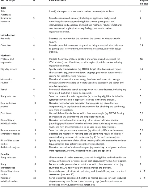
This meta-analysis was conducted according to the guideline of Preferred Reporting Items for Systematic Reviews and
Meta-Analyses (PRISMA)18 (Table S1). Because the data
included in this study were retrieved from published articles, ethical approval from ethics committees was not needed.
literature search
A comprehensive literature search was performed in PubMed, Embase, Web of Science, and Cochrane Library to identify studies evaluating IMP3 expression and clinical prognosis in solid tumors up to April 2016. The search strat-egy included the following terms through MeSH headings, keywords, and text words: “IMP3” or “Insulin-like growth factor 2 mRNA binding protein 3” or “IGF2BP3” combined with “cancer” or “carcinoma” or “neoplasm”. The references cited in the identified articles were also screened for possible inclusions. The database search and preliminary evaluation of identified studies were performed independently by two investigators (LC and YX). No language limitation existed in the process.
study selection
The inclusion criteria for selecting articles in our analysis are listed as follows: 1) studies that reported IMP3 expres-sion in cancer tissues, 2) studies analyzing the relationship between IMP3 expression level and clinical cancer outcomes, 3) studies that directly reported survival outcomes with hazard ratio (HR) and corresponding 95% confidence interval (CI) or studies that provided sufficient data for estimating HR and
95% CI by using the methods described by Tierney et al,19
and 4) studies with a median follow-up of at least 6 months. Studies were excluded if they were 1) case reports, let-ters, conference abstracts, or reviews, 2) non-human research, 3) investigations on the diagnostic role, but not the
prognostic role, of IMP3, and 4) studies with insufficient data for calculating the HR and 95% CI. If duplicate publications by the same authors were retrieved, we included only the most informative and recent study. Two independent reviewers (LC and YX) evaluated the full articles for study eligibility, and any disagreement was resolved by consensus.
Data extraction and quality assessment
Two authors (LC and YX) independently extracted data from each eligible study by using predefined item forms. The following information, if available, was recorded: first author’s name, year of publication, study country or region, type of cancer, cancer stage, number of patients, detected method, cutoff definition, percentage of high IMP3 expres-sion, follow-up period, and survival outcomes with their HRs and corresponding 95% CIs. If univariate and multivariate analyses were reported to obtain the HRs, the results of multivariate analysis were preferentially selected. If HRs and 95% CIs were not provided directly, we attempted to estimate these points with Kaplan–Meier curve or other required data in the original study by using Tierney et al’s
methods.19 Study quality was scored by two investigators
(LC and YX) using the Newcastle–Ottawa Scale, which involves three main categories: selection, comparability, and outcome ascertainment. We defined studies with scores no less than 6 as qualified to be included in the meta-analysis. Discrepancies between investigators were resolved through discussion.
statistical analysis
Pooled HRs and corresponding 95% CIs were calculated to evaluate the prognostic role of high IMP3 expression in the clinical outcomes of solid tumors. An observed HR greater than 1 implied a worse prognosis in patients with high IMP3 expression, and an HR less than 1 indicated a better prognosis. Statistical heterogeneity of combined HR was assessed using
Cochrane Q-test and Higgins I2 metrics. I2.50% was
consid-ered a measure of obvious heterogeneity.20 If no evident
het-erogeneity existed, the fixed-effect model (Mantel–Haenszel
method) was used to pool the results.21 Otherwise, the
random-effect model (DerSimonian and Laird method) was selected.22
The potential sources for heterogeneity, if significant, were further explored using a predefined subgroup analysis and meta-regression analysis (based on cancer type, ethnicity, case number, cutoff, cancer stage, HR obtained method, and analysis method). To assess the stability of the pooled results, sensitivity analysis was performed by sequential omission of each single study. Publication bias was also estimated by
OncoTargets and Therapy downloaded from https://www.dovepress.com/ by 118.70.13.36 on 25-Aug-2020
Dovepress Prognostic value of high iMP3 expression in solid tumors
visually assessing the asymmetry of the funnel plot and then
quantitatively evaluated by Begg’s and Egger’s tests.23,24 All
the abovementioned analyses were performed using STATA version 12.0 (Stata Corporation, College Station, TX, USA). All statistical tests were two sided, and statistical significance was defined as a P-value less than 0.05.
Results
search results and study characteristics
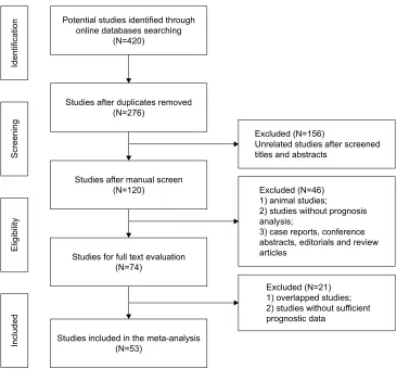
The flowchart of the literature search is shown in Figure 1. A total of 420 potentially relevant studies were retrieved from the initial literature search in the aforementioned electronic databases. A total of 144 duplicated records were excluded by a literature manager software. After carefully screening titles and abstracts of the remaining 120 records, 46 studies were excluded and 74 studies were selected for full-text assessment. Given the inclusion and exclusion criteria, 21 studies that belonged to duplicate publication or failed to offer sufficient prognostic information were excluded. Finally, 53 studies satisfied our eligibility criteria and were included in this meta-analysis.
The characteristics of these enrolled studies are summa-rized in Table 1. The 53 studies involved 8,937 patients with
different cancer types, including 6 studies of RCC,8,25–29 6 lung
cancer,9,30–34 4 oral cancer,10,35–37 4 urothelial carcinoma,38–41
4 ovarian cancer,16,17,42,43 3 hepatocellular carcinoma
(HCC),44–46 4 colorectal cancer,12,47–49 3 prostate cancer,14,15,50
3 pancreatic cancer,51–53 2 gastric cancer,11,54 2 intrahepatic
cholangiocarcinoma (ICC),55,56 and one study each of
tongue cancer,57 thyroid carcinoma,58 sacral chordoma,59
pilocytic astrocytoma and pilomyxoid astrocytoma (PA/
PMA),60 neuroblastoma,61 meningioma,62 melanoma,63 breast
cancer,64 giant cell tumor,65 bile duct carcinoma,66
esopha-geal carcinoma,67 and cervical cancer.13 A total of 25 studies
involved Caucasians and 28 involved Asians. The survival outcomes in these studies, including overall survival (OS), cancer-specific survival (CSS), disease-free survival (DFS), recurrence-free survival (RFS), progression-free survival (PFS), and metastasis-free survival (MFS), were investigated in 40, 10, 8, 7, 4, and 5 studies, respectively. HRs were reported directly in most of these studies (43/53) and were estimated indirectly in the 10 other studies. Multivariate Cox
Figure 1 Flowchart of the study selection process.
3RWHQWLDOVWXGLHVLGHQWLILHGWKURXJK RQOLQHGDWDEDVHVVHDUFKLQJ
1
6WXGLHVDIWHUGXSOLFDWHVUHPRYHG 1
([FOXGHG1
8QUHODWHGVWXGLHVDIWHUVFUHHQHG WLWOHVDQGDEVWUDFWV
([FOXGHG1 DQLPDOVWXGLHV
VWXGLHVZLWKRXWSURJQRVLV DQDO\VLV
FDVHUHSRUWVFRQIHUHQFH DEVWUDFWVHGLWRULDOVDQGUHYLHZ DUWLFOHV
([FOXGHG1 RYHUODSSHGVWXGLHV VWXGLHVZLWKRXWVXIILFLHQW SURJQRVWLFGDWD
6WXGLHVDIWHUPDQXDOVFUHHQ 1
6WXGLHVIRUIXOOWH[WHYDOXDWLRQ 1
6WXGLHVLQFOXGHGLQWKHPHWDDQDO\VLV 1
,GHQWLILFDWLRQ
6FUHHQLQJ
(OLJLELOLW\
,QFOXGHG
OncoTargets and Therapy downloaded from https://www.dovepress.com/ by 118.70.13.36 on 25-Aug-2020
Dovepress chen et al
Table 1
c
haracteristics of studies included in the meta-analysis
Author
Year
Country or region
Cancer type
Case number
Method
Cutoff
High expression
Follow-up
Outcomes
Analysis
HR obtained NOS score
Jiang et al
8 2006 U sa rcc 371 ihc
Positive vs negative*
71 (19.1%) Median 63 months O s MF s Multi r eport 9
Pei et al
26 2015 U sa rcc 346 ihc
Positive vs negative
73 (21.1%) . 10 years O s r Fs Multi r eport 8 h
offmann et al
25 2008 U sa rcc 716 ihc
Positive vs negative
213 (29.7%) 9.5 years css MF s Multi r eport 8
Park et al
27 2014 Korea rcc 148 ihc .
5% of cells stained
43 (29.1%) Median 55.5 months css Multi r eport 7
Jiang et al
28 2008 U sa rcc 317 ihc
Positive vs negative
40 (12.6%) 8.8 years O s MF s Multi r eport 9
Tantravahi et al
29 2015 U sa rcc 27 ihc .
20% of cells stained
14 (51.9%) . 2 years O s Multi r eport 6 Del g
obbo et al
34 2014 italy lung cancer 74 ihc
Positive vs negative
24 (32.4%) Mean 65.6 months O s DF s Uni r eport 7
sun et al
32 2015 c hina lung cancer 196 ihc h -score . 100 (0–300) 83 (42.3%) r
ange (16.5–69.0) months
O s DF s Multi r eport 8
Yan et al
9 2016 c hina lung cancer 95 ihc .
25% of cells stained
39 (41.1%) . 5 years O s Multi r eport 7
Zhang et al
33 2015 c hina lung cancer 186 ihc .
5% of cells stained
139 (74.7%) . 5 years O s Multi r eport 8
lin et al
30 2015 c hina lung cancer 92 ihc
Positive vs negative
62 (67.4%) . 5 years O s Multi r eport 8
Beljan Perak et al
31 2012 c roatia lung cancer 90 ihc .
10% of cells stained
61 (67.8%) . 5 years O s Uni sc 6 c
lauditz et al
35 2013 g ermany Oral cancer 145 ihc .
10% of cells stained
79 (54.5%) Mean 41.3 months O s Multi r eport 8
lin et al
37 2011 Taiwan Oral cancer 93 ihc .
25% of cells stained
51 (54.8%) Mean 44.8 months O s Multi r eport 9
li et al
36 2010 Korea Oral cancer 96 ihc
Positive vs negative
65 (67.7%) Median 73 months O s Multi r eport 9 Kim and c ha 10 2011 Korea Oral cancer 95 ihc
Positive vs negative
67 (70.5%) . 5 years O s Multi r eport 7
szarvas et al
40 2012 g ermany Urothelial carcinoma 106 ihc staining index . 7 (0–9) 17 (16.0%) Median 15 months O s css MF s Multi r eport 7
sitnikova et al
39 2008 U sa Urothelial carcinoma 214 ihc
Positive vs negative
42 (19.6%) Median 35 months PF s DF s Multi r eport 8
lee et al
41 2013 Multicenter Urothelial carcinoma 622 ihc
Positive vs negative
76 (12.2%) Median 27 months O s css r Fs Multi r eport 9 n
iedworok et al
38 2015 g ermany Urothelial carcinoma 26 ihc h -score . 100 (0–300) 7 (26.9%) Median 50 months O s PF s Uni r eport 7
Bi et al
43 2016 c hina Ovarian cancer 73 ihc .
10% of cells stained
46 (63.0%) . 5 years O s Uni sc 7
Kobel et al
16
2009
British and north
a merica Ovarian cancer 278 ihc .
5% of cells stained
147 (52.9%) . 4.6 years css Multi r eport 8 h
us et al
42 2015 Taiwan Ovarian cancer 140 ihc
The median value (
irs : 0–9) nr Median 39 months PF s Multi r eport 6 n
oske et al
17 2009 g ermany Ovarian cancer 68 ihc irs . 6 32 (47.1%) Median 37 months O s Uni sc 7 h
u et al
44 2014 c hina hcc 160 ihc
staining score (2–7 vs 0–1)
97 (60.6%) Median 36 months O s r Fs Uni sc 8
Wachter et al
45 2011 g ermany hcc 365 ihc
staining group (2–3 vs 0–1)
67 (18.4%) Mean 23.3 months O s Multi r eport 7 c
hen et al
46 2013 c hina hcc 92 ihc
Positive vs negative
65 (70.7%) . 3 years O s Multi r eport 7
Yuan et al
48 2009 Taiwan c olorectal cancer 186 ihc .
50% of cells stained
66 (35.5%) Median . 5 years O s Multi r eport 8
li et al
49 2009 c hina c olorectal cancer 203 ihc
staining score (2–7 vs 0–1)
132 (65.0%) Median 61 months O s DF s Multi r eport 9
lochhead et al
12 2012 U sa c olorectal cancer 671 ihc
intense or moderate vs weak or absent
234 (34.9%) Median 160 months O s css Multi r eport 8
lin et al
30 2013 c hina c olorectal cancer 186 ihc
Positive vs negative
143 (76.9%) . 2 years O s Multi r eport 7
ikenberg et al
15 2010 switzerland Prostate cancer 425 ihc
Positive vs negative
354 (83.3%) Median 63 months r Fs Uni r eport 9 c
hromecki et al
14 2011 U sa Prostate cancer 232 ihc .
10% of cells stained
42 (18.1%) Median 69.8 months r Fs Multi r eport 9
szarvas et al
50 2014 g ermany Prostate cancer 124 ihc .
10% of cells stained
30 (24.2%) Median 155 months O s css Uni r eport 8
Wang et al
52 2014 c hina Pancreatic cancer 50 qP cr c
utoff value based on the
r O c curve 30 (60.0%) . 2 years O s Multi r eport 7
schaeffer et al
51 2010 c anada Pancreatic cancer 127 ihc ihc score . 5 80 (63.0%) Mean 13 months O s Multi r eport 8
OncoTargets and Therapy downloaded from https://www.dovepress.com/ by 118.70.13.36 on 25-Aug-2020
Dovepress Prognostic value of high iMP3 expression in solid tumors
Morimatsu et al
53 2012 Japan Pancreatic cancer 32 ihc .
50% of cells stained
17 (53.1%) Median 33.6 months css Uni sc 6
Wang et al
54 2010 c hina g astric cancer 92 ihc
Positive vs negative
75 (81.5%) . 2 years O s Uni sc 7
Okada et al
11 2011 Japan g astric cancer 96 ihc .
10% of cells stained
71 (74.0%) Median 5.5 years O s DF s Multi r eport 8 c
hen et al
46 2013 Taiwan icc 61 ihc .
10% of cells stained
25 (41.0%) Mean 33.5 months O s DF s Uni sc 7 g
ao et al
56 2014 c hina icc 72 ihc
Positive vs negative
59 (81.9%) Median 14.9 months O s Multi r eport 8
li et al
57 2011 c hina Tongue carcinoma 65 ihc
Positive vs negative
50 (76.9%) Median 36 months css Uni sc 8 a
sioli et al
58 2010 U sa Thyroid carcinoma 103 ihc Final score . 2 (0–6) 61 (59.2%) . 5 years O s DF s MF s Multi r eport 9
Zhou et al
59 2014 c hina sacral chordoma 32 ihc
staining score (2
–7 vs 0 –1) 20 (62.5%) Median 110 months DF s Uni sc 8
Barton et al
60 2013 U sa Pa /PM a 77 ihc
Three groups (1
–2 vs 0)
24 (31.2%) Mean 8.8 years PF s Uni r eport 7 c
hen et al
61 2011 Taiwan n euroblastoma 90 ihc .
10% of cells stained
52 (57.8%) Median 39.5 months O s Multi r eport 8 h
ao et al
62 2011 U sa Meningioma 107 ihc
Positive vs negative
7 (6.5%) Median 53 months O s r Fs Multi r eport 7
sheen et al
63 2014 Taiwan Melanoma 97 ihc .
10% of cells stained
72 (74.2%) Median 5.2 years O s Multi r eport 7
Walter et al
64 2009 U sa Breast cancer 138 ihc .
10% of cells stained
45 (32.6%) Median 71.5 months O s Multi r eport 7
Zhang et al
33
2015
c
hina
g
iant cell tumor
38
ihc
staining score (3
–7 vs 0 –2) 13 (34.2%) Median 88.0 months r Fs Uni sc 6 r
iener et al
66
2009
switzerland
Bile duct carcinoma
115
ihc
intense or moderate vs weak or absent
67 (58.3%) Median 9 months css Multi r eport 8
Takata et al
67 2014 Japan esophageal carcinoma 191 ihc .
10% of cells stained
113 (59.2%) Mean 41 months O s Multi r eport 9
Wei et al
13 2014 c hina c ervical carcinoma 96 ihc .
10% of cells stained
54 (56.3%) Median 58.1 months O s Multi r eport 8 Note:
*Positive vs negative: tumor cells with any detectable staining were considered positive.
Abbreviations: CSS, cancer-specific survival; DFS, disease-free survival; HCC, hepatocellular carcinoma; HR, hazard ratio; ICC, intrahepatic cholangiocarcinoma; IHC, immunohistochemistry; IRS, immunoreactivity score; MFS, metastasis-free survival; n O s, n ewcas tle–Ottawa scale; nr , not reported; O s, overall survival; Pa /PM a , pilocytic astrocytoma and pilomyxoid astrocytoma; PF s, progression-free survival; qP cr , quantitative polymerase chain reaction; r Fs , recurrence-free survival; rcc
, renal cell carcinoma;
sc
, survival curve.
analysis was performed to evaluate the prognostic role of IMP3 in 38 studies; and univariate analysis was conducted in the other 15 studies. Immunohistochemistry (IHC) stain-ing and quantitative polymerase chain reaction (qPCR) were used to test the IMP3 expression in cancer tissues. Notably, the definition and cutoff of high IMP3 expression were heterogeneous among these studies. The majority of included studies used the percentage of positive staining cells (0%, 10%, 25%, or 50%) as the criteria, whereas in some other studies, staining scores with the percentage and intensity score were obtained as cutoff values for high IMP3 expression. The percentage of high expression in the cohort population varied in different cancer types and ranged from 6.5% to 83.3%. Quality score assessment suggested that the scores of enrolled studies ranged from 6 to 9, which were considered adequate for quantitative meta-analysis.
association of iMP3 with Os
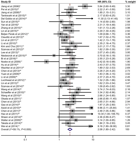
The association of IMP3 expression and OS was investigated in 40 studies containing 6,425 patients with different can-cer types. A random-effect model was selected because of
the evident interstudy heterogeneity (I2=59.1%, P=0.005).
Combined analysis revealed that high IMP3 expression was associated with the worse OS of solid tumors (HR
=2.08, 95% CI: 1.80–2.42, P,0.001, Figure 2). The effect
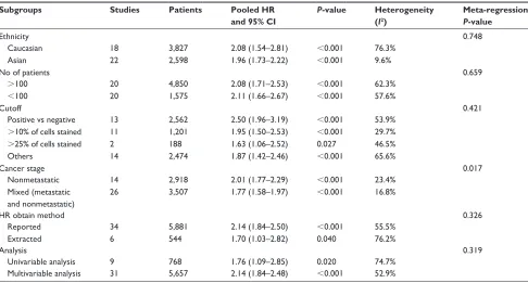
of IMP3 expression on OS was further analyzed by tumor types, and the results are presented in Figure 3A. High IMP3 expression was significantly associated with poor
OS in RCC (HR =2.80, 95% CI: 1.59–4.93, P,0.001),
lung cancer (HR =1.87, 95% CI: 1.22–2.84, P=0.004), oral
cancer (HR =1.66, 95% CI: 1.27–2.18, P,0.001),
urothe-lial carcinoma (HR =1.92, 95% CI: 1.42–2.59, P,0.001),
HCC (HR =2.25, 95% CI: 1.65–3.06, P,0.001), colorectal
cancer (HR =1.52, 95% CI: 1.23–1.90, P,0.001),
pan-creatic cancer (HR =3.54, 95% CI: 2.06–6.09, P,0.001),
gastric cancer (HR =2.67, 95% CI: 1.38–5.17, P=0.003),
and ICC (HR =2.10, 95% CI: 1.52–2.92, P,0.001) but not
in ovarian cancer (HR =1.05, 95% CI: 0.18–6.15, P=0.957).
To explore the source of heterogeneity, subgroup analysis and meta-regression were performed by the following stratifica-tion: patient ethnicity, study number, cutoff value, cancer stage, HR obtained method, and analysis style (Table 2). The results indicated that the combined HR estimates for OS in Caucasians and Asians were 2.08 (95% CI: 1.54–2.81,
P,0.001) and 1.96 (95% CI: 1.73–2.22, P,0.001),
respec-tively. Differences in the case number, cutoff value, cancer stage, HR obtained method, and analysis method did not influence the effect of IMP3 expression on the OS of solid tumors. Further meta-regression analysis revealed that cancer
OncoTargets and Therapy downloaded from https://www.dovepress.com/ by 118.70.13.36 on 25-Aug-2020
Dovepress chen et al
stage is a potential significant contributor to heterogeneity
(P=0.017), unlike other factors (P.0.05).
To assess the credibility of the pooled outcomes, we performed a sensitivity analysis through the sequential omis-sion of individual studies. The results were not obviously influenced by any single study (Figure 3C). The publication bias of all included studies was evaluated using a vertical funnel plot, Begg’s, and Egger’s tests. However, the funnel plot in Figure 3B appears asymmetrical, and the Begg’s
(P=0.015) and Egger’s tests (P=0.002) revealed existing
evidence of publication bias, which may be attributed to only seven studies that reported negative results among all the enrolled studies.
association of iMP3 with css, DFs, rFs,
PFs, and MFs
Ten studies that involved a total of 2,877 patients provided sufficient data for CSS analysis. No heterogeneity was
observed among these studies (I2=31.3%, P=0.158). Thus,
a fixed model was applied to pool the results. The combined
Figure 2 Forest plot of studies evaluating hr of high iMP3 expression in solid tumors for Os.
Notes: a pooled analysis showed that high iMP3 expression was associated with poor Os in solid tumors (hr =2.08, 95% ci: 1.80–2.42, P,0.001). Weights are from random-effects analysis.
Abbreviations: CI, confidence interval; HRs, hazard ratios; IMP3, insulin-like growth factor II mRNA-binding protein 3; OS, overall survival.
6WXG\,'
-LDQJHWDO
3HLHWDO
-LDQJHWDO
7DQWUDYDKLHWDO
'HO*REERHWDO
6XQHWDO
<DQHWDO
=KDQJHWDO
/LQHWDO
%HOMDQ3HUDNHWDO
/LQHWDO
/LHWDO
.LPDQG&KD
6]DUYDVHWDO
/HHHWDO
1LHGZRURNHWDO
%LHWDO
1RVNHHWDO
+XHWDO
:DFKWHUHWDO
&KHQHWDO
<XDQHWDO
/LHWDO
/RFKKHDGHWDO
/LQHWDO
6]DUYDVHWDO
:DQJHWDO
6FKDHIIHUHWDO
:DQJHWDO
2NDGDHWDO
&KHQHWDO
*DRHWDO
$VLROLHWDO
&KHQHWDO
+DRHWDO
6KHHQHWDO
:DOWHUHWDO
7DNDWDHWDO
:HLHWDO
2YHUDOO, 3
&ODXGLW]HWDO
+5&, ± ± ± ± ± ± ± ± ± ± ± ± ± ± ± ± ± ± ± ± ± ± ± ± ± ± ± ± ± ± ± ± ± ± ± ± ± ± ± ± ±
ZHLJKW
OncoTargets and Therapy downloaded from https://www.dovepress.com/ by 118.70.13.36 on 25-Aug-2020
Dovepress Prognostic value of high iMP3 expression in solid tumors
Figure 3 Subgroup analysis of OS stratified by tumor types, funnel plot of OS for publication bias, and sensitive analysis of OS.
Notes: (A) High IMP3 expression was significantly associated with poor OS in RCC, lung cancer, oral cancer, urothelial carcinoma, HCC, colorectal cancer, pancreatic cancer, gastric cancer, and icc but not in ovarian cancer. (B) The funnel plot for Os was asymmetric, which indicated the probability of publication bias. (C) sensitivity analysis by sequential omission of individual studies did not alter the significance, which confirmed the credibility of outcomes.
Abbreviations: CI, confidence interval; HCC, hepatocellular carcinoma; HR, hazard ratio; ICC, intrahepatic cholangiocarcinoma; In, natural logarithm; IMP3, insulin-like growth factor ii mrna-binding protein 3; Os, overall survival; rcc, renal cell carcinoma; se, standard error.
0HWDDQDO\VLVUDQGRPHIIHFWVHVWLPDWHVH[SRQHQWLDOIRUP 6WXG\RPLWWHG
-LDQJ 3HL -LDQJ 7DQWUDYDKL *REER 6XQ <DQ =KDQJ /LQ 3HUDN &ODXGLW] /LQ /L .LP 6]DUYDV /HH 1LHGZRURN %L 1RVNH +X :DFKWHU &KHQ <XDQ /L /RFKKHDG /LQ 6]DUYDV :DQJ 6FKDHIIHU :DQJ 2NDGD &KHQ *DR $VLROL &KHQ +DR 6KHHQ :DOWHU 7DNDWD :HL
$
6WXG\,'5&& /XQJFDQFHU 2UDOFDQFHU 8URWKHOLDOFDUFLQRPD 2YDULDQFDQFHU +&&
&RORUHFWDOFDQFHU 3DQFUHDWLFFDQFHU *DVWULFFDQFHU
,&&
± ± ± ± ± ± ± ± ± ±
+5&,
)XQQHOSORWZLWKSVHXGR FRQILGHQFHOLPLWV
,Q+5
6(RI,Q+5
± ±
%
&
OncoTargets and Therapy downloaded from https://www.dovepress.com/ by 118.70.13.36 on 25-Aug-2020
Dovepress chen et al
Table 2 subgroup analysis and meta-regression of the studies regarding overall survival
Subgroups Studies Patients Pooled HR
and 95% CI
P-value Heterogeneity (I2)
Meta-regression
P-value
ethnicity 0.748
caucasian 18 3,827 2.08 (1.54–2.81) ,0.001 76.3%
asian 22 2,598 1.96 (1.73–2.22) ,0.001 9.6%
no of patients 0.659
.100 20 4,850 2.08 (1.71–2.53) ,0.001 62.3%
,100 20 1,575 2.11 (1.66–2.67) ,0.001 57.6%
cutoff 0.421
Positive vs negative 13 2,562 2.50 (1.96–3.19) ,0.001 53.9%
.10% of cells stained 11 1,201 1.95 (1.50–2.53) ,0.001 29.7%
.25% of cells stained 2 188 1.63 (1.06–2.52) 0.027 46.5%
Others 14 2,474 1.87 (1.42–2.46) ,0.001 65.6%
cancer stage 0.017
nonmetastatic 14 2,918 2.01 (1.77–2.29) ,0.001 23.4%
Mixed (metastatic and nonmetastatic)
26 3,507 1.77 (1.58–1.97) ,0.001 16.8%
hr obtain method 0.326
reported 34 5,881 2.14 (1.84–2.50) ,0.001 55.5%
extracted 6 544 1.70 (1.03–2.82) 0.040 76.2%
analysis 0.319
Univariable analysis 9 768 1.76 (1.09–2.85) 0.020 74.7%
Multivariable analysis 31 5,657 2.14 (1.84–2.48) ,0.001 52.9%
Abbreviations: CI, confidence interval; HR, hazard ratio.
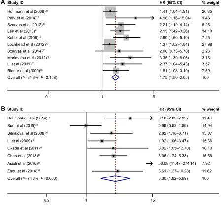
HR was 1.75 (95% CI, 1.50–2.05, P,0.001), indicating that
high IMP3 expression was associated with worse CSS in the patients with solid tumors (Figure 4A). The subgroup analysis stratified by cancer types showed that high IMP3 expression
significantly affected the RCC (HR =1.49, 95% CI: 1.11–
2.01, P=0.008) and urothelial carcinoma (HR =2.17, 95%
CI: 1.54–3.07, P,0.001). Further sensitivity analysis did not
alter the significance of combined HR, which validated the outcome credibility. Eight studies that involved 979 patients reported HRs for DFS, and the effect of high IMP3 expression is presented in Figure 4B. A combined analysis showed that high IMP3 expression was associated with poor DFS in solid
tumors (HR =3.30, 95% CI: 1.82–5.99, P,0.001).
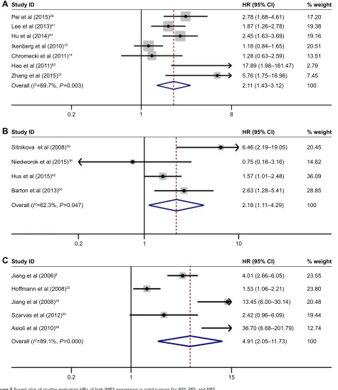
Seven studies with 1,930 patients investigated the prognostic role of IMP3 expression in the RFS of solid tumors. Pooled results demonstrated that high IMP3 adversely influenced the RFS in patients with solid
tumors (HR =2.11, 95% CI: 1.43–3.12, P,0.001, Figure 5A).
For PFS, four studies with 457 patients were included in the analysis. A forest plot of study-specific HRs for PFS is presented in Figure 5B. The combined results indicated that high IMP3 expression was significantly associated with
worse PFS in solid tumors (HR =2.18, 95% CI: 1.11–4.29,
P=0.023). In addition, five studies, including 1,613 patients,
focused on the influence of IMP3 on solid tumor metas-tasis. Meta-analysis of these studies suggested that IMP3
expression was also associated with poor MFS (HR =4.91,
95% CI: 2.05–11.73, P,0.001, Figure 5C).
Discussion
Over the past decades, increasing correlative studies describe the elevated IMP3 expression in human cancers, and various functional in vitro or in vivo studies provide strong evidence indicating that this oncofetal protein serves an essential role
in modulating tumor cell fate.6 As a molecular biomarker,
IMP3 has attracted extensive attention and can be used to distinguish different prognoses, improve prediction accuracy,
and better guide clinical decisions in different tumor types.7
Nevertheless, the relationship between IMP3 expression and oncological outcome remains controversial and requires a consensus. Consequently, we attempted to perform a sys-tematic review of published relevant studies and conduct a meta-analysis to clarify the prognostic value of IMP3 expres-sion in patients with solid tumors.
In the present research, given the inclusion criteria, 53 studies involving 8,937 patients were eligible, and the HRs of cumulative survival rates were summarized quantitatively by standard meta-analysis techniques. Our results suggested that high IMP3 expression was associated with worse OS of the solid tumors. Further subgroup analysis stratified by tumor type presented detailed results as follows. The negative prognostic effects of IMP3 on OS were specifically observed
OncoTargets and Therapy downloaded from https://www.dovepress.com/ by 118.70.13.36 on 25-Aug-2020
Dovepress Prognostic value of high iMP3 expression in solid tumors
$
6WXG\,'+RIIPDQQHWDO
3DUNHWDO
6]DUYDVHWDO
/HHHWDO
.REHOHWDO
/RFKKHDGHWDO
6]DUYDVHWDO
0RULPDWVXHWDO
/LHWDO
5LHQHUHWDO
2YHUDOO, 3
+5&,
± ± ± ± ± ± ± ± ± ± ±
ZHLJKW
%
± ± ± ± ± ± ± ± ±
+5&,
ZHLJKW
6WXG\,'
'HO*REERHWDO
6XQHWDO
6LWQLNRYDHWDO
/LHWDO
&KHQHWDO
$VLROLHWDO
=KRXHWDO
2YHUDOO, 3
2NDGDHWDO
Figure 4 Forest plot of studies evaluating hrs of high iMP3 expression in solid tumors for css and DFs.
Notes: (A) high iMP3 expression was associated with poor css in solid tumors (hr =1.75, 95% ci: 1.50–2.05, P,0.001). (B) high iMP3 expression was associated with poor DFs in solid tumors (hr =3.30, 95% ci: 1.82–5.99, P,0.001). Weights are from random-effects analysis.
Abbreviations: CI, confidence interval; CSS, cancer-specific survival; DFS, disease-free survival; HRs, hazard ratios; IMP3, insulin-like growth factor II mRNA-binding protein 3; Os, overall survival.
in RCC, lung cancer, oral cancer, urothelial carcinoma, HCC, colorectal cancer, pancreatic cancer, gastric cancer, and ICC. Besides OS, we also investigated other frequently used sur-vival outcomes, including CSS, DFS, RFS, PFS, and MFS. Similar influences were found for high IMP3 expression regarding the abovementioned end points, which provide a relatively comprehensive assessment of the value of IMP3 acting as a prognostic biomarker in solid tumors.
Accumulated literature suggests that IMP3 contributes to various aspects of cancer by promoting target genes expression by either preventing mRNA decay or stimulating mRNA translation. IMP3 knockdown in vitro can signifi-cantly inhibit the translation of IGF2 mRNA resulting in the
marked inhibition of cell proliferation.2 By using solid cancer
transcriptome data, IMP3 was also found to be correlated with HMGA2 mRNA expression in a dose-dependent manner. Additional assay for elucidating the mechanism indicated that IMP3 may function as a cytoplasmic safe house and prevents miRNA-directed mRNA decay of HMGA2 during tumor
pro-gression.4 Another recent study identified IMP3 as capable of
directly binding the mRNAs of cyclins D1, D3, and G1 in vivo and in vitro. The study also found that IMP3 can regulate the expression of these cyclins depending on their protein partner HNRNPM in six human cancer cell lines of different
origins.68 In addition, IMP3 promotes tumor cell invasion
and migration by targeting the epithelial–mesenchymal
OncoTargets and Therapy downloaded from https://www.dovepress.com/ by 118.70.13.36 on 25-Aug-2020
Dovepress chen et al
$
+5&,± ± ± ± ± ± ±
± 6WXG\,'
3HLHWDO
/HHHWDO
+XHWDO
,NHQEHUJHWDO
&KURPHFNLHWDO
+DRHWDO
=KDQJHWDO
2YHUDOO, 3
ZHLJKW
+5&,
6WXG\,'
6LWQLNRYDHWDO
1LHGZRURNHWDO
+XVHWDO
%DUWRQHWDO
2YHUDOO, 3
±
±
±
±
±
ZHLJKW
%
+5&,
6WXG\,'
-LDQJHWDO
+RIIPDQQHWDO
-LDQJHWDO
6]DUYDVHWDO
$VLROLHWDO
2YHUDOO, 3
±
±
±
±
±
±
ZHLJKW
&
Figure 5 Forest plot of studies evaluating hrs of high iMP3 expression in solid tumors for rFs, PFs, and MFs.
Notes: (A) high iMP3 expression was associated with poor rFs in solid tumors (hr =2.11, 95% ci: 1.43–3.12, P,0.001). (B) high iMP3 expression was associated with poor PFs in solid tumors (hr =2.18, 95% ci: 1.11–4.29, P=0.023). (C) high iMP3 expression was associated with poor MFs in solid tumors (hr =4.91, 95% ci: 2.05–11.73,
P,0.001). Weights are from random-effects analysis.
Abbreviations: CI, confidence interval; HRs, hazard ratios; IMP3, insulin-like growth factor II mRNA-binding protein 3; MFS, metastasis-free survival; PFS, progression-free survival; rFs, recurrence-free survival.
transition-associated molecular makers, including E-cadherin,
Slug, and vimentin.69 Overall, IMP3 plays an essential and
multifaceted role in human cancers. Hence, targeting IMP3 may serve as a potential strategy for anticancer therapy.
To our knowledge, our study is the first meta-analysis that comprehensively evaluated the association between IMP3 expression and prognosis in patients with solid tumors. However, several limitations of our study must
OncoTargets and Therapy downloaded from https://www.dovepress.com/ by 118.70.13.36 on 25-Aug-2020
Dovepress Prognostic value of high iMP3 expression in solid tumors
be acknowledged. First, we only extracted summarized population-level data rather than individual subject data from published literature. Second, different cutoff values and defi-nitions of high IMP3 expression were used in these included studies. Third, a marked study heterogeneity existed in some analyses. The subgroup analyses and meta-regression revealed that cancer stage might be a significant contributor to heterogeneity. Moreover, several potential factors such as cancer type, cutoff value, baseline characteristics (sample size, sex, age, and pathological subtype), and duration of follow-up may partially contribute to the heterogeneity. Among the enrolled studies, 10 works did not directly report the HRs. The calculated HRs, which were estimated using the methods of Tierney et al, might not be as dependable as those retrieved directly from the reported results. As such, the HRs inevitably introduced some statistical errors and may have influenced the pooled analysis. Furthermore, some studies only provided univariate analysis results, which may have introduced a bias toward overestimation of the prognostic value compared with multivariate analysis. The funnel plot and Egger’s test suggested the probability of publication bias because of fewer studies reporting negative results. However, the greater difficulty in publishing studies with insignificant results than those with significant results may be unavoidable. Finally, despite the well-recognized advantages of systematic review and meta-analysis, the results were based on the quality of the included studies. Thus, further high-quality studies with larger samples and a unified detection method are entailed to achieve a consensus on this matter.
Conclusion
The current evidence suggests that high IMP3 expression in tumor tissues is associated with adverse survival in various cancers. Hence, IMP3 might be a potential and promising biomarker that can be used to improve prognosis stratification and guide decision making in the treatment of solid tumors. Further well-designed studies are needed to confirm our find-ings and obtain more precise evaluations of the prognostic value of IMP3 in cancers.
Acknowledgments
This work was supported by the National High Tech-nology Research and Development Program of China (2014AA020607).
Disclosure
The authors report no conflicts of interest in this work.
References
1. Mueller-Pillasch F, Pohl B, Wilda M, et al. Expression of the highly conserved RNA binding protein KOC in embryogenesis. Mech Dev. 1999;88(1):95–99.
2. Liao B, Hu Y, Herrick DJ, Brewer G. The RNA-binding protein IMP-3 is a translational activator of insulin-like growth factor II leader-3 mRNA during proliferation of human K562 leukemia cells. J Biol
Chem. 2005;280(18):18517–18524.
3. Li W, Liu D, Chang W, et al. Role of IGF2BP3 in trophoblast cell invasion and migration. Cell Death Dis. 2014;5:e1025.
4. Jønson L, Christiansen J, Hansen TV, Vikeså J, Yamamoto Y, Nielsen FC. IMP3 RNP safe houses prevent miRNA-directed HMGA2 mRNA decay in cancer and development. Cell Rep. 2014;7(2):539–551.
5. Samanta S, Sharma VM, Khan A, Mercurio AM. Regulation of IMP3 by EGFR signaling and repression by ERbeta: implications for triple-negative breast cancer. Oncogene. 2012;31(44):4689–4697. 6. Lederer M, Bley N, Schleifer C, Huttelmaier S. The role of the oncofetal
IGF2 mRNA-binding protein 3 (IGF2BP3) in cancer. Semin Cancer
Biol. 2014;29:3–12.
7. Gong Y, Woda BA, Jiang Z. Oncofetal protein IMP3, a new cancer biomarker. Adv Anat Pathol. 2014;21(3):191–200.
8. Jiang Z, Chu PG, Woda BA, et al. Analysis of RNA-binding protein IMP3 to predict metastasis and prognosis of renal-cell carcinoma: a retrospective study. Lancet Oncol. 2006;7(7):556–564.
9. Yan J, Wei Q, Jian W, et al. IMP3 predicts invasion and prognosis in human lung adenocarcinoma. Lung. 2016;194(1):137–146.
10. Kim KY, Cha IH. Risk stratification of oral cancer patients using a com-bined prognostic factor including lymph node density and biomarker.
J Cancer Res Clin Oncol. 2012;138(3):483–490.
11. Okada K, Fujiwara Y, Nakamura Y, et al. Oncofetal protein, IMP-3, a potential marker for prediction of postoperative peritoneal dissemination in gastric adenocarcinoma. J Surg Oncol. 2012;105(8):780–785. 12. Lochhead P, Imamura Y, Morikawa T, et al. Insulin-like growth
factor 2 messenger RNA binding protein 3 (IGF2BP3) is a marker of unfavourable prognosis in colorectal cancer. Eur J Cancer. 2012;48(18): 3405–3413.
13. Wei Q, Yan J, Fu B, et al. IMP3 expression is associated with poor survival in cervical squamous cell carcinoma. Hum Pathol. 2014;45(11): 2218–2224.
14. Chromecki TF, Cha EK, Pummer K, et al. Prognostic value of insulin-like growth factor II mRNA binding protein 3 in patients treated with radical prostatectomy. BJU Int. 2012;110(1):63–68.
15. Ikenberg K, Fritzsche FR, Zuerrer-Haerdi U, et al. Insulin-like growth factor II mRNA binding protein 3 (IMP3) is overexpressed in pros-tate cancer and correlates with higher Gleason scores. BMC Cancer. 2010;10:341.
16. Kobel M, Xu H, Bourne PA, et al. IGF2BP3 (IMP3) expression is a marker of unfavorable prognosis in ovarian carcinoma of clear cell subtype. Mod Pathol. 2009;22(3):469–475.
17. Noske A, Faggad A, Wirtz R, et al. IMP3 expression in human ovar-ian cancer is associated with improved survival. Int J Gynecol Pathol. 2009;28(3):203–210.
18. Moher D, Liberati A, Tetzlaff J, Altman DG, Group P. Preferred reporting items for systematic reviews and meta-analyses: the PRISMA statement. Int J Surg. 2010;8(5):336–341.
19. Tierney JF, Stewart LA, Ghersi D, Burdett S, Sydes MR. Practical meth-ods for incorporating summary time-to-event data into meta-analysis.
Trials. 2007;8:16.
20. Higgins JP, Thompson SG, Deeks JJ, Altman DG. Measuring incon-sistency in meta-analyses. BMJ. 2003;327(7414):557–560.
21. Darroch JN. The Mantel-Haenszel test and tests of marginal symmetry; fixed-effects and mixed models for a categorical response, correspon-dent paper. Int Stat Rev. 1981;49(3):285–307.
22. DerSimonian R, Laird N. Meta-analysis in clinical trials. Control Clin
Trials. 1986;7(3):177–188.
23. Begg CB, Mazumdar M. Operating characteristics of a rank correlation test for publication bias. Biometrics. 1994;50(4):1088–1101.
OncoTargets and Therapy downloaded from https://www.dovepress.com/ by 118.70.13.36 on 25-Aug-2020
Dovepress chen et al
24. Egger M, Davey Smith G, Schneider M, Minder C. Bias in meta-analysis detected by a simple, graphical test. BMJ. 1997;315(7109):629–634. 25. Hoffmann NE, Sheinin Y, Lohse CM, et al. External validation of
IMP3 expression as an independent prognostic marker for metastatic progression and death for patients with clear cell renal cell carcinoma.
Cancer. 2008;112(7):1471–1479.
26. Pei X, Li M, Zhan J, et al. Enhanced IMP3 expression activates NF-small ka, CyrillicB pathway and promotes renal cell carcinoma progression.
PLoS One. 2015;10(4):e0124338.
27. Park JY, Choe M, Kang Y, Lee SS. IMP3, a promising prognostic marker in clear cell renal cell carcinoma. Korean J Pathol. 2014;48(2): 108–116.
28. Jiang Z, Lohse CM, Chu PG, et al. Oncofetal protein IMP3: a novel molecular marker that predicts metastasis of papillary and chromophobe renal cell carcinomas. Cancer. 2008;112(12):2676–2682.
29. Tantravahi SK, Albertson D, Agarwal AM, et al. Survival outcomes and tumor IMP3 expression in patients with sarcomatoid metastatic renal cell carcinoma. J Oncol. 2015;2015:181926.
30. Lin L, Zhang J, Wang Y, et al. Expression of insulin-like growth factor 2 mRNA-binding protein 3 expression and analysis of prognosis in the patients with lung squamous cell carcinoma. Xi Bao Yu Fen Zi Mian
Yi Xue Za Zhi. 2013;29(7):694–697.
31. Beljan Perak R, Durdov MG, Capkun V, et al. IMP3 can predict aggres-sive behaviour of lung adenocarcinoma. Diagn Pathol. 2012;7:165. 32. Sun X, Wei P, Shen C, et al. Prognostic value of the IASLC/ATS/ERS
classification and IMP3 expression in lung adenocarcinoma of Chinese cases. Am J Cancer Res. 2015;5(7):2266–2276.
33. Zhang J, Ou Y, Ma Y, et al. Clinical implications of insulin-like growth factor II mRNA-binding protein 3 expression in non-small cell lung carcinoma. Oncol Lett. 2015;9(4):1927–1933.
34. Del Gobbo A, Vaira V, Guerini Rocco E, et al. The oncofetal protein IMP3: a useful marker to predict poor clinical outcome in neuroendo-crine tumors of the lung. J Thorac Oncol. 2014;9(11):1656–1661. 35. Clauditz TS, Wang CJ, Gontarewicz A, et al. Expression of insulin-like
growth factor II mRNA-binding protein 3 in squamous cell carcinomas of the head and neck. J Oral Pathol Med. 2013;42(2):125–132. 36. Li S, Cha J, Kim J, et al. Insulin-like growth factor II mRNA-binding
protein 3: a novel prognostic biomarker for oral squamous cell carci-noma. Head Neck. 2011;33(3):368–374.
37. Lin CY, Chen ST, Jeng YM, et al. Insulin-like growth factor II mRNA-binding protein 3 expression promotes tumor formation and invasion and predicts poor prognosis in oral squamous cell carcinoma. J Oral
Pathol Med. 2011;40(9):699–705.
38. Niedworok C, Panitz M, Szarvas T, et al. Urachal carcinoma of the bladder: impact of clinical and immunohistochemical parameters on prognosis. J Urol. 2016;195(6):1690–1696.
39. Sitnikova L, Mendese G, Liu Q, et al. IMP3 predicts aggressive superficial urothelial carcinoma of the bladder. Clin Cancer Res. 2008; 14(6):1701–1706.
40. Szarvas T, vom Dorp F, Niedworok C, et al. High insulin-like growth factor mRNA-binding protein 3 (IMP3) protein expression is associ-ated with poor survival in muscle-invasive bladder cancer. BJU Int. 2012;110(6 pt B):E308–E317.
41. Lee DJ, Xylinas E, Rieken M, et al. Insulin-like growth factor messenger RNA-binding protein 3 expression helps prognostication in patients with upper tract urothelial carcinoma. Eur Urol. 2014;66(2):379–385. 42. Hsu KF, Shen MR, Huang YF, et al. Overexpression of the
RNA-binding proteins Lin28B and IGF2BP3 (IMP3) is associated with chemoresistance and poor disease outcome in ovarian cancer. Br J
Cancer. 2015;113(3):414–424.
43. Bi R, Shen X, Zhang W, et al. Clear cell carcinomas of the ovary: a mono-institutional study of 73 cases in China with an analysis of the prognostic significance of clinicopathological parameters and IMP3 expression. Diagn Pathol. 2016;11:17.
44. Hu S, Wu X, Zhou B, et al. IMP3 combined with CD44s, a novel predictor for prognosis of patients with hepatocellular carcinoma.
J Cancer Res Clin Oncol. 2014;140(6):883–893.
45. Wachter DL, Kristiansen G, Soll C, et al. Insulin-like growth factor II mRNA-binding protein 3 (IMP3) expression in hepatocellular carci-noma. A clinicopathological analysis with emphasis on diagnostic value.
Histopathology. 2012;60(2):278–286.
46. Chen LT, Lin LJ, Zheng LL. The correlation between insulin-like growth factor II mRNA binding protein 3 expression in hepatocellular carcinoma and prognosis. Hepatogastroenterology. 2013;60(123):553–556. 47. Lin L, Zhang J, Wang Y, et al. Insulin-like growth factor-II
mRNA-binding protein 3 predicts a poor prognosis for colorectal adenocarci-noma. Oncol Lett. 2013;6(3):740–744.
48. Yuan RH, Wang CC, Chou CC, Chang KJ, Lee PH, Jeng YM. Diffuse expression of RNA-binding protein IMP3 predicts high-stage lymph node metastasis and poor prognosis in colorectal adenocarcinoma. Ann
Surg Oncol. 2009;16(6):1711–1719.
49. Li D, Yan D, Tang H, et al. IMP3 is a novel prognostic marker that correlates with colon cancer progression and pathogenesis. Ann Surg
Oncol. 2009;16(12):3499–3506.
50. Szarvas T, Tschirdewahn S, Niedworok C, et al. Prognostic value of tissue and circulating levels of IMP3 in prostate cancer. Int J Cancer. 2014;135(7):1596–1604.
51. Schaeffer DF, Owen DR, Lim HJ, et al. Insulin-like growth factor 2 mRNA binding protein 3 (IGF2BP3) overexpression in pancreatic ductal adeno-carcinoma correlates with poor survival. BMC Cancer. 2010;10:59. 52. Wang BJ, Wang L, Yang SY, Liu ZJ. Expression and clinical
signifi-cance of IMP3 in microdissected premalignant and malignant pancreatic lesions. Clin Transl Oncol. 2015;17(3):215–222.
53. Morimatsu K, Aishima S, Yamamoto H, et al. Insulin-like growth fac-tor II messenger RNA-binding protein-3 is a valuable diagnostic and prognostic marker of intraductal papillary mucinous neoplasm. Hum
Pathol. 2013;44(9):1714–1721.
54. Wang L, Li HG, Xia ZS, Lu J, Peng TS. IMP3 is a novel biomarker to predict metastasis and prognosis of gastric adenocarcinoma: a retrospec-tive study. Chin Med J (Engl). 2010;123(24):3554–3558.
55. Chen YL, Jeng YM, Hsu HC, et al. Expression of insulin-like growth factor II mRNA-binding protein 3 predicts early recurrence and poor prognosis in intrahepatic cholangiocarcinoma. Int J Surg. 2013; 11(1):85–91.
56. Gao Y, Yang M, Jiang Z, et al. IMP3 expression is associated with poor outcome and epigenetic deregulation in intrahepatic cholangiocarci-noma. Hum Pathol. 2014;45(6):1184–1191.
57. Li HG, Han JJ, Huang ZQ, Wang L, Chen WL, Shen XM. IMP3 is a novel biomarker to predict metastasis and prognosis of tongue squamous cell carcinoma. J Craniofac Surg. 2011;22(6):2022–2025.
58. Asioli S, Erickson LA, Righi A, et al. Poorly differentiated carcinoma of the thyroid: validation of the Turin proposal and analysis of IMP3 expression. Mod Pathol. 2010;23(9):1269–1278.
59. Zhou M, Chen K, Yang H, et al. Expression of insulin-like growth factor II mRNA-binding protein 3 (IMP3) in sacral chordoma. J Neurooncol. 2014;116(1):77–82.
60. Barton VN, Donson AM, Birks DK, et al. Insulin-like growth factor 2 mRNA binding protein 3 expression is an independent prognostic factor in pediatric pilocytic and pilomyxoid astrocytoma. J Neuropathol Exp
Neurol. 2013;72(5):442–449.
61. Chen ST, Jeng YM, Chang CC, et al. Insulin-like growth factor II mRNA-binding protein 3 expression predicts unfavorable prognosis in patients with neuroblastoma. Cancer Sci. 2011;102(12):2191–2198. 62. Hao S, Smith TW, Chu PG, et al. The oncofetal protein IMP3: a novel
molecular marker to predict aggressive meningioma. Arch Pathol Lab
Med. 2011;135(8):1032–1036.
63. Sheen YS, Liao YH, Lin MH, et al. IMP-3 promotes migration and invasion of melanoma cells by modulating the expression of HMGA2 and predicts poor prognosis in melanoma. J Invest Dermatol. 2015; 135(4):1065–1073.
64. Walter O, Prasad M, Lu S, Quinlan RM, Edmiston KL, Khan A. IMP3 is a novel biomarker for triple negative invasive mammary carcinoma associated with a more aggressive phenotype. Hum Pathol. 2009; 40(11):1528–1533.
OncoTargets and Therapy downloaded from https://www.dovepress.com/ by 118.70.13.36 on 25-Aug-2020
Dovepress Prognostic value of high iMP3 expression in solid tumors
65. Zhang K, Zhou M, Chen H, Wu G, Chen K, Yang H. Expression of IMP3 and IGF2 in giant cell tumor of spine is associated with tumor recurrence and angiogenesis. Clin Transl Oncol. 2015;17(7):570–575.
66. Riener MO, Fritzsche FR, Clavien PA, et al. IMP3 expression in lesions of the biliary tract: a marker for high-grade dysplasia and an independent prognostic factor in bile duct carcinomas. Hum Pathol. 2009;40(10):1377–1383.
67. Takata A, Takiguchi S, Okada K, et al. Expression of insulin-like growth factor-II mRNA-binding protein-3 as a marker for predicting clinical outcome in patients with esophageal squamous cell carcinoma. Oncol
Lett. 2014;8(5):2027–2031.
68. Rivera Vargas T, Boudoukha S, Simon A, et al. Post-transcriptional regulation of cyclins D1, D3 and G1 and proliferation of human cancer cells depend on IMP-3 nuclear localization. Oncogene. 2014;33(22): 2866–2875.
69. Su P, Hu J, Zhang H, et al. IMP3 expression is associated with epithelial-mesenchymal transition in breast cancer. Int J Clin Exp
Pathol. 2014;7(6):3008–3017.
OncoTargets and Therapy downloaded from https://www.dovepress.com/ by 118.70.13.36 on 25-Aug-2020
Dovepress chen et al
Supplementary material
Table S1 checklist of PrisMa 2009
Section/topic # Checklist item Reported
on page # Title
Title 1 identify the report as a systematic review, meta-analysis, or both. 1
Abstract
structured summary
2 Provide a structured summary including, as applicable: background; objectives; data sources; study eligibility criteria, participants, and interventions; study appraisal and synthesis methods; results; limitations; conclusions and implications of key findings; systematic review registration number.
2
Introduction
rationale 3 Describe the rationale for the review in the context of what is already known.
3
Objectives 4 Provide an explicit statement of questions being addressed with reference to participants, interventions, comparisons, outcomes, and study design (PicOs).
3,4
Methods
Protocol and registration
5 indicate if a review protocol exists, if and where it can be accessed (eg, Web address), and, if available, provide registration information including registration number.
no
eligibility criteria 6 specify study characteristics (eg, PicOs, length of follow-up) and report characteristics (eg, years considered, language, publication status) used as criteria for eligibility, giving rationale.
4,5
information sources
7 Describe all information sources (eg, databases with dates of coverage, contact with study authors to identify additional studies) in the search and date last searched.
4
search 8 Present full electronic search strategy for at least one database, including any limits used, such that it could be repeated.
4
study selection 9 state the process for selecting studies (ie, screening, eligibility, included in systematic review, and, if applicable, included in the meta-analysis).
5
Data collection process
10 Describe method of data extraction from reports (eg, piloted forms, independently, in duplicate) and any processes for obtaining and confirming data from investigators.
5
Data items 11 List and define all variables for which data were sought (eg, PICOS, funding sources) and any assumptions and simplifications made.
5,6
risk of bias in individual studies
12 Describe methods used for assessing risk of bias of individual studies (including specification of whether this was done at the study or outcome level), and how this information is to be used in any data synthesis.
5,6
summary measures 13 state the principal summary measures (eg, risk ratio, difference in means). 5,6 synthesis of results 14 Describe the methods of handling data and combining results of studies, if
done, including measures of consistency (eg, I2) for each meta-analysis.
6
risk of bias across studies
15 specify any assessment of risk of bias that may affect the cumulative evidence (eg, publication bias, selective reporting within studies).
6
additional analyses 16 Describe methods of additional analyses (eg, sensitivity or subgroup analyses, meta-regression), if done, indicating which were pre-specified.
6
Results
study selection 17 give numbers of studies screened, assessed for eligibility, and included in the review, with reasons for exclusions at each stage, ideally with a flow diagram.
7
study characteristics
18 For each study, present characteristics for which data were extracted (eg, study size, PicOs, follow-up period) and provide the citations.
7
risk of bias within studies
19 Present data on risk of bias of each study and, if available, any outcome level assessment (see item 12).
7–14
results of individual studies
20 For all outcomes considered (benefits or harms), present, for each study: (a) simple summary data for each intervention group; (b) effect estimates and confidence intervals, ideally with a forest plot.
7–14
(Continued)
OncoTargets and Therapy downloaded from https://www.dovepress.com/ by 118.70.13.36 on 25-Aug-2020
OncoTargets and Therapy
Publish your work in this journal
Submit your manuscript here: http://www.dovepress.com/oncotargets-and-therapy-journal
OncoTargets and Therapy is an international, peer-reviewed, open access journal focusing on the pathological basis of all cancers, potential targets for therapy and treatment protocols employed to improve the management of cancer patients. The journal also focuses on the impact of management programs and new therapeutic agents and protocols on
patient perspectives such as quality of life, adherence and satisfaction. The manuscript management system is completely online and includes a very quick and fair peer-review system, which is all easy to use. Visit http://www.dovepress.com/testimonials.php to read real quotes from published authors.
Dovepress
Dovepress
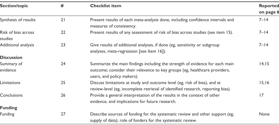
Prognostic value of high iMP3 expression in solid tumorsTable S1 (Continued)
Section/topic # Checklist item Reported
on page #
synthesis of results 21 Present results of each meta-analysis done, including confidence intervals and measures of consistency.
7–14
risk of bias across studies
22 Present results of any assessment of risk of bias across studies (see item 15). 7–14
additional analysis 23 give results of additional analyses, if done (eg, sensitivity or subgroup analyses, meta-regression [see item 16]).
7–14
Discussion
summary of evidence
24 Summarize the main findings including the strength of evidence for each main outcome; consider their relevance to key groups (eg, healthcare providers, users, and policy makers).
14,15
limitations 25 Discuss limitations at study and outcome level (eg, risk of bias), and at review-level (eg, incomplete retrieval of identified research, reporting bias).
15,16
conclusions 26 Provide a general interpretation of the results in the context of other evidence, and implications for future research.
17
Funding
Funding 27 Describe sources of funding for the systematic review and other support (eg, supply of data); role of funders for the systematic review.
none
Notes: reproduced from Moher D, liberati a, Tetzlaff J, et al, Preferred reporting items for systematic reviews and meta-analyses: the PrisMa statement. PLoS Med. 2009:6(7): e1000097.1
Reference
1. Moher D, Liberati A, Tetzlaff J, et al. The PRISMA Group (2009). Preferred Reporting Items for Systematic Reviews and Meta-Analyses: The PRISMA Statement. PLoS Med. 2009:6(7):e1000097.
OncoTargets and Therapy downloaded from https://www.dovepress.com/ by 118.70.13.36 on 25-Aug-2020