C A S E R E P O R T
Open Access
Metabolic encephalopathy secondary to
diabetic ketoacidosis: a case report
Maria Tomkins
1*, Richard McCormack
2, Karen O
’
Connell
3, Amar Agha
4and Áine Merwick
3Abstract
Introduction:Metabolic encephalopathy is a rare but potentially devastating complication of diabetic ketoacidosis (DKA). This case highlights the dramatic cognitive decline of a young man due to metabolic encephalopathy complicating DKA. The aims of this case report are to highlight metabolic encephalopathy as a complication of DKA and to explore the current research in diabetic related brain injury. The importance of investigation and treatment of reversible causes of encephalopathy is also demonstrated.
Case presentation:A 35-year-old man with a background of type 1 diabetes mellitus (T1DM) and relapsing remitting multiple sclerosis (RRMS) presented to the emergency department (ED) in a confused and agitated state. Prior to admission he worked as a caretaker in a school, smoked ten cigarettes per day, took excess alcohol and smoked cannabis twice per week. Following initial investigations, he was found to be in DKA. Despite timely and appropriate management his neurological symptoms and behavioural disturbance persisted. Neuroimaging revealed temporal lobe abnormalities consistent with an encephalopathic process. The patient underwent extensive investigation looking for evidence of autoimmune, infective, metabolic, toxic and paraneoplastic encephalopathy, with no obvious cause demonstrated.
Due to persistent radiological abnormalities a temporal lobe biopsy was performed which showed marked astrocytic gliosis without evidence of vasculitis, inflammation, infarction or neoplasia. A diagnosis of metabolic encephalopathy secondary to DKA was reached. The patient’s cognitive function remained impaired up to 18 months post presentation and he ultimately required residential care.
Conclusions:Metabolic encephalopathy has been associated with acute insults such as DKA, but importantly, the risk of cerebral injury is also related to chronic hyperglycaemia. Mechanisms of cerebral injury in diabetes mellitus continue to be investigated. DKA poses a serious and significant neurological risk to patients with diabetes mellitus. To our knowledge this is the second case report describing this acute complication.
Keywords:Diabetes mellitus, Metabolic encephalopathy, Ketoacidosis, Diabetic brain injury, Type 1 diabetes
Background
Diabetic ketoacidosis is a frequent complication of type 1 diabetes, with the UK National Diabetes Audit quoting a crude incidence rate of 3.6% among patients with type 1 diabetes [1]. Risk factors for DKA include younger age, poor glycaemic control, lower socioeconomic status and depression/psychiatric illness. Precipitants of DKA include omission of or inadequate insulin, infection, cardiovascular disease such as stroke/acute coronary syndrome, acute pan-creatitis and certain medications such as steroids, thiazide diuretics and sodium-glucose-co-transporter 2 (SGLT2)
inhibitors. Physiological stress from surgery, pregnancy or trauma has the potential to initiate a DKA due to increased release of counter-regulatory hormones [1].
Metabolic encephalopathy refers to an alteration in con-sciousness due to impaired cerebral metabolism causing dif-fuse or global brain dysfunction in the absence of primary structural dysfunction [2]. Chemical imbalance occurs in the setting of hypoxia or due to systemic organ failure (hepatic failure, renal failure, pancreatic failure) and electrolyte imbal-ance (hypercalcaemia, hypoglycaemia, hyponatraemia) result-ing in toxic neurological injury [2]. Varying presentations occur and may indicate the underlying cause, for example patients with hepatic encephalopathy may demonstrate asterixis, however in general metabolic encephalopathy pre-sents as global cerebral dysfunction with fluctuating
© The Author(s). 2019Open AccessThis article is distributed under the terms of the Creative Commons Attribution 4.0 International License (http://creativecommons.org/licenses/by/4.0/), which permits unrestricted use, distribution, and reproduction in any medium, provided you give appropriate credit to the original author(s) and the source, provide a link to the Creative Commons license, and indicate if changes were made. The Creative Commons Public Domain Dedication waiver (http://creativecommons.org/publicdomain/zero/1.0/) applies to the data made available in this article, unless otherwise stated.
* Correspondence:mariatomkins200@gmail.com
1Department of Diabetes and Endocrinology, Beaumont Hospital, Dublin, Ireland
consciousness in the absence of focal neurological signs [3,
4]. It is a very rare but potentially devastating complication of diabetic ketoacidosis (DKA). Hitherto, only one case has been reported of this complication in which the patient showed a gradual recovery with residual speech disturbance and irritability [5].
In this paper we describe a case of a patient with persist-ing neurological injury secondary to metabolic encephal-opathy associated with DKA. The aims of this case report are to highlight metabolic encephalopathy as a complica-tion of DKA and to explore the current research in dia-betic related brain injury. The importance of investigation and treatment of reversible causes of encephalopathy is also demonstrated, as this is a diagnosis of exclusion.
Case presentation
A 35-year-old man with T1DM presented to ED, having been found in an acutely confused state at home. Having not left his bedroom for 2 days, his co-habitants alerted emer-gency services who forced entry to his bedroom and found him in an unkempt, confused state. On arrival he was agi-tated, confused, unkempt and uncommunicative. The major-ity of the clinical history was provided by his parents who had seen him well 2 days previously. They described an inde-pendent 35-year-old man who had no complaints in the days leading up to his admission. They described poor engage-ment with medical services regarding his diabetes and mul-tiple sclerosis. His social and recreational history was provided by the family, who were aware of his occasional illicit drug use, excessive alcohol intake and smoking status. Additional information regarding his past medical interven-tions and treatments was available in his medical record.
His medical background was significant for T1DM, diag-nosed at age nine. He was taking basal/bolus insulin. His diabetes was complicated by background diabetic retinop-athy. He poorly engaged with diabetes services and had not attended his diabetes clinic appointments for two years prior to presentation, solely attending his general practi-tioner when repeat insulin prescriptions were required. He had a history of poor glycaemic control with HbA1c ran-ging from 67 to 99 mmol/mol (8.3 to 11.2%) over the previ-ous ten years, one previprevi-ous DKA eleven years prior which was attributed to excess alcohol intake and omission of sulin and one previous hypoglycaemic seizure following in-correct self-administration of insulin. Relapsing Remitting Multiple Sclerosis (RRMS) was diagnosed at age 26, and he was an infrequent attender of the neurology clinic, having previously been prescribed interferon beta but had self-discontinued using 5 years previous. His multiple sclerosis had been clinically and radiographically stable; with most recent MRI brain performed 2 months prior to his presen-tation (Fig.1). He had been suffering with mild to moderate depression for four years prior to admission and was taking escitalopram. He also had experienced a suspected seizure
six months prior to his presentation however, unfortu-nately, he failed to attend for investigation of this episode.
There was no significant family history. He worked as a caretaker in a school, was living independently and smoked ten cigarettes per day. He also was known to take excess alcohol, and smoked cannabis twice per week.
On examination his vital signs were normal however his Glasgow Coma Scale (GCS) was 9 (eye opening 3, verbal re-sponse 1, motor rere-sponse 5). Cardiovascular, respiratory and abdominal exams were normal. Focused neurological exam-ination including cranial nerves, fundoscopy, gait, tone and reflexes did not show a focal deficit but was limited especially in higher cortical function assessment by inability of the pa-tient to co-operate with examination.
Investigations
Biochemistry was consistent with DKA (pH 7.17, blood ke-tones 8 mmol/L and blood glucose 26 mmol/L). Alcohol levels were undetectable, urine and serum toxicology screens were negative and there was no clinical, biochem-ical or radiologbiochem-ical evidence of infection. Full blood count, urea and electrolytes, liver function, and C-reactive protein were normal. Thyroid function, iron, ferritin and B12 were normal, however he was folate deficient (2.3μg/L). HbA1c was 70 mmol/mol (8.5%). Computed tomography (CT) im-aging of the brain showed no acute pathology.
Cerebrospinal fluid (CSF) analysis on the second day of ad-mission revealed an elevated protein at 61 mg/dl with nor-mal glucose 6.3 mol/L, erythrocytes 86u/L and leucocytes 1/ uL. CSF Viral PCR for Herpes Simplex Virus 1 + 2, Varicella Zoster Virus, Enterovirus, Human Herpes Virus 6, Epstein Barr Virus DNA, John Cunningham Virus (JCV) DNA and cytomegalovirus were all negative. CSF cytology showed no evidence of abnormal or malignant cells. Serum and CSF gram stain and culture were both negative. Serum Trepo-nema pallidum, HBV, HCV and HIV 1 + 2 were negative.
Connective tissue disease screen including antibodies to nuclear factor, double stranded DNA, RNP, anti-phospholipid, Smith, Ro and La were all negative. Beta-2 glycoprotein and anticardiolipin IgG and IgM were normal. Immunoglobulins G, A and M and serum protein electro-phoresis were normal.
Serum paraneoplastic antibodies were negative. Extensive immunological screen of GFAP, anti = GAD65, anti-MOG, anti-GABABreceptor, anti-AMPA I + II, Aquaporin 4
An Electroencephalogram (EEG) was attempted repeat-edly, but unfortunately was abandoned on a number of occasions, due to severe agitation. When obtained three weeks after presentation, EEG showed global cerebral dys-function without definite epileptiform features and no electroencephalographic seizures were detected.
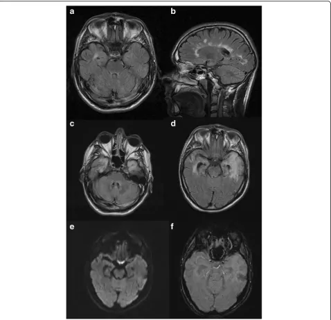
MRI brain scan showed new diffuse high signal changes in both temporal lobes and hippocampi (Fig.1c-f). He also had a number of subcortical and periventricular demyelinating pla-ques, that were stable in number and size compared with his
most recent RRMS surveillance MRI (Fig. 1a-b). Follow-up imaging at 6 weeks showed progression of these changes with high signal now extending into the insular cortex bilaterally.
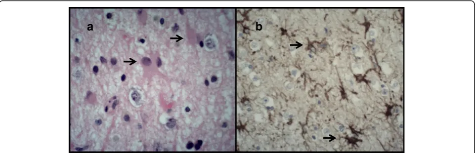
Due to these progressive radiological changes and lack of clinical improvement the patient underwent a temporal lobe biopsy which showed marked astrocytic gliosis, without evidence of vasculitis, diffuse parenchy-mal inflammation, infarction or neoplasia (Fig. 2a-b). Immunostaining for HSV1, HSV2 and SV40 (JCV marker) were all negative.
Differential diagnosis
The precipitant of DKA in this case is unclear given his absent period prior to admission, and may have been multifactorial. We postulate that it may be due to a combination of omission of insulin with or with-out alcohol misuse, as this was his previous precipi-tant. Starvation and alcohol excess may have also contributed to ketoacidosis.
There are multiple differentials of encephalopathy in this case. As the patient was not seen for two days prior to presentation he may have had an unwit-nessed traumatic brain injury however this was not apparent on imaging. Toxic brain injury was also considered however the radiological findings were inconsistent with this and toxicology screen was nega-tive. Wernicke’s encephalopathy was considered given his previous alcohol history, however there was no improvement with high dose B vitamin supplementa-tion and the clinical and imaging features would not be in keeping with that diagnosis. Infectious aetiology should always be considered in patients presenting with encephalopathy. Indeed, the finding of high sig-nal within the temporal lobes extending to the insula would fit with a viral encephalitis, such as that caused by HSV. However, the lack of diffusion restriction and microhaemorrhages on his imaging, lack of brain biopsy features as well as repeatedly negative CSF viral PCR and normal leucocyte count make this diag-nosis unlikely. Other rarer causes of encephalopathy including autoimmune, vasculitic and paraneoplastic disease were also explored and ruled out. Sub-clinical seizure activity may have caused his persistent behav-ioural disturbance in the acute phase but unfortu-nately the patient was initially unable to cooperate with EEG testing and there were no features on re-peated clinical examination by a neurologist of overt seizures. Nutritional deficiency such B vitamin / folate deficiency could have also played a role, the patient had mild folate deficiency which would not typically cause such a devastating neurological injury, however may have contributed to susceptibility to brain injury.
Cerebral oedema due to diabetic encephalopathy can cause brain injury however there was no clinical or radiographical evidence of this. Therefore, we feel that metabolic encephalopathy due to DKA is the most likely diagnosis in this case.
The differential of metabolic encephalopathy second-ary to DKA was raised approximately one week into the clinical admission following negative viral PCR and lack of improvement with thiamine and chlordiazepoxide. Initially the most likely diagnosis was a viral or toxic encephalopathy, however with no clinical improvement to treatment a wider differential was considered. A
comprehensive suite of investigations ensued to
undercover the aetiology of the patient’s behavioural disturbance, however neither imaging, laboratory or pathological specimens led to a definite cause. Follow-ing multidisciplinary discussion, the diagnosis of meta-bolic encephalopathy secondary to DKA was reached.
Treatment and clinical course
He was commenced on a DKA management protocol which consisted of aggressive intravenous fluid resus-citation, intravenous continuous insulin infusion and intravenous and oral replacement of potassium, phos-phate and magnesium. DKA management protocol was continued for forty-eight hours, at which point all biochemical markers were within normal limits and the patient was transitioned to basal-bolus insulin regimen. In addition, the patient was also given intra-venous high dose B vitamins and a reducing regimen of chlordiazepoxide over one week. He received folic acid supplementation 5 mg daily for two months. Sub-sequent treatment was largely supportive. Empiric antiviral therapy was given for HSV (later HSV PCR came back as negative, although two-week course complete). He underwent intensive rehabilitation in-volving a multidisciplinary team of occupational ther-apy, physiotherther-apy, social care and neuropsychology.
Subsequent imaging at 3, 4 and 6 months post presen-tation demonstrated persistent but stable high-signal changes in the temporal lobes bilaterally.
Unfortunately following temporal lobe biopsy the pa-tient experienced generalized tonic clonic seizures which were difficult to control, requiring three antiepileptic agents. EEG was performed at this point, ten weeks into his admission following temporal lobe biopsy. The find-ings were in keeping with an encephalopathic state with a highly epileptogenic focus in the left frontocentral region.
He remained relatively unchanged for the next 18 months with persistent severe global cognitive impair-ment with most marked deficits in attention, short-term memory and ability to learn new information. He had ongoing erratic control of his blood sugars, attributable to inconsistent oral intake. He continued to have seizures with generalised tonic-clonic events occurring approximately once per month. Behavioural issues remained a problem and he required constant supervision, ultimately requiring long term residential care.
Discussion and conclusions
From review of the literature, brain injury as a result of DKA has been well described in the paediatric popu-lation however the mechanisms of brain injury remain unclear. This uncertainty is mostly due to the myriad of
metabolic disturbances which occur during DKA [6].
There is a paucity of cases describing metabolic enceph-alopathy secondary to DKA in the adult population. Miras et al. described a very similar case in a 44 year old man [5]. Similarly, he was found collapsed and had not been contactable for 3 days prior to his admission. Inter-estingly, he also had a history of alcohol excess and poorly controlled type 1 diabetes with recurrent severe hypoglycaemic episodes. In this case the patient had sig-nificant behavioural disturbance with aggression and confusion. Neuroimaging was entirely normal, CSF examination was bland and EEG showed diffuse slowing consistent with encephalopathy. Over the following six months this patient gradually improved with neuro-psychiatric rehabilitation however had residual irritabil-ity and slow speech [5].
DKA has been shown to induce brain injury how-ever the exact pathogenesis of this injury is debated. In paediatric cases subclinical cerebral oedema is common, with frank cerebral oedema occurring in 0.5–1% of children with DKA [7]. Historically cerebral oedema secondary to DKA was thought to be due to overaggressive fluid resuscitation and loss of brain os-motic homeostasis, however recently this theory has been challenged. Cerebral hypoperfusion followed by reperfusion injury correlates with the spectrum of cytotoxic and vasogenic oedema seen in patients with cerebral oedema due to DKA and is now hypothe-sized to be the mechanism of brain injury in DKA
[7]. However, cerebral oedema in the setting of DKA
or Hyperglycaemic hyperosmolar syndrome (HHS) in adults is exceedingly rare, with a United States large
population-based study revealing a 0.03% incidence rate [8]. In the case outlined above there was no evi-dence of cerebral oedema clinically or radiologically, therefore other mechanisms of brain injury must be at play.
Interestingly Jessup et al., studied a cohort of young patients with new onset type 1 diabetes and found that patients who presented with DKA scored lower on visual cognitive tasks when compared to age-matched patients without DKA. Cognitive disparity
between the two groups remained 8–12 weeks post
discharge. The authors suggest that metabolic dysreg-ulation during DKA mediates neuroinflammation and cerebral oxidative stress causing a neuronal injury [9]. This is further supported histologically, where exam-ination of brain tissue from patients with DKA and cerebral oedema showed evidence of oxidative stress, with increased products of oxidative damage present in vulnerable brain areas compared to healthy con-trols [10]. Hoffman et al., also showed that both acute and chronic metabolic dysregulation in T1DM can affect brain function, promoting neuroinflammation, cerebral insulin resistance and reduced insulin signal-ling, culminating in increased oxidative and
inflamma-tory cerebral stress [10]. Chronic hyperglycaemia,
ketoacidosis and dehydration with superimposed acute insults, in the form of DKA, can subsequently cause
diabetic encephalopathy [10]. The long-term erratic
glycaemic control in the case outlined above may therefore have also played a role in the patient’s cog-nitive decline.
Recent research has indeed demonstrated chronic altered brain metabolism and signalling as a cause of diabetic brain dysfunction. Analysing brain metabo-lites with magnetic resonance spectroscopy (MRS)
reveals significant alterations in levels of brain
a
b
metabolites in the diabetic brain, which are consistent with and related to specific diabetic complications. Changes in these metabolites has been hypothesized to cause reduced neurotransmission, demyelination, neurodegeneration and brain atrophy. The study of brain metabolites and use of MRS in diabetic brain disease is at the initial stages. Further research and development could unveil the exact mechanism of diabetic brain injury as well as provide a new diag-nostic tool to evaluate early disease and allow inter-vention [11].
In the case outlined above there are also nutritional factors which may have increased the risk of brain in-jury. Folic acid deficiency has been linked to cognitive impairment with behavioural disturbance in young
adults [12]. Non-ketotic hyperglycaemia (NKH) can
result in multiple neurological consequences such as
seizures, hemichorea and hemianopia. In NKH,
hyperglycaemia-induced blood brain barrier perme-ability contributes to epileptogenesis [13]. Therefore, we may hypothesize that recurrent hyperglycaemia and subsequent blood brain barrier permeability may have contributed to epileptogenesis and indeed brain injury in this case. Moreover, multiple sclerosis re-lated blood-brain barrier permeability, although not the primary pathology in MS, may have contributed to reduced neuroprotection and increased risk of en-cephalopathy [14]. Factors such as nutritional defi-ciency, alcohol excess, cannabis use, depression and multiple sclerosis may have contributed to this pa-tient’s susceptibility to encephalopathy however
meta-bolic encephalopathy secondary to diabetic
ketoacidosis was the most likely diagnosis.
In summary, metabolic encephalopathy is a devastat-ing complication of DKA which can be due to both acute and chronic metabolic brain insults. To our know-ledge this is the second case report describing this acute complication. It is an area of expanding research which will hopefully lead to improved understanding, treat-ment and patient outcome.
Abbreviations
AMPA:Alpha-amino-3-hydroxy-5-methyl-4-isoxazolepropionic acid; CSF: Cerebrospinal fluid; DKA: Diabetic Ketoacidosis; DNA: Deoxyribonucleic acid; ED: Emergency Department; EEG: Electroencephalogram; FLAIR: Fluid-attenuated inversion recovery; GABA: Gamma-aminobutyric acid; GAD: Glutamate acid decarboxylase; GCS: Glasgow Coma Scale; GFAP: Glial fibrillary acidic protein; H&E: Haematoxylin and eosin; HbA1c: Haemoglobin A1c; HBV: Hepatitis B virus; HCV: Hepatitis C virus; HHS: Hyperglycaemic Hyperosmolar Syndrome; HIV: Human Immunodeficiency Virus; HSV: Herpes Simplex Virus; IgG: Immunoglobulin G; IgM: Immunoglobulin M; MOG: Myelin Oligodendrocyte Glycoprotein; MRI: Magnetic Resonance Imaging; MRS: Magnetic resonance spectroscopy; NKH: Non-ketotic hyperglycaemia; NMDA: N-methyl-D-aspartate; NMO: Neuromyelitis optica; PCR: Polymerase Chain Reaction; POLG: DNA polymerase subunit gamma; RNP: Ribonuclear protein; RRMS: Relapsing Remitting Multiple Sclerosis; SGLT2: Sodium-Glucose-Co-Transporter 2; T1DM: Type 1 diabetes mellitus; TPO: Thyroid
peroxidise; TTG: Tissue Transglutaminase; VGKC: Voltage gated potassium channel
Acknowledgements
Dr. Alan Beausang Consultant Neuropathologist, Beaumont Hospital, Dublin, Ireland and Dr. Paul Brennan Consultant Neuroradiologist, Beaumont Hospital, Dublin, Ireland and Dr. Gerard Mullins Consultant Neurophysiologist, Beaumont Hospital, Dublin, Ireland.
Authors’contributions
MT: primary author, contributed to conception, preparation and editing of report. RMC contributed to manuscript conception, reviewed and added radiology findings. KOC contributed to manuscript editing and added pathology findings. AA and AM: senior authors, contributed to manuscript conception and editing. All authors reviewed and approved the manuscript.
Funding
No funding was received for this study.
Availability of data and materials
None of the raw data pertaining to this case report is available publicly. The original investigation findings and reports are retained, as per normal procedure within the medical records of our institution.
Ethics approval and consent to participate
Not applicable.
Consent for publication
Written informed consent to publish the patient’s personal data was obtained from the patient’s next of kin and family. A copy of written consent is available for review by the Editor of this journal.
This manuscript was developed with adherence to the CARE guidelines and methodology.
Competing interests
Amar Agha is a member of the editorial board (Section Editor) of BMC Endocrine Disorders. All other authors declare that they have no competing interests.
Author details
1Department of Diabetes and Endocrinology, Beaumont Hospital, Dublin, Ireland.2Department of Rehabilitation, National Rehabilitation Hospital, Dublin, Ireland.3Department of Neurology, Beaumont Hospital, Dublin, Ireland.4Department of Diabetes and Endocrinology, Beaumont Hospital, Dublin, Ireland.
Received: 23 January 2019 Accepted: 12 June 2019
References
1. Misra S, Oliver NS. Diabetic ketoacidosis in adults. BMJ. 2015;351:h5660. 2. Hemphill JC. Chapter 60: Disorders of consciousness in systemic diseases. In:
Aminoff M, Josephson SA, editors. Aminoff’s Neurology and General Medicine. 5th ed. San Diego: Academic Press; 2014. p. 1243–61. 3. Beca J, Sidebotham D. Chapter 37: neurologic dysfunction. In: Sidebotham
D, McKee A, Gillham M, Levy J, editors. Cardiothoracic critical care. Oxford: Butterworth-Heinemann; 2007. p. 548–62.
4. Supanc V, Vargek-Solter V, Demarin V. Metabolic encephalopathies. Acta Clin Croat. 2003;42:351–7.
5. Miras AD, Ward H. Encephalopathy following diabetic ketoacidosis in a type 1 diabetes patient. Practical Diabetes Int. 2010;27(2):76–8.
6. Glaser N, Ngo C, Anderson S, Yuen N, Trifu A, O’Donnell M. Effects of hyperglycemia and effects of ketosis on cerebral perfusion, cerebral water distribution and cerebral metabolism. Diabetes. 2012;61(7):1831–7. 7. Biao SR, Agrawal S, Boney CM, Quintos JB. Rare complications of pediatric
diabetic ketoacidosis. World J Diabetes. 2015;6(1):167–74.
8. Siwakoti K, Giri S, Kadaria D. Cerebral edema among adults with diabetic ketoacidosis and hyperglycemic hyperosmolar syndrome: incidence, characteristics, and outcomes. J Diabetes. 2017;9(2):208–9.
neurocognitive function in young patients presenting with new-onset type 1 diabetes. J Clin Res Pediatr Endocrinol. 2015;7(3):203–10.
10. Hoffman WH, Siedlak SL, Wang Y, Castellani RJ, Smith M. Oxidative damage is present in the fatal brain edema of diabetic ketoacidosis. Brain Res. 2011; 1369:194–202.
11. Zhao X, Han Q, Gang X, Wang G. Altered brain metabolites in patients with diabetes mellitus and related complications–evidence from the1H MRS
study. Biosci Rep. 2018;38(5):BSR20180660.
12. Reynolds EH. Benefits and risks of folic acid in the nervous system. J Neurol Neurosurg Psychiatry. 2002;72:567–71.
13. Kim DW, Moon Y, Gee Noh H, Choi JW, Oh J. Blood-brain barrier disruption is involved in seizure and hemianopsia in nonketotic hyperglycemia. Neurologist. 2011;17(3):164–6.
14. Cramer SP, Simonsen H, Frederiksen JL, Rostrup E, Larsson HBW. Abnormal blood-brain barrier permeability in normal appearing white matter in multiple sclerosis investigated by MRI. Neuroimage Clin. 2014;4:182–9.
Publisher’s Note