RESEARCH
The fungal composition of natural
biofinishes on oil-treated wood
Elke J. van Nieuwenhuijzen
1*, Jos A. M. P. Houbraken
1, Peter J. Punt
2,3, Guus Roeselers
2,4, Olaf C. G. Adan
5and Robert A. Samson
1Abstract
Background: Biofinished wood is considered to be a decorative and protective material for outdoor constructions, showing advantages compared to traditional treated wood in terms of sustainability and self-repair. Natural dark wood staining fungi are essential to biofinish formation on wood. Although all sorts of outdoor situated timber are subjected to fungal staining, the homogenous dark staining called biofinish has only been detected on specific veg-etable oil-treated substrates. Revealing the fungal composition of various natural biofinishes on wood is a first step to understand and control biofinish formation for industrial application.
Results: A culture-based survey of fungi in natural biofinishes on oil-treated wood samples showed the common wood stain fungus Aureobasidium and the recently described genus Superstratomyces to be predominant constitu-ents. A culture-independent approach, based on amplification of the internal transcribed spacer regions, cloning and Sanger sequencing, resulted in clone libraries of two types of biofinishes. Aureobasidium was present in both biofinish types, but was only predominant in biofinishes on pine sapwood treated with raw linseed oil. Most cloned sequences of the other biofinish type (pine sapwood treated with olive oil) could not be identified. In addition, a more in-depth overview of the fungal composition of biofinishes was obtained with Illumina amplicon sequencing that targeted the internal transcribed spacer region 1. All investigated samples, that varied in wood species, (oil) treatments and expo-sure times, contained Aureobasidium and this genus was predominant in the biofinishes on pine sapwood treated with raw linseed oil. Lapidomyces was the predominant genus in most of the other biofinishes and present in all other samples. Surprisingly, Superstratomyces, which was predominantly detected by the cultivation-based approach, could not be found with the Illumina sequencing approach, while Lapidomyces was not detected in the culture-based approach.
Conclusions: Overall, the culture-based approach and two culture-independent methods that were used in this study revealed that natural biofinishes were composed of multiple fungal genera always containing the common wood staining mould Aureobasidium. Besides Aureobasidium, the use of other fungal genera for the production of biofinished wood has to be considered.
Keywords: Biofilm, Metagenomics, Mould, Wood protection, Wood staining
© The Author(s) 2017. This article is distributed under the terms of the Creative Commons Attribution 4.0 International License (http://creativecommons.org/licenses/by/4.0/), which permits unrestricted use, distribution, and reproduction in any medium, provided you give appropriate credit to the original author(s) and the source, provide a link to the Creative Commons license, and indicate if changes were made. The Creative Commons Public Domain Dedication waiver (http://creativecommons.org/ publicdomain/zero/1.0/) applies to the data made available in this article, unless otherwise stated.
Background
Microbial growth causing discolouration on surfaces of outdoor situated materials is a common phenomenon
[1–3]. Frequently these microbial stains are referred to
as biofilm, although not all commonly accepted biofilm
criteria might have been investigated [4]. Dark staining of
painted and unpainted wood is mostly attributed to fungi and generally considered as unwanted discolouration
[5, 6]. In contrast, the specific dark stain formation on
wood called biofinish is considered to be a functional
colouration [4] (Fig. 1). The colouration of a biofinish is,
together with its presumed protection and self-healing properties, an important ingredient of a sustainable
solution for a biocide free wood finish system [4, 7]. A
Open Access
*Correspondence: e.nieuwenhuijzen@cbs.knaw.nl
1 Applied and Industrial Mycology, CBS-KNAW Fungal Biodiversity Centre,
Utrecht, The Netherlands
biofinish refers to a dark pigmented layer, that covers a wood surface almost entirely without exposing underlying wood structures, contains abundant microbial mass and
that is irreversibly attached to the surface [4]. Biofinishes
have been detected on wood impregnated with olive oil or
raw linseed oil [4, 8]. The study described in this paper is
focused on the characterization of the fungal composition of these biofinishes.
Although several fungal species are associated with
outdoor wood staining [6, 9], little is known to which
extent each taxon contributes to this staining. In some studies the fungal populations on timber surfaces were
quantified [10, 11]. The study by Sailer et al. [7]
pro-vides data, particularly of interest for the biofinishes on wood. One specific sample, made of pine sapwood and impregnated with refined linseed oil dissolved in acetone, was used to study the fungal composition of a homogenous dark stained wood surface. Later, biofin-ishes were detected on other outdoor exposed oil-treated wood samples, including samples made of different wood species treated with olive oil. Analysis of these samples revealed the abundancy of the wood staining fungus
Aureobasidium, but the results also indicated that this
genus might not always dictate the fungal population of
dense dark stained wood samples [4, 12]. The wood
spe-cies, oil type and the geographical location could influ-ence the fungal community composition of a biofinish. In order to manufacture a stable biofinish and eventually apply biofinished wood in practice, a detailed composi-tion of fungi present at various stained wood surfaces is elucidated in this paper.
Different techniques are available to study a fungal community on the surface of an environmental sam-ple such outdoor exposed wood. Each technique has its advantages and disadvantages. Microscopic examination of the surface can be an easy method to study the surface
of a material [13]. The main disadvantage is that
identifi-cation and enumeration is difficult in case of moulds on
oil-treated wood surfaces [4]. A common way to
iden-tify and enumerate fungi is by culturing. A swab-based method can be used to analyse the culturable fungi that
are present on a sample surface [4, 14]. This includes the
determination of the number of colony forming units after incubation and identification based on macroscopic, microscopic and/or molecular analysis. Culture-based analysis only allows the detection of readily culturable species, which is effected by the media selection and overestimates the presence of abundantly sporulating
species [13]. Culture-independent methods based on
DNA analysis are frequently used and show
complemen-tary results [15–17]. Culture-independent approaches
that rely on Next Generation Sequencing (NGS) methods have become state of the art to study microbial
commu-nities [16, 18, 19]. Albeit that these NGS methods provide
advantages compared to earlier developed techniques, research to control and understand the biases occurring
in all steps of a NGS method is still ongoing [18–20].
The objective of the present study was to analyse the fungal composition of various biofinishes on oil-treated wood surfaces. A culture-based swab method and two non-culturing methods based on either amplicon-clon-ing followed by Sanger sequencamplicon-clon-ing or Illumina amplicon
sequencing were selected for the analysis. Wood samples without oil treatment and/or biofinish were used to com-pare the diversity and predominance of fungal genera.
Methods
Several natural biofinishes were studied with a cul-ture-based and two DNA sequencing-based methods
(Table 1). The biofinish containing samples varied in
wood species, type of oil and origin. Wood samples made of the same wood species without a biofinish, were stud-ied as well. The viable fungal composition was studstud-ied of all samples. Specific samples, which were exposed in the Netherlands, were selected for the culture-independent fungal profiling methods.
Wood samples
The different wood species tested were pine (Pinus
sylves-tris), spruce (Picea abies) and ilomba (Pycnanthus
ango-lensis). Pine samples were made totally of sapwood (sw)
or a mixture of sapwood and heartwood (hw). No specific sapwood or heartwood selection was made for spruce and ilomba. Wood blocks were impregnated with raw lin-seed, stand linseed or olive oil. Sets of impregnated and untreated wood samples were exposed outdoors at
differ-ent locations (Table 1). The sample dimensions, oil
treat-ments, outdoor exposure and handling procedures were
described in van Nieuwenhuijzen et al. [4]. All samples,
except for the samples of set 3, have been analysed with the biofinish assessment method. This method consists of observations of the dark stained surface coverage at macroscopic and microscopic scale, and
spectrophotom-eter measurements of the pigmentation [4]. In summary,
a biofinish is assigned when more than 90% of the surface is stained and does not expose structures of the wood such as annual rings or wood fibres, and the pigmentation measurements, expressed by sRGB colour space triplets, meet specific criteria (R, G and B values are below 82 and the value difference within a single RGB triplet is below 20). The presence of biofinishes on the wood samples of set 3 was only estimated with visual observations of the stain coverage as described in the biofinish assessment method, which can overestimate biofinish identification.
Culturing colonies
The concentration of colony forming units (CFU) per cm2
wood was determined for each specimen (Table 1). For
this, biomass collected with a cotton swab was analysed
as described by van Nieuwenhuijzen et al. [4]. Serial
dilu-tions were plated in duplicate (set 1 and 3–5) or triplicate (sample set 2) on dichloran 18% glycerol agar (DG18) and malt extract agar (MEA) supplemented with penicil-lin and streptomycin (P/S). The agar media was prepared
as described by Samson et al. [13]. The total number of
colonies and those phenotypically resembling
Aureoba-sidium was determined after seven and fourteen days of
incubation at 25 °C [12]. Besides the Aureobasidium
col-onies, also the predominant colonies were counted. Two or more colonies of each predominantly present colony type were transferred to new MEA plates. The isolates were deposited in the working collection of the Applied and Industrial Mycology department (DTO) housed at the CBS-KNAW Fungal Biodiversity Centre, The Nether-lands and subjected to molecular identification.
DNA extraction
DNA was extracted from cultures grown on MEA plates
according to van Nieuwenhuijzen et al. [12]. With respect
to the culture-independent methods, biomass was removed with a sterile scalpel from the upper surface of a mould stained wood sample, collected on sterile paper
and subsequently used to extract DNA [4]. In both cases
the Ultraclean Microbial DNA isolation kit (MoBio Lab-oratories, USA) was used according to manufacturer’s instructions.
PCR and Sanger sequencing of isolates
The nuclear internal transcribed spacers including the 5.8S rRNA gene (ITS) of fungal isolates were amplified
with the primer pair V9G [21] and LS266 [22]. In case
additional sequence information was needed for a proper
identification of a strain, the LSU gene was partially
amplified using the forward primer LROR (Reh)
GTAC-CCGCTTGAACTTAAGC [23] or LROR (VilU)
ACC-CGCTGAACTTAAGC [Vilgalys, unpublished] and the
reverse primer LR5 or LR7 [24]. The polymerase chain
reaction (PCR) mixtures had final concentrations of: 4%
DNA extract, 10% PCR buffer, 3% MgCl2 (25 mM), 65.8%
demineralised sterile water, 7.8% dNTP (1 mM), 5% DMSO, 2% forward primer (10 μM), 2% reverse primer and 0.4% Taq polymerase (5 U/mL, BioTaq, Bioline). The PCR program typically consisted of 1 cycle of 5 min dena-turation at 95 °C; 35 cycles of 35 s denadena-turation at 95 °C, followed by ITS-primer annealing for 30 s at 55 °C or
LSU-primer annealing at 54 °C for 50 s, and an extension
for 1.5 min at 72 °C. The PCR-products were sequenced with the same primers as used for PCR amplification using the BigDye Terminator v. 3.1 Cycle Sequencing Kit (Applied Biosystems, USA). Sequence products were analysed on an ABI PRISM 3730XL genetic analyser (Applied Biosystems, USA) and traces were assembled using Seqman Pro v. 9.0.4 (DNAstar Inc.). The sequences
were deposited in GenBank [25].
PCR, cloning and Sanger sequencing
ITS-specific clone libraries were made as described in
formed on different substrates exposed in the
Neth-erlands (Table 1, set 1): pine sapwood treated with raw
linseed oil (libraries PRL.1, PRL.2 and PRL.3) and pine sapwood treated with olive oil (PO.1, PO.2 and PO.3). The ITS region was amplified with the primers V9G and LS266 and the GoTaq Long PCR Master Mix (Progema), while using the PCR-program as described above. Puri-fied PCR products (QIAquick PCR purification kit) were
ligated and cloned (pGEM®-T Easy Vector Systems) into
an Escherichia coli plasmid library. Amplification and
Sanger sequencing of DNA from ITS containing com-petent cells was performed as described in van
Nieu-wenhuijzen et al. [12]. The sequences were deposited in
GenBank [25].
Illumina ITS1 amplicon sequencing
Internal transcribed spacer 1 region (ITS1) ampli-con libraries were made of 16 wood samples that were all exposed at one test site (The Netherlands), but
variated in wood species, treatment and the presence
or absence of a biofinish (Table 1, set 1–2). In a
pre-liminary study several ITS primer combinations and amplification approaches were tested for their suitabil-ity of generic detection of fungal genera (unpublished results). Based on these results barcoded ITS1 ampli-cons were generated using a two-step PCR approach. ITS1 regions were first amplified with the following primers: nex-ITS-BITS-F: TCGTCGGCAGCGT-CACCTGCGGARGGATCA and nex-ITS-B58S3-R:
GTCTCGTGGGCTCGGGAGATCCRTTGYTRAAA-GTT (adapted from Bokulich and Mills [26]). Each
reac-tion contained 300× purified DNA, 1X hot start PCR master mix (Thermo Scientific) and nuclease free PCR grade water to a 50 µl final reaction volume. PCR reac-tions consisted of an initial denaturation step of 95 °C for 5 min and 30 amplification cycles (95 °C for 30 s, annealing 52 °C for 45 s and elongation 72 °C for 1 min) and a final extension step (72 °C for 10 min) followed by
Table 1 Overview of the amount and type of wood samples used per type of fungal profiling method
a Biofinish assessment by Nieuwenhuijzen, van et al. [4]
b Determination based on stain coverage Sample
set Wood spe-cies Treatment Presence biofinish Location (exposure time) Number of samples
Culture method Cloning method Illumina method
1 Spruce Raw linseed oil No Utrecht, The
Nether-lands (1.5 year) 3 1
Stand linseed oil No 3 1
Olive oil Yesa 3 1
No oil No 3 1
Pine sw Raw linseed oil Yesa 3 3 2
Stand linseed oil No 3 1
Olive oil Yesa 3 3 2
No oil No 3 1
Ilomba Raw linseed oil No 3 1
Stand linseed oil No 3 1
Olive oil Yesa 3 1
No oil No 3 1
2 Pine sw Raw linseed oil Yesa Utrecht, The
Nether-lands (1.8 year) 10 2
3 Spruce Raw linseed oil No Utrecht, the
Nether-lands (1.5 year) 1
Ilomba Raw linseed oil No 1
Pine sw Raw linseed oil Yesb 1
Pine sw Olive oil Yesb 1
Pine sw No oil No 1
Pine hw Raw linseed oil Yesb 1
4 Same materials as set 3 Biofinish only on
olive oil Johannesburg, South Africa (1.7 year) 1
5 Same materials as set 3 No Dover Gardens,
Aus-tralia (1,5 year) 1 6 Same materials as set 3 Biofinish on pine
Table 2 T he pr edominan tly cultur ed c olon y t yp
es of each w
oo
d sample set
Sample set W ood species Tr ea tmen t No . of sam- ples Biofinish Number of w
ood samples with pr
edominan t c olon y t ypes Aur eoba -sidium
Black yeasts
Clad -osp orium Cr ypt o-co cc us D id ymel -lac eae Phaci -diella Pleu -rophoma Pyr eno -chaeta Cy ano der -mella Sup erstr at -omyc es Sy do wia Ta phrina 1 Spruce R. lins . oil 3 No 3 1 St. lins . oil 3 No 3 3 Oliv e oil 3 Ye s a 2 3 No oil 3 No 1 3 Pine sw R. lins . oil 3 Ye s a 1 1 2 1 St. lins . oil 3 No 3 3 Oliv e oil 3 Ye s a 3 No oil 3 No 2 3 Ilomba R. lins . oil 3 No 1 2 St. lins . oil 3 No 2 1 2 Oliv e oil 3 Ye s a 3 No oil 3 No 3 1 1 2 Pine sw R. lins . oil 10 Ye s a 3 1 1 1 10 2 3 Spruce R. lins . oil 1 No 1 Ilomba R. lins . oil 1 No 1 Pine sw R. lins . oil 1 Ye s b 1 1 Pine sw Oliv e oil 1 Ye s b 1 Pine sw No oil 1 No 1 Pine h w R. lins . oil 1 Ye s b 1 4 Spruce R. lins . oil 1 No 1 Ilomba R. lins . oil 1 No 1 Pine sw R. lins . oil 1 No 1 Pine h w R. lins . oil 1 No 1 1 5 Spruce R. lins . oil 1 No 1 Ilomba R. lins . oil 1 No 1 1 Pine sw R. lins . oil 1 No 1 Pine sw Oliv e oil 1 No 1 1 Pine sw No oil 1 No 1 Pine h w R. lins . oil 1 No 1 1 6 Spruce R. lins . oil 1 No 1 Ilomba R. lins . oil 1 No 1 1 Pine sw R. lins . oil 1 Ye s a 1 Pine sw Oliv e oil 1 No 1 Pine sw No oil 1 No 1 Pine h w R. lins . oil 1 Ye s a 1 1
a Biofinish det
ec tion b y v an N ieuw enhuijz en et al . [ 4 ]
b D
et
er
mina
tion based on stain c
ov
er
cool down (10 min at 4 °C). A negative control (blank) was included for each 24 PCR reactions. Reactions were cleaned by solid-phase reversible immobilization (SPRI) using AMPure XP SPRI beads (Beckman Coulter, Inc.). Dual barcodes (8 bp) and Illumina Sequencing adapters were attached using the Nextera XT Index Kit (Illumina, San Diego, CA) according to manufacturer’s protocols. Barcoded amplicons were quantified using the Caliper LabChip GX II system (Perkin Elmer, Hopkinton, USA), normalised to the same concentrations, pooled, and gel purified using the Qiaquick spin kit (Qiagen) and AMPure XP SPRI beads. Pooled amplicons were 250-bp paired-end sequenced using the MiSeq system (Illu-mina). Raw Illumina fastq files were deposited in the European Nucleotide Archive (accession PRJEB13755). The raw data was demultiplexed, quality filtered, and analysed using modules implemented in the Mothur
software platform [27]. Quality filtering involved a
min-imal quality score of 25 in a window of 5 and removal of reads with a length below 100 and above 500 or with ambiguous bases. In addition, chimeras detected
using UCHIME and the UNITE database (v6) [28] were
removed. Filtered raw reads were merged into paired reads. Subsequently, the relative abundance of unique sequences were calculated for each sample by dividing the number of reads of a single unique sequence by the total number of reads of the sample. Unique sequences with a total sum of relative read abundancies above 0.1% were used for further analysis.
DNA data analysis
ITS and ITS1 sequences were subjected to nucleotide
BLAST searches [29] using the non-redundant database
of GenBank [25], the Q-bank Fungi database [30] and
an internal database of the CBS-KNAW Fungal Biodi-versity Centre (Fungal Barcoding data). Identification was performed on genus level. ITS sequences obtained from non-cultured material, which resulted in hits in GenBank with an identity below 97% were marked as ‘unidentified’. Also the ITS1 sequences with query cov-erages below 90% were marked as ‘unidentified’. For the identification of culturable isolates with an unclear
identity based on only ITS sequences, LSU sequences
were also compared with the non-redundant nucleo-tide database of GenBank. ITS sequences of isolates that could not be identified on genus level were aligned with the sequences of the ITS cloning and ITS1 amplicon libraries using Nucleotide BLAST of GenBank(NCBI, Rockville Pike, USA).
Mathematical analysis
A Shannon’s diversity test was used to index the genus diversity in the Illumina amplicon data.
Results
Culturable fungal composition
After culturing biomass of 70 wood samples, the predom-inantly colony types of 68 samples could be identified
(Table 2). The total number CFU’s per square centimetre
biofinish varied between 6 × 10² and 4 × 10⁵ CFU/cm2
and the samples without biofinish, which all contained dark mould stains but less than the biofinish criteria
pre-scribed, showed similar results (2 × 101 to 4 × 10⁵ CFU/
cm2; Additional file 1: Table S1). Frequently, more than
one predominant colony type could be determined in
the plated biomass from a single wood sample (Table 2).
The isolates and sequence data obtained to identify the
predominant type of CFU are listed in Additional file 2:
Table S2.
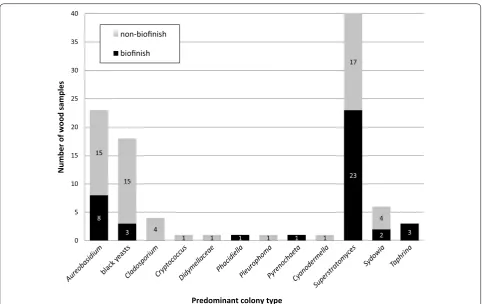
A recently described dark pycnidia producing
coelomy-cete named Superstratomyces was detected as the most
commonly occurring predominant colony type in the
biofinishes (Table 2; Fig. 2) [31]. This genus was also
pre-dominantly present on outdoor exposed samples without
a biofinish, including samples without oil (Table 2).
Inter-estingly, the detection of Superstratomyces on wood was
restricted to samples exposed in the Netherlands (sample set 1–3).
Aureobasidium was predominantly present on 8 of the
28 samples that showed a biofinish (Table 2; Fig. 2). Also
samples without a biofinish frequently showed this genus
to be one of the predominant colony types (Table 2;
Fig. 2). Aureobasidium was isolated from samples
exist-ing of all combinations of wood species and (oil) treat-ments originating from all selected outdoor locations
(Additional file 1: Table S1, Additional file 2: Table S2).
The Aureobasidium contribution to the total cultured
CFU varied largely for biofinish samples (0–97%), but the contribution per sample surface area only rarely (2 out
of 28 samples) exceeded the 50% (Additional file 1: Table
S1). The stained samples without a biofinish showed a
similar range of percentages (0–97%), and the
Aureoba-sidium contribution exceeded the 50% regurlary (13 out
of the 42 samples; Additional file 1: Table S1).
Other colonies types which were predominantly iso-lated from samples with a biofinish were black yeasts
(identfified as Exophiala, Phaeococcomyces or Knufia),
Taphrina, Sydowia, Phacidiella and Pyrenochaeta. Black
yeasts and Sydowia were also isolated as predominant
colony types from the stained samples without a
biofin-ish, expended by the genera Cladosporium,
Cryptococ-cus, Pleurophoma and Cyanodermella, and a genus in the
family Didymellaceae (Table 2).
Fungal composition of ITS clone libraries
either raw linseed or olive oil. Each library contained
61–71 clones (Additional file 3: Table S3). In all libraries
several genera were identified always including
Aureoba-sidium (Fig. 3).
Aureobasidium was predominantly present in the
cloned DNA of biofinishes on pine sapwood treated with raw linseed oil. In each of the three clone libraries
more than 50% of the clones were identified as
Aureoba-sidium. The investigated biofinishes on pine sapwood
treated with olive oil did not show this predominance.
Their clone libraries had a much lower Aureobasidium
percentage, varying from 2 to 13%. Interestingly, the number of sequences which could not be assigned to the genus level was high in these samples. These diver-gent sequences all differed from the molecular identi-fied strains obtained with the culturing method. Many of them had more than one best hit with sequences in the database while showing identity scores of 82% compared to sequences in GenBank that were named
Aureobasidium, Sarcinomyces and Rhizosphaera. The
other divergent sequences showed identity coverages varying from 80 to 96% compared to the best hits, which represented up to five genus names for each library
(Additional file 3: Table S3). Furthermore, analysis of
sequences obtained from all biofinish samples revealed
the presence of other dark pigmented fungi, with
Lapi-domyces as a major contributor (6–27% per library). The
in general more sparsely occurring genera were
Cap-ronia, Cladosporium, Exophiala, Phaeococcomyces and
Epicoccum.
8 3
1 1
23
2 3
15
15
4
1 1 1 1
17
4
0 5 10 15 20 25 30 35 40
Aureobasidiumblack
yeasts
Cladosporiu m
Cryp tococ
cus
Didym ellacea
e
Phacidiell a
Pleurophom a
Pyren ocha
eta
Cyanodermella Supers
tratom yces
Sydowi a
Taphrin a
Number of wood
samples
Predominant colony type non-biofinish
biofinish
Fig. 2 Identified colony types predominantly isolated from 28 outdoor exposed wood samples with a biofinish
pine_
rawlin
s.(PRL
.1)
pine_
rawlin
s.(PRL
.2)
pine_
rawlin
s.(PRL
.3)
pine_
olive
(PO.1)
pine_
olive
(PO.2)
pine_
olive
(PO.3)
0 20 40 60 80 100
Perce
nt
ag
eo
fc
lo
ne
s(
%)
Aureobasidium Capronia Cladosporium Epicoccum Exophiala Lapidomyces Phaeococcomyces Taphrina unidentified unidentified genus
(82% ident.Sarcinomyces/
Aureobasidium/Rhizosphaera)
Fungal composition of Illumina ITS1 libraries
Application of the Illumina amplicon method to ana-lyse the fungal biofinish community of the 16 selected wood samples resulted in a total of 2.17 million filtered ITS1 reads. The amount of reads passing the occur-rence threshold was 2.02 million. Each wood sample
had 6.5 × 10⁴ up to 4.6 × 10⁵ reads with a mean length
of 171 nucleotides. In total 400 unique sequences were detected with no nucleotide variation among the reads
of a single unique sequence (Additional file 4: Table
S4). Most of these unique sequences could be identi-fied to genus-level, but some sequences represented multiple genera or represented an unidentified genus
(Fig. 4; Additional file 4: Table S4). The read percentage
of this latter category varied for the biofinish samples between 3% till 22% of the total reads. Some of these sequences showed 100% similarity with the unidenti-fied sequences obtained from the clone libraries, while none of these sequences were highly similar to the ITS sequences derived from any of the fungal isolates cul-tured from biofinishes. The number of different iden-tified genera for the biofinish samples ranged from 26 to 34 and for the non-biofinish wood samples (that all contained dark mould stains but less than the biofin-ish criteria prescribed) from 27 to 35. The calculated Shannon’s diversity indices were generally lower for the
biofinish samples (average 0.8) compared to the samples
without a biofinish (average 1.4; Fig. 5).
The Illumina amplicon method revealed the presence of two predominant genera in the amplicon sequencing libraries of the eight samples that contained a biofinish:
Aureobasidium and Lapidomyces (Fig. 4). Both genera
were determined in the DNA extractions of all samples. In the amplicon sequence libraries of six biofinish
con-taining samples the predominance of Aureobasidium
was determined, including all four pine sapwood samples treated with raw linseed oil. These four samples had the
highest contribution of Aureobasidium reads per sample
(more than 56% for each sample). However, the predomi-nance of this genus was only determined for half of the olive oil-treated samples that contained a biofinish. The
other half of the samples contained Lapidomyces as the
predominant genus. With respect to the analysed wood samples without biofinish, not one sample showed the
predominance of Aureobasidium in the amplicon library,
while most of them had Lapidomyces as predominantly
present genus. In line with these results the percentage
of Aureobasidium reads was higher for the biofinish
sam-ples [average 58%, standard deviation (SD) 20%] than for a non-biofinish sample (average 19%, SD 12%), despite
the variation in substrates (Fig. 4). The two types of oil
that were used for biofinish substrates differed in the
per-centages of Aureobasidium sequences. The biofinishes on
wood treated with olive oil showed a lower percentage
for Aureobasidium (31–55%) than the biofinishes on raw
linseed oil (59–89%).
Although Cladosporium was detected in all amplicon
sequencing libraries of the biofinish samples, the results showed a relative low contribution (average 1%, SD 1%) of this genus to the total reads of a library. The
contribu-tion of Cladosporium to the amplicon sequence libraries
of samples without a biofinish was higher (average 7%, SD 5%). Similar results were found for the black yeast
Phaococcomyces (biofinishes: average 0.2%, SD 0.2%;
non-biofinishes: average 6%, SD 7%).
Discussion
The fungal composition of biofinishes
The fungal compositions generated with the culture-based, cloning and Illumina sequencing approach showed overlapping and partly complementary results.
For example Aureobasidium was detected in biofinishes
with all three techniques. In contrast, the genus
Super-stratomyces, detected by culturing as a predominant
colony type, was absent in the data generated with the culturing-independent approaches. The monotypic genus
Lapidomyces, represented by the rock-inhabiting
spe-cies L. hispanicus [32], was detected in high numbers in
the cloning and Illumina libraries, while it was absent bio
finish p
ine_ra w (PR L.1) biofinis h p ine_ra w (PR L.2) biofin ish pin e_raw (PR L.4,se t 2) bio finis h p ine_ raw (PR L.5,se t 2) biofinis h p ine _oliv e (PO.1
) biofinis h p ine _olive (PO .2) biofinis
h spru ce_oliv
e (SO.1) bio finis h ilo mba_o live (IO .1) pin
e (Ppin.1)e_ stand (PSL.1 ) spru ce_raw (SR L.1) sp ruce_s tan d (S SL.1) spruce (S.1) ilom ba_raw (RL.1) ilom ba_sta nd (ISL.1) ilom ba(I.1) 0 10 20 30 40 50 60 70 80 90 100 Percentage of re ad s( %) Alternaria Aureobasidium Catenulostroma Cladosporium Devriesia Epicoccum Lapidomyces Microdiplodia mult. genera in Ascomycota mult. genera in Didymellaceae mult. genera in Dothideomycetes Neostagonospora Phaeococcomyces Phaeosphaeria Pyrenochaeta other genera unidentified
in the predominantly cultured isolates. The culturing-independent approaches generated overlapping results,
since Aureobasidium was detected with both techniques
as predominant genus in the biofinishes on pine treated
with raw linseed oil from set 1, with Lapidomyces as the
secondly abundant genus. However, the results generated by these two methods of the biofinishes on pine sapwood treated with olive oil from set 1 were more complemen-tary. Although both methods determined the presence
of Lapidomyces, it was only predominant in the Illumina
amplicon libraries, while the clone libraries had a remark-able high number of unidentified genera. In general the Illumina approach showed a larger diversity of genera (up to 30) compared to the clone approach (up to 7). Despite the complementary results of the three used techniques, each technique showed that biofinishes contain several
genera including Aureobasidium.
The variation in the composition of the fungal com-munity on a specific habitat with culture-dependent and culture-independent methods has been frequently
reported [17, 33, 34]. Because each method is selective,
variation in results seems inevitable. Firstly, only viable fungal propagules that are able to grow in specific lab con-ditions are identified in the culturing method, while in the case of the methods based on direct DNA extractions also non-culturable fungi can be detected. This explains why
Lapidomyces, detected as one of the predominant
gen-era in biofinishes, could only be found with the culture-independent techniques. One of the characteristics of this genus is the slow growth at low temperatures, such as 6 or
15 °C, and its inability to grow at 24 °C [32], while the
incu-bation temperature used in this study was above 24 °C. Secondly, although a DNA-based method seems more complete than a culture-based, selection may happen already during the DNA extraction, since there is no equal efficiency of DNA extraction between all fungal species
and/or cell structures [35, 36]. Also primers and PCR
pro-grams are known to selectively influence the profile of the
microbial community [18, 19, 26]. Besides selection
dur-ing DNA extraction and amplification, the possibility of variation in community composition due to low sampling numbers should be recognised in the case of the labour intensive cloning approach. Namely, the highest number of identified ITS-fragments represented not more than
(71 clones/6,25 cm2 sampling area =) 11 fungal units per
cm2 biofinish, while based on the CFU count the numbers
in this study, only the culturable fungi can already be up to
4 × 10⁵ fungal units per cm2.
Fungal identification in this study was primarily based on ITS sequences and therefore restricted to genus level.
The ITS locus, the formal fungal barcode [28, 37], is not
necessarily unique for each species [38, 39] and
particu-larly when only the ITS1 region is analysed. In case of
Aureobasidium, the ITS sequences obtained from type
strains [12] do show differences between species, but
not when the ITS1 sequences are compared. Besides limited species discrimination, the taxonomic reliability of ITS sequences in a database can also be questioned
[40], especially when updates of the data based on
mod-ern taxonomic revisions within, such as proposed for 0,00
0,50 1,00 1,50 2,00 2,50
Shannon's
diver
si
ty
inde
x
Biofinish samples Non-biofinish samples
Aureobasidium [41], are lacking. In order to obtain more accurate species identifications multi-locus sequencing
should be applied [12].
The wood staining fungi Aureobasidium, Lapidomyces
and Superstratomyces mainly contributed to the fungal
biofinish composition
Aureobasidium was frequently detected with all tree
tech-niques as predominant genus in natural biofinishes on oil-treated wood, which indicates the importance of this genus.
Although the culturing method also revealed Aureobasidium
among the predominant isolates of the stained wood samples that did not meet the specific biofinish criteria, the results of the Illumina approach used in this study showed that this predominance is not as easily detected as it may seem. All eight selected samples without a visible biofinish contained
Aureobasidium in their amplicon library, but did not reveal
its predominance , whereas six of the eight biofinishes did
contain Aureobasidium as predominant detected genus. A
few other fungal quantification studies of outdoor substrates, concerning the surface population of grapes, leaves of grape-vines, apples and plasticised polyvinyl chloride, also indicate
Aureobasidium to be predominantly present [42–45]. Other
studied substrates, such as decomposing spruce logs [46],
Scots pine needles [47], leaves/leaf litter [48], residential
sur-faces [49], public restroom floors [50] and the oral
microbi-ome [51] contain Aureobasidium, but not as predominant
fungus. In various substrates, even in fungal populations
present in wood [52–55], Aureobasidium was not detected
at all [33, 34, 56–59]. Apparently substrates and exposure
conditions are selective and the surface of outdoor situated oil-treated wood samples that enables biofinish formation
has favourable conditions for Aureobasidium.
The importance of genera other than Aureobasidium to
outdoor biofinish formation has to be considered,
espe-cially of Superstratomyces and Lapidomyces. As expected
from the previous results in the study by van
Nieuwen-huijzen et al. [4], Aureobasidium was not always detected
as the predominant genus of a biofinish. The culture-based method of the current study enabled the
identifi-cation of at least five other predominant genera (Fig. 2)
with Superstratomyces as the most commonly
occur-ring predominant colony type isolated from biofinishes.
The detection of Superstratomyces at outdoor exposed
untreated wood samples showes that the presence of this genus is not limited to biofinishes or oil-treated substrates. In the clone libraries as well as the amplicon sequence libraries of biofinishes on pine sapwood treated
with raw linseed oil, the predominance of Aureobasidium
was determined. This is in line with the earlier finding
of Aureobasidium as the only predominant genus in the
fungal DNA extracted from a single specific sample with
homogeneous dark staining published by Sailer et al. [7].
However, biofinishes on pine treated with olive oil
con-tained more Lapidomyces than Aureobasidium sequences
in their clone and Illumina amplicon libraries. The
Illu-mina amplicon approach resulted in Lapidomyces as
predominant genus, while sequences of an unidentified
genus (top hits: Sarcinomyces/Aureobasidium/Rhizospha
era, maximum identity scores: 82%) were predominant in
the clone libraries of these biofinishes.
In contrast to the potential importance of abundantly present genera in biofinishes, the importance of some of the detected genera can be questioned. For example, the results of the Illumina amplicon approach indicated a
lower contribution of Cladosporium reads to the libraries
of biofinish samples compared to the libraries of stained samples that did not meet the biofinish criteria(7%), but also a relative low abundance in total (1%). Also the black
yeast Phaeococcomyces was far less represented in the
libraries of samples with a biofinish than in the libraries of wood samples without a biofinish. These results are in line with the calculated Shannon’s diversity indices that indi-cated a lower diversity in the fungal population of biofin-ish samples than of wood samples without a biofinbiofin-ish.
Competitive advantage of Aureobasidium in natural biofinishes
The detection of the structural presence and frequent
predominance of Aureobasidium in biofinishes analysed
in this study, confirms the important role of
Aureobasid-ium in biofinish formation. As concluded from van
Nieu-wenhuijzen et al. [12] the development of Aureobasidium
on oil-treated wood starts in the first few days of outdoor exposure. Since many parameters define the conditions on the surface of a material outdoors, it is likely that mul-tiple factors are further responsible for the establishment
of Aureobasidium in biofinishes on oil-treated wood.
In particular the production of melanin seems to be
involved in the survival of Aureobasidum in biofinishes
dur-ing outdoor exposure. Melanin plays a role in the protection
against UV-light and other environmental stresses [60, 61].
Melanin production is observed in cultures of
Aureobasid-ium [62, 63]. However, other genera are also known to
pro-duce melanins [60, 61]. Also the results in this study showed
the presence of several other melanin producing fungi on
weathered wood surfaces such as Cladosporium and
Exo-phiala. Correlations between the pigmentation of fungal
genera, the type and amount of melanins and their specific protective functions need to be investigated to understand
the competitive advantage of Aureobasidium.
Also the presence of oil in wood, an essential ingredi-ent for biofinish formation, is thought to play an impor-tant role at least due to its provision of carbon sources
for Aureobasidium growth ([12], unpublished data).
addition of oil to a specific wood species is related to an
increase in the amount of Aureobasidium biomass on
the wood surface. However, the use of oil or it deriva-tives as nutrient for growth is also known for species
of other genera such as Exophiala (unpublished data)
and Malassezia [64, 65]. To enhance the applicability of
biofinishes in wood protection more studies are required on the availability of oil (components) at the wood sur-face and their role in fungal growth.
In addition, the water conditions on the surface of oil-treated wood samples that enables biofinish formation
might favour growth of Aureobasidium. At first sight,
the surfaces of wood samples treated with oils seem to be dry, unless it rains. However, dew drops have been noticed frequently on the hydrophobic surfaces during outdoor exposure. Although a few fungal species
includ-ing Aureobasidium sp. and Cladosporium spp. are known
to survive periods of low relative humidity [67, 68], fungal
structures of Aureobasidium might benefit more than
oth-ers from the considerably wet conditions. For example, this genus quickly formed visible colonies after inoculation of the biomass on MEA plates (water activity 0.99), while other genera needed more time to appear (data not shown). A detailed study on the water conditions on oil-treated wood samples and the impact of different water conditions on growth of wood-inhabiting genera should be performed.
Conclusions
The presence of the common wood stain fungus
Aureobasidium in biofinishes on oil-treated wood with
both culturing- and DNA-based techniques is dem-onstrated. Moreover, the frequent predominance of
Aureobasidium emphasises the importance of this genus
as biofinish component. The importance of other genera,
such as Lapidomyces and Superstratomyces, in biofinish
formation has to be considered, but is not recognized for all other detected wood inhibiting fungi.
Additional files
Additional file 1: Table S1. The total CFU concentration and the
Aureobasidium CFU percentage of each wood sample (r. lins. oil = raw
linseed oil, st. lins. oil = raw linseed oil, underlined numbers = estimated
number, CFU count below 10).
Additional file 2: Table S2. Fungal isolates of each colony type with their culture collection numbers and GenBank accession numbers for the sequenced loci.
Additional file 3: Table S3. ITS specific clones inferred from biofinish DNA, their identification and GenBank accession number.
Additional file 4: Table S4. Illumina amplicon sequences identified by a BLAST search against the database of GenBank, Q-bank and CBS (type strains of the CBS Barcoding database were selected from data gener-ated until September 2015). Sequences were identified to genus level when possible and marked as ‘unidentified genus’ when BLAST resulted in hits with an identity below 97%. The number of reads of each unique sequence is listed for each sample.
Authors’ contributions
PJP, OCGA and RAS are guarantors of this manuscript. EJvN, PJP, OCGA and RAS designed the outline of the study. The culturing and cloning of fungal fragments was done at CBS-KNAW. The Illumina sequencing was carried out at TNO and EJvN, PJP and GR analysed the resulting sequence data. EJvN and JAMP drafted the manuscript with specific contributions from GR, PJP and RAS. All authors read and approved the final manuscript.
Author details
1 Applied and Industrial Mycology, CBS-KNAW Fungal Biodiversity Centre,
Utrecht, The Netherlands. 2 TNO, Microbiology and Systems Biology, Zeist, The
Netherlands. 3 Present Address: Dutch DNA Biotech BV, Zeist, The
Nether-lands. 4 Present Address: Danone Nutricia Research, Utrecht, The Netherlands.
5 Department of Applied Physics, Section Transport in Permeable Media,
University of Technology Eindhoven, Eindhoven, The Netherlands.
Acknowledgements
The authors acknowledge Karl Rumbold (University of the Witwatersrand), Manon Timmermans (Life Without Barriers) and Lone Ross Gobakken (NIBIO, Norwegian Institute of Bioeconomy Research) for the outdoor exposure of wood samples. The authors thank Leo Smit for his technical assistance, Michel de Vries for his assistance with the CBS-database and Jan Dijksterhuis (CBS-KNAW Fungal Biodiversity Centre) for the introduction to the Shannon Diversity Index.
Competing interests
The authors declare that they have no competing interests.
Availability of data and materials
The datasets generated and/or analysed during the current study are available in the European Nucleotide Archive repository (http://www.ebi.ac.uk/ena), or are included in this published article (and its additional files).
Funding
This research is supported by the Dutch Technology Foundation STW within the Netherlands Organisation for Scientific Research (NWO) partly funded by the Ministry of Economic Affairs. Partners of this STW research project are CBS-KNAW Fungal Biodiversity Centre, Eindhoven University of Technology, TNO, Lambert van den Bosch, Stiho, Touchwood and Regge Hout.
Received: 11 November 2016 Accepted: 7 January 2017
References
1. Gaylarde CC, Morton LHG, Loh K, Shirakawa MA. Biodeterioration of external architectural paint films: a review. Int Biodeterior Biodegrad. 2011;658:1189–98.
2. Gobakken LR, Vestol GI. Surface mould and blue stain fungi on coated Norway spruce cladding. Int Biodeterior Biodegrad. 2012;75:181–6. 3. Villa F, Stewart PS, Klapper I, Jacob JM, Cappitelli F. Subaerial biofilms
on outdoor stone monuments: changing the perspective toward an ecological framework. Bioscience. 2016;664:285–94.
4. van Nieuwenhuijzen EJ, Sailer MF, Gobakken LR, Adan OCG, Punt PJ, Sam-son RA. Detection of outdoor mould staining as biofinish on oil treated wood. Int Biodeterior Biodegrad. 2015;105:215–27.
5. Bussjaeger S, Daisey G, Simmons R, Spindel S, Williams S. Joint Coatings Forest Prod C. Mildew and mildew control for wood surfaces. J Coat Technol. 1999;71890:67–9.
6. Gobakken LR, Westin M. Surface mould growth on five modified wood substrates coated with three different coating systems when exposed outdoors. Int Biodeterior Biodegrad. 2008;624:397–402.
7. Sailer MF, van Nieuwenhuijzen EJ, Knol W. Forming of a functional biofilm on wood surfaces. Ecol Eng. 2010;362:163–7.
9. Schmidt O. Wood and tree fungi: biology, damage, protection, and use. Berlin Heidelberg: Springer; 2006.
10. Uzunovic A, Yang DQ, Gagne P, Breuil C, Bernier L, Byrne A, et al. Fungi that cause sapstain in Canadian softwoods. Can J Microbiol. 1999;4511:914–22.
11. Kelley J, Kennedy R, Kinsey G, Springle WR. Statistical methods applied to field colonisation of coatings by fungi. Part 3: ecology analysis. Surf Coat Int A Coat J. 2006;89(2):91–5.
12. van Nieuwenhuijzen EJ, Houbraken J, Meijer M, Adan OCG, Samson RA.
Aureobasidium melanogenum: a native of dark biofinishes on oil treated
wood. Antonie Van Leeuwenhoek. 2016;1095:661–83.
13. Samson RA, Houbraken J, Thrane U, Frisvad JC, Andersen B. Food and Indoor fungi. Utrecht: CBS-KNAW Fungal Biodiversity Centre; 2010. 14. Pitt JI, Hocking AD. Fungi and food spoilage. New York: Springer; 2009. 15. Pitkaranta M, Meklin T, Hyvarinen A, Paulin L, Auvinen P, Nevalainen
A, et al. Analysis of fungal flora in indoor dust by ribosomal DNA sequence analysis, quantitative PCR, and culture. Appl Environ Microbiol. 2008;741:233–44.
16. Su C, Lei LP, Duan YQ, Zhang KQ, Yang JK. Culture-independent methods for studying environmental microorganisms: methods, application, and perspective. Appl Microbiol Biotechnol. 2012;933:993–1003.
17. Rämä T. Diversity of marine wood-inhabiting fungi in North Norway. Tromsø: Tromsø University Museum; 2014.
18. Lindahl BD, Nilsson RH, Tedersoo L, Abarenkov K, Carlsen T, Kjoller R, et al. Fungal community analysis by high-throughput sequencing of amplified markers: a user’s guide. New Phytol. 2013;1991:288–99.
19. Tedersoo L, Anslan S, Bahram M, Polme S, Riit T, Liiv I, et al. Shotgun metagenomes and multiple primer pair-barcode combinations of amplicons reveal biases in metabarcoding analyses of fungi. Mycokeys. 2015;10:1–43.
20. van Dijk EL, Jaszczyszyn Y, Thermes C. Library preparation methods for next-generation sequencing: tone down the bias. Exp Cell Res. 2014;3221:12–20.
21. de Hoog GS, van den Ende A. Molecular diagnostics of clinical strains of filamentous Basidiomycetes. Mycoses. 1998;415–6:183–9.
22. Masclaux F, Gueho E, Dehoog GS, Christen R. Phylogenetic relationships
of human-pathogenic Cladosporium (Xylohypha) species inferred from
partial LS rRNA sequences. J Med Vet Mycol. 1995;335:327–38.
23. Rehner SA, Samuels GJ. Taxonomy and phylogeny of Gliocladium
ana-lyzed from nuclear large subunit ribosomal DNA-sequences. Mycol Res. 1994;98:625–34.
24. Vilgalys R, Hester M. Rapid genetic identification and mapping of
enzy-matically amplified ribosomal DNA from several Cryptococcus species. J
Bacteriol. 1990;1728:4238–46.
25. Agarwala R, Barrett T, Beck J, Benson DA, Bollin C, Bolton E, et al. Database resources of the national center for biotechnology information. Nucleic Acids Res. 2016;44D1:D7–19.
26. Bokulich NA, Mills DA. Improved selection of internal transcribed spacer-specific primers enables quantitative, ultra-high-throughput profiling of fungal communities. Appl Environ Microbiol. 2013;798:2519–26. 27. Schloss PD, Westcott SL, Ryabin T, Hall JR, Hartmann M, Hollister EB, et al.
Introducing mothur: open-source, platform-independent, community-supported software for describing and comparing microbial communi-ties. Appl Environ Microbiol. 2009;7523:7537–41.
28. Koljalg U, Nilsson RH, Abarenkov K, Tedersoo L, Taylor AFS, Bahram M, et al. Towards a unified paradigm for sequence-based identification of fungi. Mol Ecol. 2013;2221:5271–7.
29. Altschul SF, Gish W, Miller W, Myers EW, Lipman DJ. Basic local alignment search tool. J Mol Biol. 1990;2153:403–10.
30. Bonants P, Edema M, Robert V. Q-bank, a database with information for identification of plant quarantine plant pest and diseases. EPPO Bull. 2013;432:211–5.
31. van Nieuwenhuijzen EJ, Miadlikowska JM, Houbraken JAMP, Adan OCG, Lutzoni FM, Samson RA. Wood staining fungi revealed taxonomic novel-ties in Pezizomycotina: new order Superstratomycetales and new species
Cyanodermella oleoligni. Stud Mycol. 2016;85:107–24.
32. Egidi E, de Hoog GS, Isola D, Onofri S, Quaedvlieg W, de Vries M, et al. Phylogeny and taxonomy of meristematic rock-inhabiting black fungi in
the Dothideomycetes based on multi-locus phylogenies. Fungal Divers.
2014;651:127–65.
33. Pangallo D, Buckova M, Krakova L, Puskarova A, Sakova N, Grivalsky T, et al. Biodeterioration of epoxy resin: a microbial survey through culture-independent and culture-dependent approaches. Environ Microbiol. 2015;172:462–79.
34. Stefani FOP, Bell TH, Marchand C, de la Providencia IE, El Yassimi A, St-Arnaud M, et al. Culture-dependent and-independent methods capture different microbial community fractions in hydrocarbon-contaminated soils. PLoS ONE. 2015;10(6):e0128272.
35. Karakousis A, Tan L, Ellis D, Alexiou H, Wormald PJ. An assessment of the efficiency of fungal DNA extraction methods for maximizing the detection of medically important fungi using PCR. J Microbiol Methods. 2006;651:38–48.
36. Fredricks DN, Smith C, Meier A. Comparison of six DNA extraction meth-ods for recovery of fungal DNA as assessed by quantitative PCR. J Clin Microbiol. 2005;4310:5122–8.
37. Schoch CL, Seifert KA, Huhndorf S, Robert V, Spouge JL, Levesque CA, et al. Nuclear ribosomal internal transcribed spacer (ITS) region as a universal DNA barcode marker for Fungi. Proc Natl Acad Sci USA. 2012;10916:6241–6. 38. Geiser DM, Klich MA, Frisvad JC, Peterson SW, Varga J, Samson RA. The
current status of species recognition and identification in Aspergillus. Stud
Mycol. 2007;59:1–10.
39. Woudenberg JHC, Groenewald JZ, Binder M, Crous PW. Alternaria
rede-fined. Stud Mycol. 2013;75:171–212.
40. Nilsson RH, Ryberg M, Kristiansson E, Abarenkov K, Larsson KH, Koljalg U. Taxonomic reliability of DNA sequences in public sequence databases: a fungal perspective. PLoS ONE. 2006;1(1):e59.
41. Gostincar C, Ohm RA, Kogej T, Sonjak S, Turk M, Zajc J, et al. Genome
sequenc-ing of four Aureobasidium pullulans varieties: biotechnological potential,
stress tolerance, and description of new species. BMC Genom. 2014;15(1):1. 42. Webb JS, Nixon M, Eastwood IM, Greenhalgh M, Robson GD, Handley PS.
Fungal colonization and biodeterioration of plasticized polyvinyl chloride. Appl Environ Microbiol. 2000;668:3194–200.
43. Prakitchaiwattana CJ, Fleet GH, Heard GM. Application and evaluation of denaturing gradient gel electrophoresis to analyse the yeast ecology of wine grapes. FEMS Yeast Res. 2004;48:865–77.
44. Vero S, Garmendia G, Gonzalez MB, Garat MF, Wisniewski M.
Aureoba-sidium pullulans as a biocontrol agent of postharvest pathogens of apples
in Uruguay. Biocontrol Sci Technol. 2009;1910:1033–49.
45. Pinto C, Pinho D, Sousa S, Pinheiro M, Egas C, Gomes AC. Unravelling the diversity of grapevine microbiome. PLoS ONE. 2014;9(1):e85622. 46. Ottosson E, Kubartova A, Edman M, Jonsson M, Lindhe A, Stenlid J, et al.
Diverse ecological roles within fungal communities in decomposing logs of Picea abies. FEMS Microbiol Ecol. 2015;91(3):fiv012.
47. Millberg H, Boberg J, Stenlid J. Changes in fungal community of Scots
pine (Pinus sylvestris) needles along a latitudinal gradient in Sweden.
Fungal Ecol. 2015;17:126–39.
48. Voriskova J, Baldrian P. Fungal community on decomposing leaf litter undergoes rapid successional changes. ISME J. 2013;73:477–86. 49. Adams RI, Miletto M, Taylor JW, Bruns TD. The diversity and distribution of
fungi on residential surfaces. PLoS ONE. 2013;8(11):e78866.
50. Fouquier J, Schwartz T, Kelley ST. Rapid assemblage of diverse environ-mental fungal communities on public restroom floors. Indoor Air. 2016. doi:10.1111/ina.12279.
51. Ghannoum MA, Jurevic RJ, Mukherjee PK, Cui F, Sikaroodi M, Naqvi A, et al. Characterization of the oral fungal microbiome (Mycobiome) in healthy individuals. PLoS Pathog. 2010;6(1):e1000713.
52. Ding S, Hu H, Gu JD. Fungi colonizing wood sticks of Chinese fir
incu-bated in subtropical urban soil growing with Ficus microcarpa trees. Int J
Environ Sci Technol. 2015;1212:3781–90.
53. Rama T, Norden J, Davey ML, Mathiassen GH, Spatafora JW, Kauserud H. Fungi ahoy! Diversity on marine wooden substrata in the high North. Fungal Ecol. 2014;8:46–58.
54. Hoppe B, Purahong W, Wubet T, Kahl T, Bauhus J, Arnstadt T, et al. Linking molecular deadwood-inhabiting fungal diversity and community dynam-ics to ecosystem functions and processes in Central European forests. Fungal Divers. 2016;771:367–79.
• We accept pre-submission inquiries
• Our selector tool helps you to find the most relevant journal
• We provide round the clock customer support
• Convenient online submission
• Thorough peer review
• Inclusion in PubMed and all major indexing services
• Maximum visibility for your research
Submit your manuscript at www.biomedcentral.com/submit
Submit your next manuscript to BioMed Central
and we will help you at every step:
56. Yuan ZL, Chen YC, Yang Y. Diverse non-mycorrhizal fungal endophytes
inhabiting an epiphytic, medicinal orchid (Dendrobium nobile): estimation
and characterization. World J Microbiol Biotechnol. 2009;252:295–303. 57. Redou V, Navarri M, Meslet-Cladiere L, Barbier G, Burgaud G. Species
rich-ness and adaptation of marine fungi from deep-subseafloor sediments. Appl Environ Microbiol. 2015;8110:3571–83.
58. Langarica-Fuentes A, Fox G, Robson GD. Metabarcoding analysis of home composts reveals distinctive fungal communities with a high number of unassigned sequences. Microbiol SGM. 2015;161:1921–32.
59. Zhang T, Wang NF, Zhang YQ, Liu HY, Yu LY. Diversity and distribution of fungal communities in the marine sediments of Kongsfjorden, Svalbard (High Arctic). Sci Rep. 2015;5:14524.
60. Kogej T, Stein M, Volkmann M, Gorbushina AA, Galinski EA,
Gunde-Cimer-man N. Osmotic adaptation of the halophilic fungus Hortaea werneckii:
role of osmolytes and melanization. Microbiol SGM. 2007;153:4261–73. 61. Pal AK, Gajjar DU, Vasavada AR. DOPA and DHN pathway orchestrate
melanin synthesis in Aspergillus species. Med Mycol. 2014;521:10–8.
62. Gniewosz M, Duszkiewicz-Reinhard W. Comparative studies on pul-lulan synthesis, melanin synthesis and morphology of white mutant
Aureobasidium pullulans B-1 and parent strain A: p.-3. Carbohydr Polym.
2008;723:431–8.
63. Hernandez VA, Evans PD. Technical note: melanization of the
wood-stain-ing fungus Aureobasidium pullulans in response to UV radiation. Wood
Fiber Sci. 2015;471:120–4.
64 Satow MM, Attili-Angelis D, de Hoog GS, Angelis DF, Vicente VA. Selective factors involved in oil flotation isolation of black yeasts from the environ-ment. Stud Mycol. 2008;61:157–63.
65 Nazzaro Porro MN, Passi S, Caprilli F, Nazzaro P, Morpurgo G. Growth
requirements and lipid metabolism of Pityrosporum orbiculare. J Invest
Dermatol. 1976;663:178–82.
66 Velegraki A, Cafarchia C, Gaitanis G, Iatta R, Boekhout T. Malassezia
infec-tions in humans and animals: pathophysiology, detection, and treatment. PLoS Pathog. 2015;11(1):e1004523.
67 Park D. Phylloplane fungi: tolerance of hyphal tips to drying. Trans Br Mycol Soc. 1982;79AUG:174–8.