R E S E A R C H A R T I C L E
Open Access
Quantifying cervical spondylosis: reliability
testing of a coherent CT-based scoring
system
Eric Rydman
1,3*, Sara Bankler
2, Sari Ponzer
1and Hans Järnbert-Pettersson
1Abstract
Background:Grading of degeneration of the cervical spine is of great clinical value, considering the vast amount of radiological investigations that are being done with this query. Despite the fact that Computed Tomography (CT) is frequently used in clinical practice there is today no user-friendly and reliable scoring system for assessment of cervical spondylosis on CT-scans available. The aim of this study was to establish a scoring system for cervical spondylosis based on CT-scans and to test it for reliability.
Methods:Twenty adult patients undergoing CT of the cervical spine due to neck pain following a motor vehicle accident were included in the study. Three independent raters, i.e. one orthopedic surgeon and two radiologists, assessed their CT-scans. Two of the raters repeated the assessments after three months. A radiographic-based scoring system for cervical disc degeneration, addressing disc height, osteophytes and endplate sclerosis, was applied on CT and tested for reliability. A pre-existing, reliable CT-based scoring system for facet joint degeneration, considering joint space narrowing, osteophytes and irregularity of the articular surface was modified and
reevaluated. This in order to develop a coherent CT-based total degeneration score for cervical spondylosis. Results:The scoring systems for cervical disc degeneration and facet joint degeneration both exhibited an acceptable or better level of strength of agreement regarding intra- and interrater agreement. The total disc degeneration score showed a moderate level of inter-rater reliability with a kappa-value of 0.47 and a good intra-rater agreement with intra-class correlation coefficients (ICC) of 0.67 and 0.60 for the two intra-raters performing the assessments. The total facet joint degeneration score showed a moderate level of inter-rater reliability (kappa 0.54) and an excellent intra-rater agreement with ICC 0.75 for one of the raters and fair for the other rater (ICC 0.54). When the total disc and facet joint degeneration score were classified into a three-point total degeneration score the inter-rater agreement was 0.695 and the ICC 0.82 and 0.73 respectively.
Conclusions:This coherent scoring system assessing both disc degeneration and facet joint degeneration on CT-scans of the cervical spine was shown to meet the standards of reliability.
Keywords:Disc degeneration, Neck pain -facet joint degeneration - computed tomography, Cervical spine, Scoring system
© The Author(s). 2019Open AccessThis article is distributed under the terms of the Creative Commons Attribution 4.0 International License (http://creativecommons.org/licenses/by/4.0/), which permits unrestricted use, distribution, and reproduction in any medium, provided you give appropriate credit to the original author(s) and the source, provide a link to the Creative Commons license, and indicate if changes were made. The Creative Commons Public Domain Dedication waiver (http://creativecommons.org/publicdomain/zero/1.0/) applies to the data made available in this article, unless otherwise stated. * Correspondence:[email protected]
1Department of Clinical Science and Education, Södersjukhuset, Karolinska
Institutet, Stockholm, Sweden
3Department of Orthopedics, Södersjukhuset, SE-118 83 Stockholm, Sweden
Background
Cervical spondylosis is considered to be related to a complex process of pathophysiological and biomechan-ical factors [1–3]. It can often be asymptomatic and to date there is limited evidence that shows an association between subjective symptoms of unspecific neck pain and radiographic findings of spinal degeneration [4–6].
The discs and the facet joints are important for the biomechanical shift of stress on the cervical spine, as well as for mobility [1,2]. The most common source of pain in post-traumatic chronic neck pain is suggested to be the facet joints [7]. However, other anatomical struc-tures, including intervertebral discs may also be involved [8]. It is assumable that cervical spondylosis may have an adverse effect on the prognosis after neck trauma, al-though this has yet not been confirmed [9]. Previous studies investigating association between pre-existing cervical degeneration and outcome after trauma have based their assessments on plain radiographs or MRI with methods not tested for reliability [10–13].
Computed tomography (CT) scanning is a common method for detecting fractures and edema in an emer-gency department setting. Furthermore, CT is occasion-ally used in the medical investigations of patients with non-specific neck pain in general practice for detecting degenerative changes in intervertebral discs and facet joints. However, MRI is considered to be superior in evaluating degenerative changes in the cervical spine and is gold standard in evaluation of spondylosis [14]. The accuracy of detecting facet joint degeneration has contradicting been shown to be higher when using CT instead of MRI [15].
The radiological assessment of cervical spondylosis is often arbitrary regarding grading of degenerative findings. Reliable scoring systems already exist for degenerative changes in facet joints [16,17]. However for degenerative discs there are so far only MRI-based [18] or radiographic-based scoring systems [19,20]. With CT advancing its role over radiography in the diagnostics of acute spinal trauma [21] and its superiority in detecting degenerative changes [22], a coherent scoring system for both aspects of cervical spondylosis on CT is important. No such system is in broad clinical use today. Consequently, there is a lack of user-friendly and reliable scoring system based on CT-scans for coherent degeneration of the cervical spine.
The aim of this study was to establish an objective nu-merical scoring system for cervical spondylosis based on CT scans. The aims were to investigate whether a preex-isting radiographic scoring system for cervical disc de-generation was applicable on CT and to reliability test the existing scoring system of cervical facet joint degen-eration on CT. Both scoring systems have initially been developed by Walraevens et al. and exhibit satisfactory inter-rater agreement [16].
Methods
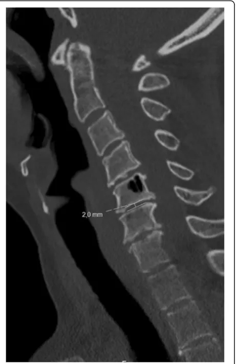
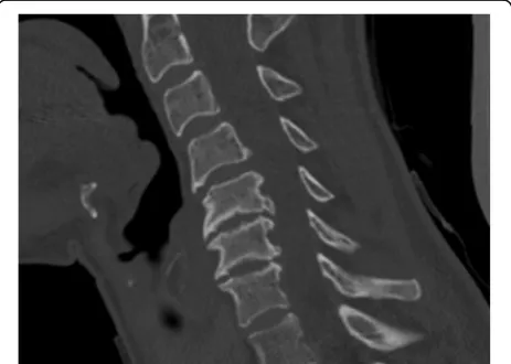
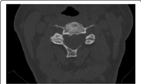
The scoring system for cervical disc degeneration used in this study is an adaption from an existing scoring sys-tem based on lateral radiographs. We used CT scans to determine the grade of degeneration. The scoring system consists of three variables: height loss, anterior osteo-phytes and endplate sclerosis (Table1). As the degenera-tive process is suggested to begin with disc desiccation and height loss [1], this factor is ascribed the most im-portance and thus the largest impact on the total degen-eration score. For all variables, the segment with the highest level of degeneration was chosen. Height loss was defined as the middle disc height measured in a mid-sagittal slice as compared to a normal (or least degenerated) disc height at any segment of the cervical spine as shown in Fig. 1. Anterior osteophytes were measured where the length was the greatest and com-pared to the anteroposterior diameter of the correspond-ing vertebral body as measured in the mid-sagittal slice (Fig. 2). Endplate sclerosis was ascribed on one of three discernible grades; no sclerosis, detectable sclerosis or definite sclerosis (Fig. 3). Finally, all variables were summed to an overall disc degeneration score (Table1).
The scoring system for cervical facet joint degener-ation was developed for CT. It contains three variables: joint space narrowing, osteophytes and irregularity of the articular surface (Table1). The original scoring sys-tem [16] also included facet joint hypertrophy, which we chose to exclude as it was the variable with the lowest inter-rater agreement in the previous study and offered limited additional information on the degree of facet joint degeneration. Joint space narrowing was assessed on sagittal scans and if any of the facet joints were nar-rowed, this variable was ascribed as 1 point (Fig. 4). Osteophytes and joint space irregularity were assessed on axial scans. If any osteophytes were present, the vari-able was ascribed as 1 point. Similarly, the presence of joint space irregularity received 1 point whereas smooth articular surfaces received 0 points (Fig. 5). Finally, the variables were summed to achieve an overall facet joint degeneration score.
For the total degeneration score, all variables were summed up to achieve an overall degeneration score for cervical spondylosis and were categorized as 0–1 p–no degeneration, 2–6 p –moderate degeneration and 7–12 p–severe degeneration.
Procedure
sternal tip. The expanded FOV was compensated with a low radiation dose profile with CTDIvolof around 3.8.
Patients aged > 18 years that were admitted to the emergency department at Södersjukhuset, Stockholm, Sweden for neck pain after a motor vehicle accident were included in the study. Those requiring medical imaging in the emergency setting according to the Canadian C-spine rules [23] underwent a CT of the cervical spine. The patients whom the examining physician deemed not to require medical imaging were later contacted by the research team and offered to participate in the study. If they accepted, they were also examined with a CT of the cervical spine. All patients had been contacted and gave their informed consent prior to the CT scan.
Statistical analysis
The statistical software package SPSS 22 (SPSS Inc., Chi-cago, IL) was used for analysis of both intra-rater and
Table 1Scoring system of cervical disc and facet joint degeneration. AP = anteroposterior
Disc degeneration
Height loss 0% 0
points
≤25% 1
point
> 25-≤50% 2
points
> 50%-≤75% 3
points
> 75% 4
points
Anterior osteophytes No osteophytes 0
points
≤1/8 AP diameter 1 point
> 1/8 -≤1/4 AP diameter 2 points
> 1/4 AP diameter 3 points
Endplate sclerosis No sclerosis 0
points
Detectable 1
point
Definite 2
points
Overall degree of disc degeneration 1 + 2 + 3
0 points (no degeneration)
0 points
1–3 points (mild degeneration)
1 point
4–6 points (moderate degeneration)
2 points
7–9 points (severe degeneration)
3 points
Facet joint degeneration
Joint space narrowing Normal 0
points
Narrowed 1
point
Osteophytes No osteophytes 0
points
Yes 1
point
Irregularity of articular surface Normal 0 points
Irregular 1
point
Overall degree of facet joint degeneration (1 + 2 + 3)
0 points (no degeneration)
0 points
1 point (mild degeneration)
1 point
2 points (moderate degeneration)
2 points
3 points (severe degeneration)
3 points
inter-rater agreement. An additional SPSS macro was used to compute the inter-rater agreement level for cat-egorical data between multiple observers [24]. The Kappa values were considered significant ifp< 0.05. The intra-rater agreement was assessed with intra-class cor-relation coefficient (ICC).
Interpretation
The interpretation guideline established by Landis & Koch [25] was used to evaluate the strength of inter-observer agreement using the kappa statistic.
Kappa values of > 0.40 were considered representing clinically acceptable level of strength of agreement for a scoring system [26].
The standards for strength of intra-rater agreement proposed by Fleiss et al. [27] were used as a basis for
interpretation of the magnitude of the ICC values obtained.
Results
Inter-rater reliability
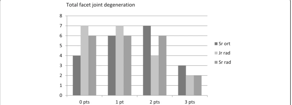
Figures 6, 7 and 8 illustrate the variance in degener-ation scores as assessed by the three raters regarding discs, facet joints and total degeneration, respectively (Figs. 6, 7 and 8).
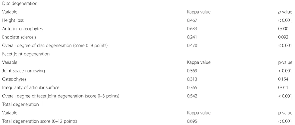
As shown in Figs. 6, 7 and 8, the senior orthopedic surgeon had a consistent tendency to assign the vari-ables a more severe degree of degeneration than both the radiologists. A statistical analysis of the agreement in scorings by the three raters generated kappa values for the variables (Table 2). Anterior osteophytes generated the highest kappa value, i.e. the raters tended to rate the presence and size of anterior osteophytes similarly. Re-garding facet joint degeneration, in terms of joint space narrowing the raters were closest to agreement.
Intra-rater reliability
To test the intra-rater reliability and thus reproducibility of the scoring system, the senior orthopedic surgeon and the junior radiologist repeated their assessments of the 20 patients after a minimum of 3 months (Table3). The analysis showed excellent agreement between the two assessments for the total degeneration and at least fair agreement for all parameters with ICC spanning from 0.538 to 0.820.
Discussion
Despite the vast amount of CT-scans of the cervical spine that are made addressing degenerative changes, to date there is a lack of clinical standardized rating models. Cervical spondylosis is a common radiological Fig. 2Most prominent anterior vertebral osteophyte. 4.4 mm/16.4 mm = 0.27= > 1/4 resulting in 3 points
finding and the association to disability and pain is still unclear. This might be partly due to lack of consensus in grading models for degeneration why it is of importance to obtain reliable assessment models. The effort of this study was to contribute to establishing such a scoring system and validate it in the aspects of inter-rater and intra-rater reliability. Focus was put in creating a user-friendly system for clinical implementation.
Inter-rater reliability
The kappa value for the overall degree of degeneration showed a substantial agreement. However, this value represents the agreement between the raters when add-ing their degeneration scores on the separate variables and then dividing the subjects into three separate cat-egories (no degeneration, moderate degeneration or se-vere degeneration). When grouped together, the rate of disagreement on the separate variables is masked and
the agreement when only three categories exist is pre-sumably consequently higher than it would be if more than three categories of degeneration were eligible. This becomes apparent when analyzing the separate variables, where kappa values are considerably lower. In two of the variables the null-hypothesis could not be rejected (end-plate sclerosis and facet joint osteophytes). The only variable where the strength of agreement was substantial was anterior osteophytes. This variable is weighted to contribute less to the disc degeneration score than height loss, which only reached a moderate strength of agreement. The fact that agreement for the total level of degeneration was higher than for the separate variable could be explained by compensation mechanisms of the individual rater. For example, a borderline case of facet joint osteophytes could have been neglected with a com-pensatory affirmation of borderline irregularity of the ar-ticular surface.
Our study showed similar results with those of the study of Walraevens et al. [16] concerning facet joint degeneration, even though our classification criteria differed, with low strength of agreement on osteo-phytes and irregularity of the articular surface but slightly higher strength of agreement for joint space narrowing.
However, when applying the radiograph-based scoring system for disc degeneration to CT there seemed to have been a slight loss of reliability compared to Walraevens et al. [16]. They showed“good”or“excellent”agreement on the disc degeneration variables apart from endplate sclerosis which was low in both studies, whereas our re-sults ranged from “moderate” to “substantial” with a slightly lower level of agreement overall. However, the trend is clear; assessing endplate sclerosis, facet joint Fig. 4Facet joint degeneration as exhibited by one of the study patients. To the left, an axial slice through the C5-C6 segment shows joint space narrowing, osteophytes and marked irregularities of the right facet joint space leading to 3 points. The right image illustrates the facet joint space narrowing at the same segment as compared to normal joint spaces on adjacent levels on a lateral view
osteophytosis and irregularity of facet joint articular sur-faces is more complex than the three other variables.
Considering a cut-off limit of 0.40 for strength of agree-ment, which is arbitrarily set, many of our obtained kappa values indicate an acceptable or good level of agreement. However, several Kappa values were below 0.40. There are a few reasons for the relatively low values that must be considered. First, the relatively small sample size could have affected the level of agreement. Another factor might have been the multi-segment assessment. Determining the spinal segment with the highest level of degeneration is an assessment by itself. It is plausible that the raters were in fact reviewing different segments and consequently asses-sing them differently. Lack of training among the raters might also affect the level of agreement. In this material, the raters deliberately had no joint training session of the scoring system prior to the assessment procedure. This was to simulate a clinical setting to a high extent.
The goal of developing a scoring system that is easily applicable and experience- and discipline independent is of importance. However, we believe minor modifica-tions could be done to improve the scoring system while still keeping it user-friendly. For example, one source of disagreement on the height loss-variable may have been presence of endplate compression affecting the disc height.
Intra-rater reliability
The ICC-values obtained all indicated fair, good or ex-cellent intra-rater agreement, with total degeneration scores having the strongest agreement for both raters. However, the confidence intervals were large and the true ICC-values thus hard to discern. They are inter-preted to originate from the variation between exam-iners using an ordinal scale on a relatively small material. Only two of the raters participated in the intra-Fig. 6Total disc degeneration score (0–9 points) as assessed by the three raters; the senior orthopedic surgeon (Sr ort), junior radiologist (Jr rad) and the senior radiologist (Sr rad) respectively
rater reliability part of the study. As in the inter-rater analysis, the agreement of the total degeneration score was higher when summing disc degeneration and facet joint degeneration scores.
In comparison with other scoring scales in the field the agreement is regarded equivalent. Considering inter-rater reliability of the assessment of disc degeneration, previous scales vary from 0.41–0.78 [16, 28, 29] intra-rater reliability of the discs vary from 0.71–0.86 [16,29]. In the material reviewed, the inter-rater agreement for facet joint degeneration the agreement varied from 0.43–0.49 [15, 16] and the intra-rater agreement from 0.57–0.72 [15,16]. When comparison is made, one must consider the different radiologic modalities that are used in previous materials.
In summary, our results indicate a well acceptable level of agreement regarding both inter-rater and intra-rater reliability of a CT based scoring system, especially
addressing facet joint degeneration and overall degener-ation. The findings enable a role for this scoring system in both future research and clinical practice. However, when analyzing individual parameters in the scores, the agreements were lower than in the total scores. Hence, we recommend the system to be clinically applied in its aggregated form to assess disc degeneration, facet joint degeneration and overall degeneration.
This study has a few limitations. First, the sample size is rather small and for wide clinical implication, future studies with larger material are required to confirm the results.
Second, the study population in this this material con-sists exclusively of post-traumatic patients. This makes it less representable for the general population and is nei-ther to be considered an asymptomatic cohort nor a co-hort with non-specific neck pain. We welcome further investigations in a different clinical setting to validate the scoring system.
Fig. 8Total degeneration score (0–12 points) as assessed by the three raters
Table 2Inter-rater agreement between the three raters regarding the degree of degeneration
Disc degeneration
Variable Kappa value p-value
Height loss 0.467 < 0.001
Anterior osteophytes 0.633 0.000
Endplate sclerosis 0.241 0.092
Overall degree of disc degeneration (score 0–9 points) 0.470 < 0.001
Facet joint degeneration
Variable Kappa value p-value
Joint space narrowing 0.569 < 0.001
Osteophytes 0.313 0.154
Irregularity of articular surface 0.365 0.011
Overall degree of facet joint degeneration (score 0–3 points) 0.542 < 0.001
Total degeneration
Variable Kappa value p-value
Conclusions
To our knowledge, this is the first study evaluating a co-herent scoring system for degeneration of the cervical spine based on CT. It confirms that a preexisting scoring system for cervical facet joint degeneration has an ac-ceptable level of strength of agreement for the overall degeneration. This study also showed that a radiographic scoring system for cervical disc degeneration is applic-able on CT, achieving a moderate degree of strength of agreement for the overall degeneration. Both scoring systems meet the standards for a clinically accepted level. Combined or individually, they make a reliable, co-herent and objective scoring system readily applicable in both research and in clinical settings where it can sim-plify and objectify the assessment of presence and degree of cervical degeneration.
Abbreviations
CT:Computed Tomography; CTDIvol: Volume Computed Tomography Dose Index; FOV: Field of View; ICC: Intraclass Correlation Coefficient
Acknowledgements
Erling Englund for kind assistance with the macro to the Fleiss Kappa-analysis.
Authors‘contributions
ER and SB participated in the design of the study. ER and SB were two of the assessors of the radiographs. ER and HJP carried out the statistical analysis. All authors participated in the process of writing the manuscript. All authors read and approved the final manuscript.
Funding
This study was supported by the Stockholm County through grants for the Department of Orthopedics for research, development and education (FoUU-medel). The department had no role in the design, execution or writing process of this study.
Availability of data and materials
The data and materials in this article can be made available upon request by sending an e-mail to the corresponding author.
Ethics approval and consent to participate
Ethical approval for the study was obtained from the Regional Ethical Review Board. (Dnr 2015/1121–31/4) and all patients gave their informed written consent before inclusion.
Consent for publication Not applicable.
Competing interests
The authors declare that they have no competing interests.
Author details
1Department of Clinical Science and Education, Södersjukhuset, Karolinska
Institutet, Stockholm, Sweden.2Department of Radiology, Södersjukhuset, Stockholm, Sweden.3Department of Orthopedics, Södersjukhuset, SE-118 83 Stockholm, Sweden.
Received: 15 December 2017 Accepted: 21 May 2019
References
1. Shedid D, Benzel EC. Cervical spondylosis anatomy: pathophysiology and biomechanics. Neurosurgery. 2007;60(1):7–13.
2. Kelly JC, Groarke PJ, Butler JS, Poynton AR, O'Byrne JM. The natural history and clinical syndromes of degenerative cervical spondylosis. Adv Orthop. 2012;2012:393642.
3. Ferrara LA. The biomechanics of cervical spondylosis. Adv Orthop. 2012; 2012:493605.
4. Bannister G, Amirfeyz R, Kelley S, Gargan M. Whiplash injury. J Bone Joint Surg Br. 2009;91(7):845–50.
5. Marchiori DM, Henderson CN. A cross-sectional study correlating cervical radiographic degenerative findings to pain and disability. Spine (Phila Pa 1976). 1996;21(23):2747–51.
6. Alli S, Anderson I, Khan S. Cervical spondylotic myelopathy. Br J Hosp Med (Lond). 2017;78(3):C34–c7.
7. Persson M, Sorensen J, Gerdle B. Chronic whiplash associated disorders (WAD): responses to nerve blocks of cervical zygapophyseal joints. Pain Med. 2016;17(12):2162–75.
8. Schofferman JMD, Bogduk NMD, Slosar PMD. Chronic whiplash and whiplash-associated disorders: an evidence-based approach. J Am Acad Orthop Surg. 2007;15(10):596–606.
9. Scholten-Peeters GG, Verhagen AP, Bekkering GE, van der Windt DA, Barnsley L, Oostendorp RA, et al. Prognostic factors of whiplash-associated disorders: a systematic review of prospective cohort studies. Pain. 2003; 104(1–2):303–22.
10. Eck JC, Hodges SD, Humphreys SC. Whiplash: a review of a commonly misunderstood injury. Am J Med. 2001;110(8):651–6.
11. Norris SH, Watt I. The prognosis of neck injuries resulting from rear-end vehicle collisions. J Bone Joint Surg Br. 1983;65(5):608–11.
12. Cote P, Cassidy JD, YongHing K, Sibley J, Loewy J. Apophysial joint degeneration, disc degeneration, and sagittal curve of the cervical spine -can they be measured reliably on radiographs? Spine. 1997;22(8):859–64. 13. Daimon K, Fujiwara H, Nishiwaki Y, Okada E, Nojiri K, Shimizu K, et al. A
20-year prospective longitudinal MRI study on cervical spine after whiplash injury: follow-up of a cross-sectional study. J Orthop Sci. 2018. [Epub ahead of print]
14. Manelfe C. Imaging of degenerative processes of the spine. Curr Opin Radiol. 1992;4(1):63–70.
15. Xu C, Ding ZH, Xu YK. Comparison of computed tomography and magnetic resonance imaging in the evaluation of facet tropism and facet arthrosis in degenerative cervical spondylolisthesis. Genet Mol Res. 2014;13(2):4102–9. 16. Walraevens J, Liu B, Vander Sloten J, Goffin J. Qualitative and quantitative assessment of degeneration of cervical intervertebral discs and facet joints. Eur Spine J. 2009;18(3):358–69.
17. Park MS, Lee YB, Moon SH, Lee HM, Kim TH, Oh JB, et al. Facet joint degeneration of the cervical spine: a computed tomographic analysis of 320 patients. Spine (Phila Pa 1976). 2014;39(12):E713–8.
18. Griffith JF, Wang YX, Antonio GE, Choi KC, Yu A, Ahuja AT, et al. Modified Pfirrmann grading system for lumbar intervertebral disc degeneration. Spine (Phila Pa 1976). 2007;32(24):E708–12.
19. Kettler A, Wilke HJ. Review of existing grading systems for cervical or lumbar disc and facet joint degeneration. Eur Spine J. 2006;15(6):705–18. 20. Kellgren JH, Lawrence JS. Osteo-arthrosis and disk degeneration in an urban
population. Ann Rheum Dis. 1957;17:388–97.
21. Hol PK. Imaging in whiplash. Cephalalgia. 2008;28 Suppl 1:25–7. 22. Pathria M, Sartoris DJ, Resnick D. Osteoarthritis of the facet joints:
accuracy of oblique radiographic assessment. Radiology. 1987;164(1): 227–30.
Table 3Intra-rater agreement between the two assessments made by the junior radiologist and the senior orthopedic surgeon, respectively
Intra-rater agreement between the two assessments
Junior radiologist Senior orthopedic surgeon
ICC 95% CI ICC 95% CI
Disc degeneration 0.672 0.103–0.875 0.598 0.023–0.838
Facet joint degeneration 0.538 −0.284-0.833 0.750 0.361–0.902
Total degeneration 0.820 0.542–0.929 0.727 0.076–0.905
23. Stiell IG, Wells GA, Vandemheen KL, Clement CM, Lesiuk H, De Maio VJ, et al. The Canadian C-spine rule for radiography in alert and stable trauma patients. J Am Med Assoc. 2001;286(15):1841–8.
24. Nicholls D. NSKAPPA, SPSS:http://imaging.mrc-cbu.cam.ac.uk/statswiki/FAQ/ kappa/multiple
25. Landis JR, Koch GG. The measurement of observer agreement for categorical data. Biometrics. 1977;33(1):159–74.
26. Sim J, Wright CC. The kappa statistic in reliability studies: use, interpretation, and sample size requirements. Phys Ther. 2005;85(3):257–68.
27. Fleiss JL. Reliability of measurement. Hoboken, NJ. USA: John Wiley & Sons, Inc.; 1999.
28. Wierzbicki V, Pesce A, Marrocco L, Piccione E, Colonnese C, Caruso R. How old is your cervical spine? Cervical spine biological age: a new evaluation scale. Eur Spine J. 2015;24(12):2763–70.
29. Cote P, Cassidy JD, Yong-Hing K, Sibley J, Loewy J. Apophysial joint degeneration, disc degeneration, and sagittal curve of the cervical spine. Can they be measured reliably on radiographs? Spine (Phila Pa 1976). 1997; 22(8):859–64.
Publisher’s Note