R E S E A R C H
Open Access
Effect of three lactobacilli with
strain-specific activities on the growth
performance, faecal microbiota and ileum
mucosa proteomics of piglets
Yating Su
1, Xingjie Chen
2, Ming Liu
3and Xiaohua Guo
1*Abstract
Background:The beneficial effects ofLactobacillusprobiotics in animal production are often strain-related. Different strains from the same species may exert different weight-gain effect on hosts in vivo. Most lactobacilli are selected based on their in vitro activities, and their metabolism and regulation on the intestine based on strain-related characters are largely unexplored. The objective of the present study was to study the in vivo effects of the three lactobacilli on growth performance and to compare the differential effects of the strains on the faecal microbiota and ileum mucosa proteomics of piglets.
Methods:Three hundred and sixty piglets were assigned to one of four treatments, which included an antibiotics-treated control and three experimental groups supplemented with the three lactobacilli,L. salivariusG1-1,L. reuteri G8-5 andL. reuteriG22-2, respectively. Piglets were weighed and the feed intake was recorded to compare the growth performance. The faecal lactobacilli and coliform was quantified using quantitative PCR and the faecal microbiota was profiled by denaturing gradient gel electrophoresis (DGGE). The proteomic approach was applied to compare the differential expression of proteins in the ileum mucosa.
Results:No statistical difference was found among the threeLactobacillus-treated groups in animal growth performance compared with the antibiotics-treated group (P> 0.05). Supplementation of lactobacilli in diets significantly increased the relative 16S rRNA gene copies ofLactobacillusgenus on both d 14 and d 28 (P< 0.05)., and the bacterial community profiles based on DGGE from the lactobacilli-treated groups were distinctly different from the antibiotics-treated group (P< 0.05). The ileum mucosa of piglets responded to allLactobacillus
supplementation by producing more newly expressed proteins and the identified proteins were all associated with the functions beneficial for stabilization of cell structure. Besides, some other up-regulated and down-regulated proteins in differentLactobacillus-treated groups showed the expression of proteins were partly strain-related.
Conclusions:All the three lactobacilli in this study show comparable effects to antibiotics on piglets growth performance. The three lactobacilli were found able to modify intestinal microbiota and mucosa proteomics. The regulation of protein expression in the intestinal mucosa are partly associated with the strains administrated in feed.
Keywords:Faecal microbiota, Growth,Lactobacillus, Mucosa proteomics, Probiotics
* Correspondence:[email protected] 1
Provincial Key Laboratory for Protection and Application of Special Plants in Wuling Area of China, College of Life Science, South-Central University for Nationalities, No. 182, Minyuan Road, Hongshan District, Wuhan, Hubei Province 430074, China
Full list of author information is available at the end of the article
Background
As living microorganisms, probiotics act in the intes-tine to modulate the host microbiota [1]. Among the strains of probiotics, lactic acid bacteria (LAB), espe-cially from Lactobacillus and Bifidobacteria species, are recognized as one of the main sources and are widely used in food, drugs and feed additives as in-testinal flora improvers [2]. In animal production, probiotics are expected to improve performance and to produce high-qualified meat without drug residues as an alternative to antibiotics [3].
Generally, Lactobacillus species selected for probio-tics are highly diverse in the phenotypic and genetic characteristics [4]. Different strains may exert differ-ent weight-gain effect on hosts in vivo even if in the same species. Million et al. assessed the effect of lactobacilli-containing probiotics on weight based on 51 studies on farm animals and suggested that the weight-gain effect was greatly associated with strains of the genus [3]. Simon et al. showed similar results after summarizing above 20 published papers on lactobacilli used in feed additives [5]. The phenomena suggest that Lactobacillus strains may benefit their hosts through different mechanisms and more work should be done to explore the relationship between the choice of strains and their in vivo behaviours [6].
Nowadays the selection ofLactobacillusis often based on the strains’ activities in vitro, which is expected to show corresponding effectiveness in vivo. The strains with bacteriocin-producing activity showed specific anti-infective effect in the gut [7]. The strains with enzyme activities including amylase, protease and α -galactosidase had the potential to stimulate feed diges-tion [8–10]. However, the gut ecosystem was so compli-cated and the in vivo activities often depended on the strains’ survival and metabolism in the gastrointestinal tract (GIT) [10]. In our previous studies, three Lactoba-cillus strains (Lactobacillus salivarius G1-1, Lactobacil-lus reuteri G8-5 and Lactobacillus reuteri G22-2) were selected from swine faeces for probiotic use. They shared strarelated in vitro functional properties, in-cluding antimicrobial activity, amylolytic activity and bile-salt-hydrolase activity, respectively [11]. Meanwhile, from the in vivo studies in rats, the three Lactobacillus
species showed some similar beneficial effects and some of the functionalities to rats were strain-specific [12]. When used in swine nutrition, the lactobacilli were hy-pothesized to interact with the intestinal flora and with the host mucosa, which might be associated with the mechanism of lactobacilli as probiotics. The objective of the present study was to study the in vivo effects of the three lactobacilli on growth performance and to com-pare the differential effects of the strains on the faecal microbiota and ileum mucosa proteomics of piglets.
Methods
Lactobacillusstrains and freeze-dried powder preparation
Three strains,L. salivariusG1-1, L. reuteri G8-5 and L. reuteri G22-2 were isolated for probiotics based on the strain-specific functional properties in vitro [11]. All strains were incubated in DeMan Rogosa Sharp broth under anaerobic conditions at 37 °C for about 24 h. The microbial cells were collected by centrifugation at 11,000×g for 10 min, washed twice and mixed with pro-tective additives for freeze-drying. The freeze-dried sam-ple was smashed and diluted with dextrin as a carrier. The concentration of viable cells from each strain was determined by agar-plate assay and adjusted to 0.5 × 10⁹ colony forming unit per gram (CFU/g) by the carrier before animal trial.
Animals, diets, experimental design and sampling
Three hundred and sixty castrated male, crossbred (Landrace × Large White) piglets, 35–40 days old, were randomly assigned to one of four treatments, which in-cluded an antibiotics control (Group A) and three ex-perimental groups supplemented withL. salivariusG1-1 (Group B), L. reuteri G8-5 (Group C) and L. reuteri
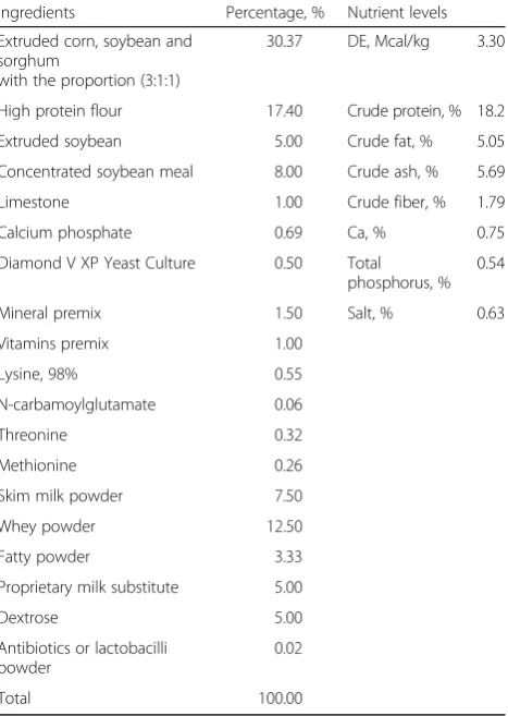
G22-2 (Group D), respectively. The piglets were housed with 15 piglets per pen and six pens of piglets received each treatment (n =6). The pigs had free access to feed and water throughout the feeding trial with the environ-mental temperature 25–28 °C. The diet composition was listed in Table 1. The diet in the antibiotics control was supplemented with 200 mg/kg flavomycin. The three ex-perimental diets consisted of the basal diet supple-mented with 200 mg/kgLactobacilluspowder (109CFU/ kg of feed) from each strain.
DNA extraction, real-time quantitative PCR and PCR-DGGE analyses
The total genomic DNA was extracted from faeces (about 1.0 g) based on the method of bead-beating and following phenol-chloroform extraction [13, 14]. Total lactobacilli and coliform were detected by real-time quantitative PCR, re-spectively. The lactobacilli were quantified using primer Lac1 (5′-AGCAGTAGGGAATCTTCCA-3′), and Lab0677 (5′- CACCGCTACACATGGAG −3′) [15]. Two primers, EcoliFimH2F (5′-AGCAGTAGGGAATCTTCCA-3′) and EcoliFimH2R (5′- TCATCCCTGTTATAGTTGYYGGTCT-3′) were used to amplify 16S rRNA gene of coliform [16]. The reverse transcription PCR (RT-PCR) system was quan-tified using the ABI 7500 system (Applied Biosystems, US). The optimum thermal cycles were performed as fol-lows: pre-denaturation at 95 °C for 10 min, 40 cycles of 95 °C for 15 s and 60 °C for 1 min, and followed by the stage of melting curve. The relative 16S rRNA gene copies were calculated through the 2−ΔΔCT method according to the report of Livak [17]. The
results were compared based on the three paralleled values of faeces from each treatment.
A set of universal primers, U968-GC (5′-CGCCCG GGGCGCGCCCCGGGCGGGGCGGGGGCACGGGG GGAACGCGAAGAACCTTAC-3′), L1401 (5′-CGGTGT GTACAAGACCC-3′) [18], Bact 1369 F (5′-CGGTGAA TACGTTCYCGG-3′), and 1492R (5′-GGWTACCTT GTTACGACTT-3′) [19] were employed to amplify the total bacteria. The amplicons were separated by DGGE ac-cording to the specification as described previously [20]. Briefly, DGGE was performed in 8% polyacrylamide gels (acrylamide-bis, 37.5:1). The gels with a 38–51% denatur-ing gradient was used for the separation of PCR products based on the primers U968-GC and L1401, while gra-dients of 30–45% were applied for the separation of the Bact 1369 F and 1492R generated amplicons. The electrophoresis procedures were performed at 70 V for 16 h at 60 °C and the gel was finally stained with SYBR Green I for 30 min after electrophoresis. The DGGE gels were scanned using an image scanner and analysed with Bio-rad gel imaging system through Quantity One software (Version 4.6.2).
The similarities among DGGE profiles were determined by Dice coefficient based on the unweighted pair group method with arithmetic average (UPGMA) clustering al-gorithm [21]. The faecal samples from the antibiotics group were evenly mixed and conducted for DGGE pro-files used as the control band. The bands from three paralled faecal samples of each Lactobacillusgroup were profiled and compared with the control band (n =3).
2-dimensional electrophoresis (2-DE), image analysis and protein identification
Isoelectric focusing (IEF) was performed using immobi-lized pH gradient (IPG) Strips (pH 4–7; 7 cm long; Phar-macia Biotech.). Samples were diluted with IEF buffer containing 7 mol/L urea, 2 mol/L thiourea, 4% CHAPS, 20 mmol/L Tris–HCl, pH 8.5, 20 mmol/L DTT, 0.5% carrier ampholyte (pH 4–7) and a trace of bromophenol blue. The desired protein amount in buffer was 50 μg. After equilibration, the immobilized pH gradient strips were loaded onto 12.5% (w/v) homogeneous acrylamide gels and sealed with 1% (w/v) agarose. The electrophor-etic separation of proteins was conducted as described previously [22, 23]. Upon completion of 2-dimensional SDS-PAGE, the gels were stained by silver or Coomassie Brilliant Blue G-250. The high-resolution gel images (200 dpi) from silver-stained gels were obtained using an image scanner (Powerlook1100, UMAX) for image ana-lysis. The gels stained by silver were run in triplicate, and spots that appeared consistently in all three runs were selected for analysis. Spot detection and analysis were performed using the PDQuest version 6.1 software (Bio-Rad) according to the protocols provided by the
Table 1Basal diet formula and nutrient levels
Ingredients Percentage, % Nutrient levels
Extruded corn, soybean and sorghum
with the proportion (3:1:1)
30.37 DE, Mcal/kg 3.30
High protein flour 17.40 Crude protein, % 18.2
Extruded soybean 5.00 Crude fat, % 5.05
Concentrated soybean meal 8.00 Crude ash, % 5.69
Limestone 1.00 Crude fiber, % 1.79
Calcium phosphate 0.69 Ca, % 0.75
Diamond V XP Yeast Culture 0.50 Total
phosphorus, % 0.54
Mineral premix 1.50 Salt, % 0.63
Vitamins premix 1.00
Lysine, 98% 0.55
N-carbamoylglutamate 0.06
Threonine 0.32
Methionine 0.26
Skim milk powder 7.50
Whey powder 12.50
Fatty powder 3.33
Proprietary milk substitute 5.00
Dextrose 5.00
Antibiotics or lactobacilli powder
0.02
Total 100.00
a
Vitamins provided per kilogram diets: vitamin A, 8000 IU; vitamin D3,
1800 IU; vitamin E, 30 IU; vitamin K3, 3.56 mg; vitamin B1, 1.8; vitamin B2,
6 mg; vitamin B6, 1.26 mg; vitamin B12, 0.02 mg; folic acid, 0.3 mg; biotin,
0.44 mg; niacin, 32 mg; pantothenic acid, 15 mg
b
manufacturer. Some differentially expressed protein spots with 3.0-fold differences in volume detected by the soft-ware were selected for protein identification. The protein spots of interest were confirmed in the Coomassie Bril-liant Blue stained gels and manually excised for the treat-ment of digestion by trypsin. The matrix-assisted laser desorption/ionization time of flight mass spectrometry (MALDI-TOF MS) was used for protein identification as described by early reports [24, 25]. The peptide fragments produced from each protein spot were employed to pro-duce peptide-mass mapping (PMM) data. The protein identification was carried out by peptide mass fingerprint-ing (PMF) analysis through the MASCOT server (www.matrixscience.com; Matrix Science, UK). The search parameters were as follows, database: Swiss-Prot Sus (34361 sequences); species: sus; enzyme: trypsin; fixed modifications: carbamidomethylation; variable modifica-tions: oxidation (M). The gene name, accession code and function of each protein were determined using the Mascot V2.1 software protein database search engine and the Swiss-Prot Sus protein database.
Statistical analyses
All quantitative data were expressed as the mean and standard deviation of replicates. The differences among antibiotics-treated and lactobacilli-treated groups were con-sidered statistically significant at P< 0.05 using one-way analysis of variance (One-way ANOVA) through JMP soft-ware (JMP; SAS Institute Inc., Cary, NC). 0.5 <P< 0.1 was considered a trend towards significance.
Results
Growth performance
Over the 4-week feeding trial, there was no statistical difference in ADG, ADFI and F:G between piglets sup-plemented with lactobacilli and the antibiotics group (Table 2). Among the three Lactobacillus groups, the diet containing L. reuteri G8-5 tended to show lower ADG and ADFI than that of the other twoLactobacillus
groups (0.5 <P< 0.1).
Relative 16S rRNA gene copies by RT-PCR
A comparison of the relative 16S rRNA gene copies of
Lactobacillus and coliform in faeces on d 14 and d 28 was shown in Fig. 1. Supplementation of lactobacilli in diets significantly increased the counts of Lactobacillus
genus on both d 14 and d 28 compared with the antibi-otics group (P< 0.05). However, no significant difference in the relative 16S rRNA gene copies of coliform was ob-served in all groups (P> 0.05).
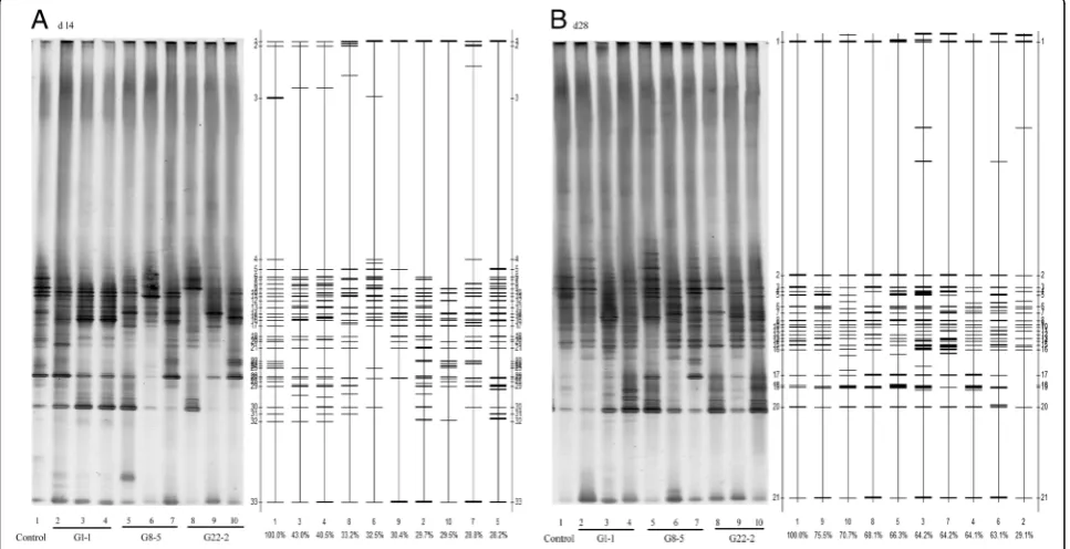
PCR-DGGE profiles
The representative DGGE profiles were presented in Fig. 2. The DGGE patterns were transformed into graphs by the Bio-Rad Quantity OneTM software, which calculated the Dice similarity among lanes (Fig. 2). The similarities among four treatments on d 14 and d 28 were listed in Table 3. On d 14, the dendrogram based on the banding patterns showed low similarities and the bacterial community profiles form the lactobacilli were distinctly different from the antibiotics group (P< 0.05). Meanwhile, the similarities in L. reuteri G8-5 group were significantly lower than those in L. salivariusG1-1 group (P< 0.05). On d 28, the percentage of similarity in all Lactobacillus
groups increased but was still significantly lower than that of antibiotics group. There was no marked difference in similarities in all Lactobacillus-treated groups on d 28 (P> 0.05).
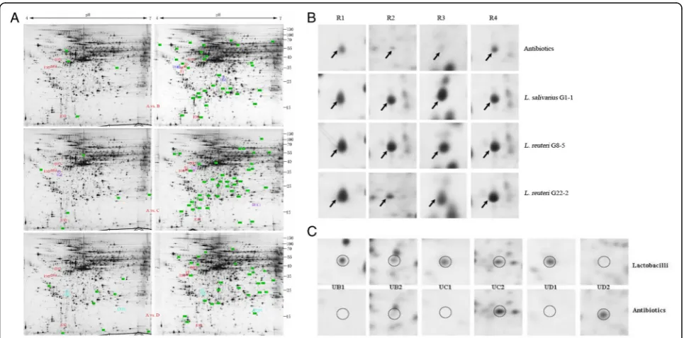
2-DE profiles of differentially expressed proteins
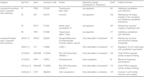
By comparing the 2-DE profiles of differentially expressed proteins in the ileum of piglets between the antibiotics-treated and Lactobacillus-treated groups, supplementation of lactobacilli significantly increased the counts of newly expressed proteins. 4, 6 and 8 new proteins were expressed only in the antibiotics group compared with the three Lactobacillus groups, respect-ively. Nevertheless, 32, 40 and 27 new proteins only existed in the three Lactobacillusgroups compared with the antibiotics group, respectively (Fig. 3a). Among the differentially expressed proteins, 4 protein spots which were up-regulated in all theLactobacillus-treated groups were selected for the identification by MALDI-TOF.
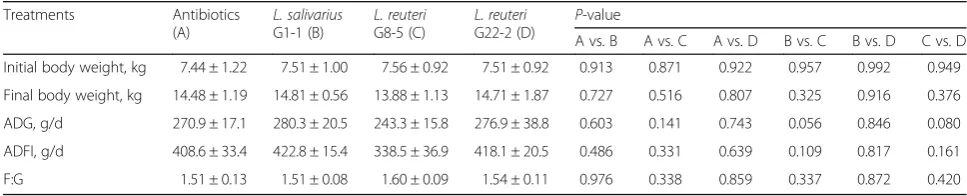
Table 2The effect of three lactobacilli on the growth performance of weaned piglets during a 4-week feeding trial
Treatments Antibiotics
(A)
L. salivarius G1-1 (B)
L. reuteri G8-5 (C)
L. reuteri G22-2 (D)
P-value
A vs. B A vs. C A vs. D B vs. C B vs. D C vs. D
Initial body weight, kg 7.44 ± 1.22 7.51 ± 1.00 7.56 ± 0.92 7.51 ± 0.92 0.913 0.871 0.922 0.957 0.992 0.949
Final body weight, kg 14.48 ± 1.19 14.81 ± 0.56 13.88 ± 1.13 14.71 ± 1.87 0.727 0.516 0.807 0.325 0.916 0.376
ADG, g/d 270.9 ± 17.1 280.3 ± 20.5 243.3 ± 15.8 276.9 ± 38.8 0.603 0.141 0.743 0.056 0.846 0.080
ADFI, g/d 408.6 ± 33.4 422.8 ± 15.4 338.5 ± 36.9 418.1 ± 20.5 0.486 0.331 0.639 0.109 0.817 0.161
F:G 1.51 ± 0.13 1.51 ± 0.08 1.60 ± 0.09 1.54 ± 0.11 0.976 0.338 0.859 0.337 0.872 0.420
These proteins included tropomyosin beta chain (TPM2, Spot R1), vimentin (VIM, Spot R2), keratin type I cyto-skeletal 19 (KRT19, Spot R3), tropomyosin alpha-1 chain (TPM1, Spot R4) (Table 4; Fig. 3b). Other six protein spots were chosen because they were specifically affected by different Lactobacillus strains (Table 4; Fig. 3c). The proteins in L. salivarius G1-1 group included the up-regulation of phosphatidylinositol 4,5-bisphosphate 3-kinase catalytic subunit gamma isoform (PIK3CG, Spot UB1) and
cofilin-1 (CFL1, Spot UB2), which were only detectable in
L. salivariusG1-1-treated group. The proteins expressed in L. reuteri G8-5 group included the up-regulation of Rho GDP-dissociation inhibitor 2 (ARHGDIB, Spot UC1; only detectable in lactobacilli group) and the down-regulation of nucleophosmin (NPM1, Spot UC2). The proteins in L. reuteri G22-2 group in-cluded the up-regulation of Rho GDP-dissociation inhibitor 2 (ARHGDIB, Spot UD1; only detectable in
A
B
Control G1-1 G8-5 G22-2 Control G1-1 G8-5 G22-2
Fig. 1Effect of threeLactobacillusstrains on the fecal relative 16S rRNA gene copies of lactobacilli (a) andE. coli(b), respectively on d 14 and d 28.abmean in the same column from the result on d 14 with different scripts differ significantly (
P< 0.05);ABmeans in the same column from the result on d 28 with different scripts differ significantly (P< 0.05)
lactobacilli-treated group) and the down-regulation of actin cytoplasmic 1 (ACTB, UD2; only detectable in antibiotics group).
Discussion
The supplementation of lactobacilli in animal diets af-fects gastrointestinal tract health and growth perform-ance of piglets [1, 5]. However, different Lactobacillus
strains used as probiotics may achieve the beneficial ef-fects on hosts through different mechanisms [12, 26]. The present study was conducted to compare the differ-ent efficacies among three lactobacilli with strain-specific activities in growth performance, faecal micro-biota and ileum mucosa proteomics of piglets.
No significant differences in growth performance among Lactobacillus-treated groups were observed
compared with the antibiotics-treated group. The result showed that all the three lactobacilli had the same po-tential as alternative to antibiotics in feed. However, among the three lactobacilli, the supplementation of L. reuteri G8-5 was the least effective in enhancing the growth performance of piglets, which was in line with the previous study in the rat experiment [12]. The rea-son is probably associated with the strain’s lower anti-microbial activity compared with the other two strains, which was reported in the previous study [11].
Increased lactobacilli in faeces from lactobacilli-treated piglets on both d 14 and d 28 in this study verified the ability of the three lactobacilli to maintain the balance of microbiota, which was one of the possible mechanisms of lactobacilli as probiotics in vivo [27]. Meanwhile, the modulation of intestinal microbiota by lactobacilli might be strain-insensitive since all the three lactobacilli used in the study showed the same ability as intestinal flora improvers. No difference in coliform counts was ob-served in whole feeding period compared with the anti-biotics group. The result suggested the antianti-biotics used in the study and lactobacilli had the similar resistance to pathogens and kept them in low level in the gastrointes-tinal tract. It is assumed that the increasing intesgastrointes-tinal microbial abundance caused by antibiotics or lactobacilli has more power to resist the disruption of microbial bal-ance [28, 29]. Further analysis on the microbiota in the gastrointestinal tract treated by lactobacilli and antibi-otics by PCR-DGGE was investigated for the comparison
Table 3Effect of threeLactobacillusstrains on the similarities among digitalized DGGE profiles of PCR-amplified 16S rRNA from fecal DNA after Bio-Rad Quantity One software comparison
Similarity, %
Treatments d 14 d 28
Antibiotics 100.00 ± 0.00a 100.00 ± 0.00a
L. salivariusG1-1 37.73 ± 7.07b 52.47 ± 20.24b L. reuteriG8-5 29.83 ± 2.33c 64.53 ± 1.63b L. reuteriG22-2 31.03 ± 1.93bc 71.43 ± 3.75b
Values are means ± S.D,n =3.a, b, c
Mean in a same column with different superscripts differ significantly (P< 0.05)
of microbial diversity. On d 14 and d 28, the similarities in all Lactobacillus-treated groups were significantly dif-ferent from the antibiotics-treated group. The results suggested that the mechanisms of antibiotics and lacto-bacilli on regulating intestinal microbiota were through different ways and lactobacilli contributed to compara-tively complex bacterial community. Some similar re-sults were also shown in other reports [30, 31]. The results in Table 4 and Fig. 2 showed the discrepancy in similarities between lactobacilli and antibiotics treat-ments tended to decrease from d 14 to d 28. This indi-cates the bacterial diversity tended to be stable and not sensitive to extraneous drugs or introduced bacteria dur-ing animals’growth. The significantly lower Dice similar-ity in L. reuteriG8-5 compared with L. salivarius G1-1 was observed in this study, and the result was in line with that in the growth performance.
Proteomics play an important role in the assessment of specific health-promoting activities exerted by Lacto-bacillus species [32, 33]. The ileum mucosa samples were collected to compare the differentially expressed proteins through 2-DE profiles. From the result in Table 4, the supplementation of lactobacilli all greatly in-creased the number of expressed protein spots com-pared with the antibiotics group. Similar result was also observed in the study of Wang et al. [32]. Up-regulation of four proteins including TPM2, VIM, KRT19 and TPM1 in all threeLactobacillusgroups are all associated
with the functions of maintaining and stabilizing cell structure and stabilization. The four proteins were in-ferred to be Lactobacillus-insensitive, and the mutual mechanisms for Lactobacillus as probiotics were to enhance the expression of proteins beneficial for stabilization of cell structure. Both TPM1 and TPM2 bind to actin filaments and up-regulation of the two pro-teins benefit to stabilizing cytoskeleton actin filaments [34]. Meanwhile, increased level of VIM is responsible for maintaining cell shape, integrity of the cytoplasm, and stabilizing cytoskeletal interactions [35]. The up-regulation of KRT19 is responsible for the structural in-tegrity of epithelial cells [36]. The increased expression of KRT19 in lactobacilli groups can contribute to more opportunities for living cells to adhere to the epithelial and exclusively inhibit pathogen infection [37, 38]. Simi-lar result was also observed in the study of Wang et al. [32], in which KRT10 was higher in the intestinal mu-cosa of piglets supplemented with L. fermentum I5007 compared with that in antibiotics piglets [32]. Both KRT10 and KRT19 belong to the keratin family which are intermediate filament proteins responsible for the structural integrity of epithelial cells [36].
There were six extra proteins differently expressed in differentLactobacillusgroups, which were inferred to be
Lactobacillus-related. The different expression of protein might be caused by the characters of specific strains. In the groups of L. salivarius G1-1, two proteins, PIK3CG
Table 4Differentially expressed proteins in the ileum mucosa of piglets supplemented by three lactobacilli in diets compared with antibiotics
Category Spot No.a
Gene Accession code Protein Expression change (Lactobacilli VS. Antibiotics)
Scoreb
Putative function
Lactobacilli-insensitive spots compared with antibiotics
R1 TPM2 F1SG00 Tropomyosin beta chain
Up-regulation 261 Stabilizing cytoskeleton actin filaments
R2 VIM P02543 Vimentin Up-regulation 138 Maintaining cell shape, integrity of the cytoplasm, and stabilizing cytoskeletal interactions
R3 KRT19 F1S0J8 Keratin type I cytoskeletal 19
Up-regulation 185 Maintaining structural integrity of epithelial cells
R4 TPM1 F2Z5B6 Tropomyosin alpha-1 chain
Up-regulation 277 Stabilizing cytoskeleton actin filaments
Lactobacilli-related spots compared with antibiotics
UB1(G1-1) PIK3CG O02697 Phosphatidylinositol 4,5-bisphosphate 3-kinase catalytic subunit
Only detectable in lactobacilli 137 Maintaining structural and functional integrity of epithelia
UB2(G1-1) CFL1 P10668 Cofilin-1 Only detectable in lactobacilli 121 Regulation of cell morphology and cytoskeletal organization
UC1(G8-5) ARHGDIB F1SQW8 Rho GDP-dissociation inhibitor 2
Only detectable in lactobacilli 109 Small GTPase regulator activity receptor binding;
UC2(G8-5) NPM1 F1RRY2 Nucleophosmin Down-regulation 99 Ribosome biogenesis and transport
UD1(G22-2) ARHGDIB F1SQW8 Rho GDP-dissociation inhibitor 2
Only detectable in lactobacilli 116 Small GTPase regulator activity receptor binding;
UD2(G22-2) ACTB Q6QAQ1 Actin cytoplasmic 1 Only detectable in antibiotics 139 Involved in cell motility, structure, and integrity
a
Spot No. refers to protein spot numbers that were labeled in Fig.3
b
and CFL1, detected only in Lactobacillus group were also associated with cell structure and stability.
ARHGDIB was only detectable in the ileum mucosa of piglets in response to the supplementation of bothL. reu-teriG8-5 andL. reuteriG22-2. The high expression of the protein enhances the recycling and distribution of acti-vated Rho GTPases in the cell and play a role in regulating cell motility through the modulation of Rho proteins [39]. NPM1 help cells survive environmental stresses, such as drug attack [40]. Up-regulation of NPM1 in antibiotics might be associated with the intake of flavomycin. The in-crease in ACTB found in vivo would indicate drastic oxi-dative modification leading to functional impairments [41], which might be the side-effect of antibiotics supple-mented in feed. More experiments are needed in order to document the potential beneficial effects of the lactobacilli strains for the piglets, notably in terms of mucosal health.
Conclusions
In conclusion, this study provides a comprehensive com-parison of three lactobacilli with strain-specific activities through the supplementation in piglet diets. All the three lactobacilli show the potential as alternatives to an-tibiotics and no statistical difference in animal growth performance compared with the antibiotics group. Sup-plementation of lactobacilli in diets could significantly increase the relative 16S rRNA gene copies of lactobacilli genus on both d 14 and d 28, and the bacterial commu-nity profile based on PCR-DGGE from the lactobacilli are distinctly different from the antibiotics group. The ileum mucosa piglets respond to all lactobacilli supple-mentation by more newly expressed proteins and the identified proteins are all associated with the functions beneficial for stabilization of cell structure. Besides, some other up-regulated and down-regulated proteins in different Lactobacillus groups showed the expression of proteins were partly strain-related.
This comparative study helps to explore the mutual mechanisms for Lactobacillus as probiotics on altering intestinal abundance of microbiota and expression of mucosa proteins in piglets and provides information for strain-specific screening in application.
Abbreviations
2-DE:2-dimensional electrophoresis; ACTB: Actin cytoplasmic 1; ADFI: Average daily feed intake; ADG: Average daily weight gain; ARHGDIB: Rho GDP-dissociation inhibitor 2; CFU: Colony forming unit; DGGE: Denaturing gradient gel electrophoresis; DNA: Deoxyribose nucleic acid; F:G: Feed conversion Ratio; GIT: Gastrointestinal tract; IEF: Isoelectric focusing; IPG: Immobilized pH gradient; KRT19: Keratin type I cytoskeletal 19; LAB: Lactic acid bacteria; MALDI-TOF MS: Matrix-assisted laser desorption/ ionization time of flight mass spectrometry; NPM1: Nucleophosmin; PCR: Polymerase chain reaction; PIK3CG: Phosphatidylinositol 4,5-bisphosphate 3-kinase catalytic subunit gamma isoform; PMF: Peptide mass fingerprinting; PMM: Peptide-mass mapping; rRNA: Ribosomal ribonucleic acid; RT-PCR: Reverse transcription-polymerase chain reaction; SDS-PAGE: Sodium dodecyl sulfate-polyacrylamide gel electrophoresis; TPM1: Tropomyosin alpha-1 chain; TPM2: Tropomyosin beta chain;
UPGMA: Unweighted pair group method with arithmetic average; VIM: Vimentin
Acknowledgements
We would like to thank Jingjing Yang, Huang Ren and Xiaoping Fu for their lab analysis.
Funding
This work was financially supported by the National Natural Science Foundation of China (No. 31372348; No. 31672455) and the Fundamental Research Funds for the Central Universities (CZY15026).
Availability of data and materials
The datasets generated during and/or analysed during the current study are available from the corresponding author on reasonable request.
Authors’contributions
XHG was involved in the study design, data analysis, interpretation and drafting the manuscript; YTS participated in all laboratory analyses; XJC and ML performed the animal management. All authors read and approved the final manuscript.
Competing interests
The authors declare that they have no competing interests.
Consent for publication
All authors read and approved the final manuscript.
Ethics approval
All procedures for this study followed the protocols approved by Experimental Animal Care and Use Guidelines and approved by Ethical Committee of College of Life Science of South-Central University for Nationalities.
Author details
1Provincial Key Laboratory for Protection and Application of Special Plants in Wuling Area of China, College of Life Science, South-Central University for Nationalities, No. 182, Minyuan Road, Hongshan District, Wuhan, Hubei Province 430074, China.2Guangxi Yang-Xiang Animal Husbandry Co. Ltd., Guigang, Guangxi Province 537100, China.3Beijing China-agri Hong-Ke Biotechnology Co., Ltd., Beijing 102206, China.
Received: 13 March 2017 Accepted: 18 May 2017
References
1. Fuller R, Gibson GR. Modification of the intestinal microflora using probiotics and prebiotics. Scand J Gastroenter. 1997;32:28–31.
2. Naidu A, Bidlack W, Clemens R. Probiotic spectra of lactic acid bacteria (LAB). Crit Rev Food Sci. 1999;39:13–126.
3. Million M, Angelakis E, Paul M, Armougom F, Leibovici L, Raoult D. Comparative meta-analysis of the effect ofLactobacillusspecies on weight gain in humans and animals. Microb Pathog. 2012;53:100–8.
4. Klaenhammer TR. Functional activities ofLactobacillusprobiotics: genetic mandate. Int Dairy J. 1998;8:497–505.
5. Simon O, Jadamus A, Vahjen W. Probiotic feed additives-effectiveness and expected modes of action. J Anim Feed Sci. 2001;10:51–67.
6. Abbott A. Microbiology: gut reaction. Nature. 2004;427:284–6. 7. Corr SC, Li Y, Riedel CU, O’Toole PW, Hill C, Gahan CG. Bacteriocin
production as a mechanism for the antiinfective activity ofLactobacillus salivariusUCC118. P Natl Acad Sci USA. 2007;104:7617–21.
8. Lee H, Gilliland S, Carter S. Amylolytic cultures ofLactobacillus acidophilus: potential probiotics to improve dietary starch utilization. J Food Sci. 2001;66:338–44. 9. LeBlanc JG, Piard JC, Sesma F, de Giori GS.Lactobacillus fermentumCRL 722
is able to deliver activeα-galactosidase activity in the small intestine of rats. FEMS Microbiol Lett. 2005;248:177–82.
11. Guo XH, Kim JM, Nam HM, Park SY, Kim JM. Screening lactic acid bacteria from swine origins for multistrain probiotics based onin vitrofunctional properties. Anaerobe. 2010;16:321–6.
12. Guo XH, Zhao ZD, Nam HM, Kim JM. Comparative evaluation of three Lactobacilli with strain-specific activities for rats when supplied in drinking water. Anton Leeuw Int J G. 2012;102:561–8.
13. Zoetendal EG, Akkermans AD, De Vos WM. Temperature gradient gel electrophoresis analysis of 16S rRNA from human fecal samples reveals stable and host-specific communities of active bacteria. Appl Environ Microbiol. 1998;64:3854–9.
14. Su Y, Yao W, Perez-Gutierrez ON, Smidt H, Zhu WY. 16S ribosomal RNA-based methods to monitor changes in the hindgut bacterial community of piglets after oral administration ofLactobacillus sobriusS1. Anaerobe. 2008;14:78–86. 15. Su Y, Yao W, Perez-Gutierrez ON, Smidt H, Zhu WY. Changes in abundance
ofLactobacillusspp. andStreptococcus suisin the stomach, jejunum and ileum of piglets after weaning. FEMS Microbiol Ecol. 2008;66:546–55. 16. Li J, Wang J, Wang F, Wang A, Yan P. Evaluation of gaseous concentrations,
bacterial diversity and microbial quantity in different layers of deep litter system. Asian Australas J Anim Sci. 2016;30:275–83.
17. Livak KJ, Schmittgen TD. Analysis of relative gene expression data using real-time quantitative PCR and the 2−ΔΔCTmethod. Methods. 2001;25:402–8. 18. Nübel U, Engelen B, Felske A, Snaidr J, Wieshuber A, Amann RI, et al.
Sequence heterogeneities of genes encoding 16S rRNAs inPaenibacillus polymyxadetected by temperature gradient gel electrophoresis. J Bacteriol. 1996;178:5636–43.
19. Suzuki MT, Taylor LT, DeLong EF. Quantitative analysis of small-subunit rRNA genes in mixed microbial populations via 5′-nuclease assays. Appl Environ Microbiol. 2000;66:4605–14.
20. Muyzer G, de Waal EC. Determination of the genetic diversity of microbial communities using DGGE analysis of PCR-amplified 16S rDNA. Microbial Mats. 1994;35:207–14.
21. Wu X, Ma C, Han L, Nawaz M, Gao F, Zhang X, et al. Molecular characterisation of the faecal microbiota in patients with type II diabetes. Curr Microbiol. 2010;61:69–78.
22. Echan LA, Tang HY, Ali‐Khan N, Lee K, Speicher DW. Depletion of multiple high‐abundance proteins improves protein profiling capacities of human serum and plasma. Proteomics. 2005;5:3292–303.
23. Izquierdo E, Horvatovich P, Marchioni E, Aoude‐Werner D, Sanz Y, Ennahar S. 2‐DE and MS analysis of key proteins in the adhesion ofLactobacillus plantarum, a first step toward early selection of probiotics based on bacterial biomarkers. Electrophoresis. 2009;30:949–56.
24. Cordero H, Morcillo P, Cuesta A, Brinchmann MF, Esteban MA. Differential proteome profile of skin mucus of gilthead seabream (Sparus aurata) after probiotic intake and/or overcrowding stress. J Proteomics. 2016;132:41–50. 25. Yang F, Wang J, Li X, Ying T, Qiao S, Li D, et al. 2-DE and MS analysis of
interactions betweenLactobacillus fermentumI5007 and intestinal epithelial cells. Electrophoresis. 2007;28:4330–9.
26. Du Toit M, Franz C, Dicks L, Schillinger U, Haberer P, Warlies B, et al. Characterisation and selection of probiotic lactobacilli for a preliminary minipig feeding trial and their effect on serum cholesterol levels, faeces pH and faeces moisture content. Int J Food Microbiol. 1998;40:93–104. 27. Brashears M, Jaroni D, Trimble J. Isolation, selection, and characterization of
lactic acid bacteria for a competitive exclusion product to reduce shedding ofEscherichia coliO157: H7 in cattle. J Food Prot. 2003;66:355–63. 28. Wohlgemuth S, Haller D, Blaut M, Loh G. Reduced microbial diversity and
high numbers of one singleEscherichia colistrain in the intestine of colitic mice. Environ Microbiol. 2009;11:1562–71.
29. Kim HB, Borewicz K, White BA, Singer RS, Sreevatsan S, Tu ZJ, et al. Microbial shifts in the swine distal gut in response to the treatment with antimicrobial growth promoter, tylosin. P Natl Acad Sci USA. 2012;109:15485–90. 30. Sung V, Hiscock H, Tang ML, Mensah FK, Nation ML, Satzke C, et al. Treating
infant colic with the probioticLactobacillus reuteri: double blind, placebo controlled randomised trial. BMJ. 2014;348:g2107.
31. Pieper R, Janczyk P, Urubschurov V, Hou Z, Korn U, Pieper B, et al. Effect of Lactobacillus plantarumon intestinal microbial community composition and response to enterotoxigenicEscherichia colichallenge in weaning piglets. Livest Sci. 2010;133:98–100.
32. Wang X, Yang F, Liu C, Zhou H, Wu G, Qiao S, et al. Dietary supplementation with the probioticLactobacillus fermentumI5007 and the antibiotic aureomycin differentially affects the small intestinal proteomes of weanling piglets. J Nutr. 2012;142:7–13.
33. Ruiz L, Hidalgo C, Blanco-Míguez A, Lourenço A, Sánchez B, Margolles A. Tackling probiotic and gut microbiota functionality through proteomics. J Proteomics. 2016;147:28–39.
34. Perry SV. Vertebrate tropomyosin: distribution, properties and function. J Muscle Res Cell M. 2001;22:5–49.
35. Wang N, Stamenovic D. Mechanics of vimentin intermediate filaments. J Muscle Res Cell M. 2002;23:535–40.
36. Bragulla HH, Homberger DG. Structure and functions of keratin proteins in simple, stratified, keratinized and cornified epithelia. J Anat. 2009;214:516–59. 37. Dhanani AS, Bagchi T.Lactobacillus plantarumCS24. 2 preventsEscherichia
coliadhesion to HT‐29 cells and also down‐regulates enteropathogen‐ induced tumor necrosis factor‐αand interleukin‐8 expression. Microbiol Immunol. 2013;57:309–15.
38. Li X, Yue L, Guan X, Qiao S. The adhesion of putative probiotic lactobacilli to cultured epithelial cells and porcine intestinal mucus. J Appl Microbiol. 2008;104:1082–91.
39. Garcia-Mata R, Boulter E, Burridge K. The‘invisible hand’: regulation of RHO GTPases by RHOGDIs. Nat Rev Mol Cell Bio. 2011;12:493–504.
40. Yang YX, Hu HD, Zhang DZ, Ren H. Identification of proteins responsible for the development of adriamycin resistance in human gastric cancer cells using comparative proteomics analysis. BMB Rep. 2007;40:853–60. 41. Li G, Chang M, Jiang H, Xie H, Dong Z, Hu L. Proteomics analysis of
methylglyoxal‐induced neurotoxic effects in SH‐SY5Y cells. Cell Biochem Funct. 2011;29:30–5.
• We accept pre-submission inquiries
• Our selector tool helps you to find the most relevant journal • We provide round the clock customer support
• Convenient online submission • Thorough peer review
• Inclusion in PubMed and all major indexing services • Maximum visibility for your research
Submit your manuscript at www.biomedcentral.com/submit