*Corresponding Author: Department of Food Hygiene and Quality Control, Faculty of Veterinary Medicine, Urmia University, West Azarbaijan, Iran. Tel: +98-044-31942633,Fax: +98-044-32771926, E-mail:m.moradi@urmia.ac.ir; moradi.mehran@yahoo.com
Biofilm Removal and Antimicrobial Activities of Agar Hydrogel Containing
Colloid Nano-Silver against
Staphylococcus
aureus
and
Salmonella
typhimurium
Leyla Sadat Bouryabaf
,
Mehran Moradi
*,
Hossein Tajik,
Armen Badali
Department of Food Hygiene and Quality Control, Faculty of Veterinary Medicine, Urmia University, West Azarbaijan, Iran.
Please cite this paper asBouryabaf LS, Moradi M, Tajik H, Badali A.Biofilm Removal and Antimicrobial Activities of Agar Hydrogel Containing Colloid Nano-Silver against Staphylococcus aureus and Salmonella typhimurium. J Med Bacteriol. 2017; 6 (3, 4): pp.51-58.
ARTICLE INFO ABSTRACT
Article type:
Original Article
Background: Antibacterial and biofilm removal effects of agar hydrogel incorporating silver nanoparticles (SNP) at various concentrations were studied against Staphylococcus aureus and
Salmonella typhimuriumin vitro.
Methods: The minimum inhibitory concentrations (MIC) of SNP was determined by agar dilution method. Then, hydrogels were prepared by mixing of 0.5% w/v agar and SNP (1/2 MIC, MIC, and 2 MIC) and their inhibitory efficacies against planktonic and biofilm forms of bacteria were measured using agar spot and microtiter test, respectively.
Results: The MIC value was 125 µg/ mL for both bacteria. All SNP hydrogels represented antibacterial activity against Staphylococcus aureus and S. typhimurium on agar culture, which was significant compared to control group (silver sulfadiazine cream). The developed biofilm of S. aureus
and S. typhimurium were strongly (85% reduction) and modernly affected (60% reduction) by SNP hydrogels during 15 min contact time, respectively. A dose-dependent biofilm reduction was not demonstrated when different SNP concentrations were tested. Moreover, the results from this study confirmed the moderate sanitizing ability of SNP loaded hydrogel against planktonic forms of both bacteria, which SNP (2MIC) hydrogel decreased only 2.3 log10 CFU/ mL in a primary population of
S. typhimurium during 15 min exposure time.
Conclusion: We recommended SNP incorporated agar hydrogel as an effective biofilm removal sanitizer.
Article history:
Received: 18 Feb 2017 Revised: 25 Mar 2017 Accepted: 11 Sep 2017 Published: 15 Oct 2017
Keywords:
J Med Bacteriol. Vol. 6, No. 3, 4 (2017): pp.51-58 jmb.tums.ac.ir
52
Introduction
Hydrogels are a three-dimensional network formed from cross-linked hydrophilic polymers. Each hydrogel has its own characteristics, which should be considered when using them as a carrier of functional compounds. Polymer selection for hydrogel preparation depends on 1) the amount of water to be absorbed by the hydrogel and 2) the method of polymer chains connection within the gel network (1, 2). For hydrogel preparation, a variety of natural and synthetic polymer compounds may be used separately or in combination with each other (3). Anionic polymers (carrageenan and pectin), cationic (chitosan and poly-L-lysine), amphoteric (collagen and gelatin) and neutral (dextran and agar) are among the most important compounds used for the preparation of hydrogel (4, 5). The ability of hydrogels to control
microorganisms may be improved by
incorporating antimicrobial compounds. Many antimicrobials have been reported to improve antimicrobial effectiveness of gels (6-9).
Silver nanoparticles (SNPs) are widely known as highly toxic to 16 major species of bacteria due to their multiple mechanisms of action. The small size and high surface to the volume ratio of SNPs, allowing them to contact with microorganisms, and represent antimicrobial activity (6, 10). In this case, SNPs attack cellular membranes and interrupt membrane normal performance. Metal nanoparticles also interacting with sulfur-containing proteins and damage phosphorus-containing compounds like DNA (11).
Currently, research dealing with the use of gel in sanitation schedule are on the increase. Due to heavy microbial contamination, food plants can be recognized as the target industry to use
gel-sanitation technology for cleaning and
disinfection. The technology offers many advantages over traditional sanitation methods, including, easy and safe operation, less use of detergents or sanitizers and the high speed and performance. Contrary to the foam sanitation which needs the physical addition of air into sanitizing solution, gel formation resulting from
internal interactions that occur by adding water into a gel-producing material. This polymeric network could be used as a place to incorporate sanitizer or detergent (12).
The aim of this study was to assess the antibacterial and biofilm removal properties of agar hydrogel prepared by incorporation of different concentrations of SNPs. We also investigated the sanitizing activity of SNP hydrogels against Staphylococcus aureus and
Salmonella typhimurium.
Materials and Methods
Materials
Ready-to-use nanoparticles solution (4000 µg/ mL with the size of 35 nm) was purchased from Pars Nano Nasb Co (Tehran, Iran). The stock solution was sterilized by filtration through 0.22 µm filters. Luria-Bertani (LB) broth and agar, Mueller Hinton agar, peptone water, phosphate buffered saline (PBS) and Agar-agar were obtained from Sigma Chemical Co (St. Louis, MO., USA) while all chemicals were acquired
from Merck (Darmstadt, Germany).
Staphylococcus aureus ATTC 25923 and
Salmonella typhimurium ATTC 14028 were supplied by the Department of Food Hygiene and Quality Control, Urmia University, Urmia, Iran.
Preparation of bacterial suspension
Ten microliters of frozen stocks (at -20 °C) of S. aureus and S. typhimurium were transferred to 10 mL of LB broth and incubated at 37±1 °C for 24 h. The CFU/mL of the bacterial suspensions was adjusted via spectrophotometer (Amersham Pharmacia Biotech Inc., Buckinghamshire, UK) at 600 nm and confirmed by plating and counting on LB after 24 h at 37±1 °C.
Antimicrobial testing of SNP
J Med Bacteriol. Vol. 6, No. 3, 4 (2017): pp.51-58 jmb.tums.ac.ir
53
SNP against S. aureus and S. typhimurium. Firstly, serial two-fold dilution of the SNP was dispensed in cooled (48±1 °C) molten agar to achieve final concentrations ranging from 7.8 to 200 µg/ mL. Tubes were mixed well before pouring into Petri dishes and then the agar allowed to dry. Two microliters of bacterial suspension (105 CFU/mL per drop) was dropped on the surface of the agar. The inoculum was allowed to be absorbed before incubation at 37±1 °C for 24 h. The MIC was reported as the lowest SNP concentration at which there is no visible growth of the bacteria.
Hydrogel preparation
Agar hydrogel was prepared according to Incoronato et al. (2011) (13) with modification. Hydrogel developed by adding 5 grams agar into one liter distilled water. The final solution was autoclaved at 121 °C for 15 min. SNP at different concentrations was poured in molten agar at 50 °C achieving final hydrogels with ½ MIC, MIC, and 2 MIC of SNP. Control hydrogels were developed identically but without the addition of SNP.
Antimicrobial testing of hydrogels
Antimicrobial activity of hydrogels was performed by spot test. Mueller Hinton agar plates were swabbed with a bacterial suspension (106 CFU/ mL) and then, 10 µL of hydrogels containing 0, 1/2 MIC, MIC and 2 MIC concentrations of SNP was placed on inoculated agar. After incubation at 37±1 °C for 24 h, the zone surrounding the droplets was measured. Silver sulfadiazine cream was used as control (14). The same steps were performed with 1/2 MIC, MIC and 2 MIC concentrations of SNP to investigate antibacterial activity of SNP in alone.
Biofilm removal properties of hydrogels
Biofilm properties of SNP hydrogels were examined by Microtiter plate (MTP)-based systems using according to the procedure
described by Mahdavi et al. (2007) (15) Reduction percent of biofilms was according to this formula:
𝑅𝑒𝑑𝑢𝑐𝑡𝑖𝑜𝑛 𝑝𝑒𝑟𝑐𝑒𝑛𝑡 =(𝐶 − 𝐵) − (𝑇 − 𝐵) (C − B) × 100
C= OD540nm of control wells, B= OD540nm of negative controls and T= OD540nm of treated wells.
The same steps were performed with 1/2 MIC, MIC and 2 MIC concentrations of SNP to investigate the biofilm removal activity of SNP in alone.
Sanitizing properties of hydrogels
Sanitizing effects of SNP- agar hydrogels were investigated according to Phongphakdee & Nitisinprasert (2015), (16) with some adaptations. Bacterial cultures (1×10⁷ CFU/ mL) (0.5 mL) were
added to tubes containing 4.5 mL of either agar hydrogel or hydrogel with SNP at a final concentration of 1/2 MIC, MIC, and 2 MIC. Tubes without hydrogel were used as a control. All tubes were kept at 23± 2 °C for 15 min and then samples from each tube were taken, serially diluted in 0.1 % peptone water, and plated on LB agar. All plates were then incubated for 24 h at 37 °C, and CFU were counted. The same steps were performed with 1/2 MIC, MIC and 2 MIC concentrations of SNP to investigate the sanitizing activity of SNP in alone.
Statistical analysis
Each experiment was replicated three times and data were expressed as means ± S.E. Data were analyzed by analysis of variance (ANOVA, P<0.05) with Newmane Keuls posttest using GraphPad Prism version 5.00 for Windows, GraphPad Software, San Diego California USA
J Med Bacteriol. Vol. 6, No. 3, 4 (2017): pp.51-58 jmb.tums.ac.ir
54
Results
Antimicrobial activity of SNP
In the current study, and in terms of applicability and relevance, the agar dilution method was preferred to broth dilution for the rapid antimicrobial susceptibility. The MIC of SNP against both S. aureus and S. typhimurium was 125 µg mL-1, which suggest that Gram-positive is as susceptible to SNP as Gram negative bacteria.
Antimicrobial properties of hydrogels
Images of agar hydrogel and SNP hydrogels dispersed on the surface of stainless steel are given in Figure 1. SNP hydrogels were yellowish in color and translucent with no signs of grittiness. Antimicrobial effectiveness of hydrogels was observed in terms of the zone of inhibition conducted according to the spot method. The zone of inhibition increased with the increasing SNP concentration in a hydrogel with no statistic significant trend (Table 1) (Figure 2).
According to criteria proposed by Villanueva et al. (2016) (7) the inhibition zone of all hydrogels prepared in this study stand as good, display the sufficient release of SNP to the media. The property of “good” applied for hydrogels if the inhibitory zone is observed both around and under the spotted gel.
Biofilm removal properties of hydrogels
The results showed that the SNP loaded hydrogels had a more effect on biofilm than SNP under similar conditions. It is clear from Figure 3 that increase of the concentration of SNP, for one- day-old biofilm of both bacteria, resulted in no increase in biofilm activity. In the case of S.
typhimurium, by increasing the concentration of SNP, the biofilm removal properties of SNP were significantly decreased (P < 0.05). This could be shown by a decrease from 52 % to 40 % when the concentration was increased from ½ MIC to 2 MIC. However, in contrast to the biofilm removal
activity of SNP, by increasing the concentration of SNP in hydrogel the biofilm removal effect did not decrease.
Sanitizing properties of hydrogels
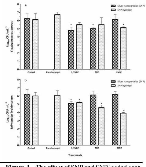
Figure 4 show the susceptibility of S. aureus and
S. typhimurium to different concentrations of SNP and SNP-hydrogels during 15 min contact time. Statistically significant difference between control and treated groups were shown. In contrast to solid medium, increasing concentrations reduced the antimicrobial effects of SNP on both bacteria in a liquid medium. At 1/2MIC concentration, SNP exhibited up to 1.5 and 1.1 log reductions in 15 min on S. aureus and S. typhimurium population, respectively, while the 2 MIC concentration did not change the viable population of both bacteria after 15 min.
J Med Bacteriol. Vol. 6, No. 3, 4 (2017): pp.51-58 jmb.tums.ac.ir
55
Discussion
SNP are proposed as a well-known and very interesting antimicrobial compounds with high effectiveness on a broad range of microbes at low doses used. The results of antimicrobial activity of SNP (Table 1) are in agreement with Zarei et al. (2014) (17) who reported the MIC values of similar
SNP against Listeria monocytogenes, S.
typhimurium, Escherichia coil O157:H7 and
Vibrio parahaemolyticus in the range of 3.12- 6.25 µg/ mL according to microdilution method in Tryptic soy broth. Moreover, SNPs manifest distinguished antibacterial mechanisms on microorganisms by adsorption of SNP onto the cell and formation of ‘pits’ which resulting in the destruction of bacterial cell integrity. The production of free radicals may also physically damage the cell membrane and cause the killing of a bacterial population (10, 18). It has been revealed Figure 2. Zone of inhibition of different
concentration of SNP loaded agar hydrogel according to spot test. MIC: minimum inhibitory concentrations.
Figure 3. The effect of SNP and SNP loaded agar hydrogel on reducing one-day old biofilm of
Staphylococcusaureus (a) and S. typhimurium (b)
developed on polystyrene surface with 15 min contact time.* indicates a significant difference (P < 0.05) among SNP and SNP loaded agar hydrogel treatment at each concentration. MIC: minimum inhibitory concentrations.
Figure 4. The effect of SNP and SNP loaded agar hydrogel on the survival of S. aureus (a) and S.
typhimurium (b) during 15 min exposure time .*
J Med Bacteriol. Vol. 6, No. 3, 4 (2017): pp.51-58 jmb.tums.ac.ir
56
in several reports that antibacterial efficacy of SNP varied between different prepared SNPs. Given the fact that factors such as the type of bacteria, the media of inoculation, type and the size of nanoparticle and the method of preparation must be considered when compare the antibacterial activity of different nanoparticles (19-22).
An increase in activity of SNP hydrogels may be due to the migration of SNP from the agar matrix and interact with lipid layer of membrane (23). The zone of inhibition in S. aureus was similar to S. Typhimurium (Table 1). This is in contrast with the results of Zakaria et al. (2016) (8) which reported the susceptibility of Gram-negative strains into gel formulations containing cefotaxime than Gram-positive. In addition, the efficacy of SNP hydrogels against S. aureus and S. typhimurium was significantly higher than the well-known standard (silver sulfadiazine cream) (P < 0.05), particularly at a high concentration. The similarity of inhibition zones of SNP with SNP loaded hydrogel unveiled the fact that incorporation of antimicrobial compounds into hydrogels does not decrease its effectiveness against both Gram-positive and Gram-negative strains, this entirely associated with spreadability characteristic of hydrogel (8). Additionally, the neat hydrogel showed no antibacterial activity against tested bacteria. According to Alshehri (2016) (24) the AgNPs– hydrogel prepared from carboxymethyl cellulose, polyvinyl alcohol, followed by the incorporation of AgNPs by microwave radiation revealed higher antibacterial activity against E. coli, Klebsiella pneumoniae, Pseudomonas aeruginosa, Proteus vulgaris, S. aureus and Proteus mirabilis
compared to the neat hydrogel (24). The nature of the base gel, antimicrobial agents, and the environment display a substantial role in the release of nanoparticles from the gel into medium (25).
Observed variation in biofilm removal activity of SNP or SNP loaded hydrogels on tested strains (Figure 3) may be related to the surface characteristics of Gram positive and Gram negative bacteria. There could be several reasons for decreasing antimicrobial effectiveness by
increasing SNP concentration. SNP aggregation which occurs at a high concentration of SNP reduces the contact surface of nanoparticles with bacteria and thus diminish its biological activity. SNP reveal agglomeration after adding into the media. When SNP is suspended in nutritious media such as LB, rather than the pure solution, the aggregation was very noticeable. A work performed by Zhou et al. (2015) (26) support our results and verified that SNP aggregation depends on the media and antimicrobial concentration. Perez-Diaz et al. (2016) (6) studied the biofilm removal activity of SNP loaded chitosan gel on oxacillin-resistant P. aeruginosa and S. aureus. In this research, gels had a greater effect on MRSA, by 6 log reduction at the concentration of 100 ppm of SNP and 3.3 log reduction of P. aeruginosa at a concentration of 1000 ppm.
Due to antibacterial property, SNP is widely used in medicine as a disinfectant and antibacterial agents. It is believed that this novel material can be used as a new technology for the production of new sanitizing agents. Zakaria et al. (2016) (8) studied the cefotaxime gel formulations on bacterial pathogens survival. In this research, the antibacterial activity of cefotaxime loaded gels were worthwhile, 4 to 5-log reduction against P. aeruginosa and E. coli after 24 h incubation. Observed variation in cell reduction (Figure 4) may be related mainly to exposure time and also to the type and concentration of gel base, type, and concentration of loaded antimicrobial agents and the type of bacteria strains.
J Med Bacteriol. Vol. 6, No. 3, 4 (2017): pp.51-58 jmb.tums.ac.ir
57
Bacteria Silver nanoparticle SNP-hydrogels Control
½ MIC MIC 2MIC ½ MIC MIC 2MIC Silver sulfadiazine cream Chloramphenicol 30 µg Ampicillin 10 µg
S.aureus 1.00±0.09 1.20±0.03 1.50±0.1 0.86±0.05 1 ±0.00 1.13±0.05 0.8±0.00 28.40±0.5 23.75±0.7
S. Typhimurium 1.20±0.07 1.20±0.1 1.30±0.1 1 ±0.1 1.10±0.1* 1.13±0.05 0.8±0.00 10.25±0.24* 18.74±0.27*
* indicates a significant difference (P < 0.05) among different treatments in a column.
Conclusion
To our knowledge, this is the first report of the antimicrobial and biofilm removal activity of agar hydrogel incorporated with SNP. The results of the current study revealed that antimicrobial property of SNP loaded hydrogel is mainly affected by the nature of the medium (broth or agar), the concentration of nanoparticles and somewhat by the type of bacteria. In general terms, the SNP hydrogel activity has proved more effective than SNP in alone in the liquid and solid medium. On the other hand, the decrease in antimicrobial effects of SNP with increasing concentration, by loading SNP into hydrogel nanoparticles. SNP hydrogel showed also displayed biofilm removal activity against both tested bacteria in an SNP concentration-dependent manner.
Acknowledgements
This research was financially supported by the
Faculty of Veterinary Medicine, Urmia
University.
Conflict of interest
Authors declare no conflict of interest in this study.
References
1. Bhattarai N, Gunn J, Zhang M.
Chitosan-based hydrogels for controlled, localized drug delivery. Adv Drug Deliv Rev 2010; 62(1): 83-99.
2. Caló E, Khutoryanskiy VV. Biomedical applications of hydrogels: A review of patents and commercial products. Eur Polym J 2015; 65: 252-67.
3. Ahmed EM. Hydrogel: Preparation,
characterization, and applications: A review.
J Adv Res 2015; 6(2): 105-21.
4. Hoffman AS. Hydrogels for biomedical applications. Adv Drug Deliv. Rev 2002; 54(1): 3-12.
5. Shewan HM, Stokes JR. Review of
techniques to manufacture micro-hydrogel particles for the food industry and their applications. J Food Eng 2013; 119(4): 781-92.
6. Pérez-Díaz M, Alvarado-Gomez E, Magaña-Aquino M, et al. Anti-biofilm activity of chitosan gels formulated with silver nanoparticles and their cytotoxic effect on human fibroblasts. Mater Sci Eng C 2016; 60: 317-23.
7. Villanueva ME, Diez AM, González JA, et al. Antimicrobial activity of starch hydrogel incorporated with copper nanoparticles. ACS Appl Mater Interfaces 2016; 8(25): 16280-8. 8. Zakaria AS, Afifi SA, Elkhodairy KA.
Newly developed topical cefotaxime sodium hydrogels: Antibacterial activity and in vivo Table 1. Diameters of inhibition zones (mm) of silver nanoparticle (SNP) and hydrogels against S.
J Med Bacteriol. Vol. 6, No. 3, 4 (2017): pp.51-58 jmb.tums.ac.ir
58
evaluation. BioMed Res Int 2016; 2016: 15. 9. Emtiazi G, Shahrokh Esfahani S. Flow
Cytometry detection of bacterial cell entrapment within the chitosan hydrogel and antibacterial property of extracted chitosan.
J Med Bacterial 2016; 5 (3, 4): pp.9-14. 10.Prabhu S, Poulose EK. Silver nanoparticles:
Mechanism of antimicrobial action,
synthesis, medical applications, and toxicity effects. Int Nano Lett 2012; 2(1): 32. 11.Allahverdiyev AM, Kon KV, Abamor ES, et
al. Coping with antibiotic resistance: Combining nanoparticles with antibiotics and other antimicrobial agents. Expert Rev Anti Infect 2011; 9(11): 1035-52.
12.Stanga M. Sanitation; cleaning and disinfection in the food industry: Wiley-VCH; 2010.
13.Incoronato AL, Conte A, Buonocore GG, et al. Agar hydrogel with silver nanoparticles to prolong the shelf life of fior di latte cheese. J Dairy Sci 2011; 94(4): 1697-704. 14.Tülin A, Serap D, Banu M. Comparative
evaluation of antibacterial activity of caffeic acid phenethyl ester and plga nanoparticle
formulation by different methods.
Nanotechnology 2016; 27(2): 025103. 15.Mahdavi M, Jalali M, Kasra Kermanshahi R.
The effect of nisin on biofilm forming foodborne bacteria using microtiter plate method. Res Pharm Sci 2007; 2(2): 113-8.
16.Phongphakdee K, Nitisinprasert S.
Combination inhibition activity of nisin and ethanol on the growth inhibition of pathogenic gram negative bacteria and their application as disinfectant solution. J Food Sci 2015; 80(10): M2241-M6.
17.Zarei M, Jamnejad A, Khajehali E. Antibacterial effect of silver nanoparticles
against four foodborne pathogens.
Jundishapur J Microbiol 2014; 7(1): e8720. 18.Chernousova S, Epple M. Silver as
antibacterial agent: Ion, nanoparticle, and metal. Angew Chem Int Ed 2013; 52(6): 1636-53.
19.Gurunathan S, Han J, Kwon DN, et al. Enhanced antibacterial and anti-biofilm activities of silver nanoparticles against gram-negative and gram-positive bacteria.
Nanoscale Res Lett 2014; 9(1): 373.
20.Hajipour MJ, Fromm KM, Ashkarran AA,
et al. Antibacterial properties of nanoparticles. Trends Biotechnol 2012; 30(10): 499-511.
21.Le Ouay B, Stellacci F. Antibacterial activity of silver nanoparticles: A surface science insight. Nano Today 2015; 10(3): 339-54. 22.Shameli K, Ahmad M, Jazayeri S, et al.
Investigation of antibacterial properties silver nanoparticles prepared via green method. Chem Cent J 2012; 6(1): 73. 23.Swaroop K, Francis S, Somashekarappa
HM. Gamma irradiation synthesis of ag/pva hydrogels and its antibacterial activity.
Mater Today 2016; 3(6): 1792-8.
24.Alshehri SM, Aldalbahi A, Al-hajji AB, et
al. Development of carboxymethyl
cellulose-based hydrogel and nanosilver composite as antimicrobial agents for uti pathogens. Carbohydr Polym 2016; 138: 229-36.
25.Deen G, Chua V. Synthesis and properties of new “stimuli” responsive nanocomposite hydrogels containing silver nanoparticles.
Gels 2015; 1(1): 117.
26.Zhou Y, Kong Y, Kundu S, et al. Antibacterial activities of gold and silver nanoparticles against escherichia coli and
bacillus calmette-guérin. J