AN COMPLETE OVERVIEW OF ALVEOLAR CLEFT BONE DEFECTS AND ITS MANAGEMENT
1Consultant Oral & Maxillofacial Surgeon
2Consultant oral & maxillofacial Radiologist
A R T I C L E I N F O
INTRODUCTION
A better understanding of anatomy, embryology, and the growth of facial structures has dramatically revolutionized the management of alveolar clefts over the past century. Treatment goals have been refined and therapeutic options established only to unleash an array of controversies surrounding the timing and type of management. This article provides an overview of alveolar clefts, providing a background on embryology and etiology, along with past, present and future treatment modalities.
Embryology
Craniofacial development in an embryo commences during the middle of the third week. The overall palate is formed during weeks 4 to 12 of gestation from the frontonasal and maxillary prominences. With the incisive foramen providing the anatomical landmark, the
prominences divides the palate into two parts: the primary and secondary palates1.
The primary palate: the lip and hard palate anterior to the incisive foramen is formed during weeks 4 to 7 of gestation. Variations from normal developme
period may result in clefting of the primary palate, manifesting as incomplete or complete and unilateral or bilateral cleft alveolus.
The secondary palate: posterior to the incisive foramen and the soft palate. This is formed during weeks
gestation as a continuation of anterior palate.
International Journal of Current Advanced Research
ISSN: O: 2319-6475, ISSN: P: 2319-6505,
Available Online at www.journalijcar.org
Volume 7; Issue 2(C); February 2018
DOI: http://dx.doi.org/10.24327/ijcar.2018
Article History:
Received 14th November, 2017 Received in revised form 5th December, 2017
Accepted 3rd January, 2018
Published online 28th February, 2018
Key words:
Sabg - secondary alveolar bone grafting Clcp - cleft lip and cleft palate Bmp- bone morphogenic protein
GTR - Guided Tissue Regeneration
Nasoalveolar Molding (NAM) Gingivoperiosteoplasty (GPP)
Copyright©2018 Payak A.P and Bhadouria .P. This is an open access article distributed under the Creative Commons Attribution License, which permits unrestricted use, distribution, and reproduction in an
*Corresponding author: Payak A.P
Consultant Oral & Maxillofacial Surgeon Dental Experts (2/13 Chatrasal Nagar Phase-2) J.K Road Bhopal 462023
AN COMPLETE OVERVIEW OF ALVEOLAR CLEFT BONE DEFECTS AND ITS MANAGEMENT
Payak A.P
1and Bhadouria .P
2Maxillofacial Surgeon Dental Experts (2/13 Chatrasal Nagar Phase J.K Road Bhopal 462023
Consultant oral & maxillofacial Radiologist
A B S T R A C T
A better understanding of anatomy, embryology, and the growth revolutionized the management of alveolar clefts over the past refined and therapeutic options established only to unleash an timing and type of management. This article provides an overview background on embryology and etiology, along with past, present
A better understanding of anatomy, embryology, and the growth of facial structures has dramatically revolutionized the management of alveolar clefts over the past century. Treatment goals have been refined and therapeutic options established only h an array of controversies surrounding the timing and type of management. This article provides an overview of alveolar clefts, providing a background on embryology and etiology, along with past, present and future treatment modalities.
iofacial development in an embryo commences during the middle of the third week. The overall palate is formed during weeks 4 to 12 of gestation from the frontonasal and maxillary prominences. With the incisive foramen providing the anatomical landmark, the junction of these prominences divides the palate into two parts: the primary
The primary palate: the lip and hard palate anterior to the incisive foramen is formed during weeks 4 to 7 of gestation. Variations from normal development during this period may result in clefting of the primary palate, manifesting as incomplete or complete and unilateral or
The secondary palate: posterior to the incisive foramen and the soft palate. This is formed during weeks 7 to 12 of gestation as a continuation of anterior palate.
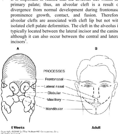
It is important to realize that the alveolus
primary palate; thus, an alveolar cleft is a result of divergence from normal development during frontonasal prominence growth, contact, and fusion. Therefore, alveolar clefts are associated with cleft lip but not with isolated cleft palate deformities. The cleft in the alveolus is typically located between the lateral incisor and the canine, although it can also occur between the central and lateral incisors1.
Fig 1 Diagram of a six-week-old embryo. The frontonasal process will give
rise to the central lip and premaxilla, the lateral nasal process will develop into the alae of the nose, and the maxillary processes will produce the lateral lip
and maxillary segments.
International Journal of Current Advanced Research
6505, Impact Factor: SJIF: 5.995
www.journalijcar.org
2018; Page No. 9758-9764
//dx.doi.org/10.24327/ijcar.2018.9764.1627
Molding (NAM) Gingivoperiosteoplasty (GPP)
This is an open access article distributed under the Creative Commons Attribution License, which permits unrestricted use, distribution, and reproduction in any medium, provided the original work is properly cited.
Consultant Oral & Maxillofacial Surgeon Dental Experts 2) J.K Road Bhopal 462023
AN COMPLETE OVERVIEW OF ALVEOLAR CLEFT BONE DEFECTS AND ITS MANAGEMENT
Dental Experts (2/13 Chatrasal Nagar Phase-2)
growth of facial structures has dramatically past century. Treatment goals have been an array of controversies surrounding the overview of alveolar clefts, providing a present and future treatment modalities.
It is important to realize that the alveolus lies within the primary palate; thus, an alveolar cleft is a result of divergence from normal development during frontonasal prominence growth, contact, and fusion. Therefore, alveolar clefts are associated with cleft lip but not with deformities. The cleft in the alveolus is typically located between the lateral incisor and the canine, although it can also occur between the central and lateral
old embryo. The frontonasal process will give e to the central lip and premaxilla, the lateral nasal process will develop into the alae of the nose, and the maxillary processes will produce the lateral lip
and maxillary segments.
Research Article
Incidence
Among the many congenital birth defects, there have been more than 250 different types of facial clefting disorders described in the literatures. The most frequently reported defect is clefting of the palatine bone and the alveolar process of the maxilla. In 75% of the cleft lip and palate occurrences, the cleft runs through the alveolar ridge2.
Etiology
It is currently believed that less than 40% of the clefts of the lip and palate are of genetic origin.
Mutations in specific collagen genes in Stickler syndrome, homeodomain-containing protein PAX3 in Waarden burg’s syndrome, and sonic hedgehog in midline craniofacial defects are examples of direct genetic correlation with cleft lip and palate.
Environmental factors play a clear role in gene expression, which affects the phenotype. For example, Hwang et al report direct correlation between maternal smoking and clefting. Antiepileptic drugs also have been linked to clefting. Maternal alcohol consumption has been under extensive debate as an etiologic factor. Infections (rubella and toxoplasmosis) and growth factor deficiency are among other environmental factors2.
Treatment goals of Alveolar Bone Grafting
Close vestibular and palatal oronasal fistula.
Restore physiologic continuity of the dental arch to enable oral and dental health to be maintained.
Provide bone for stability and continuity of the dental arch and pre maxilla.
Allow eruption of the permanent teeth or placement of dental implants through bone graft.
Provide support for the lateral ala of the nose.
Allow for the orthodontic alignment of the teeth.
Facilitate nasolabial muscle and soft tissue reconstruction.
Establish functional nasal airway.
Provide support for the lip3.
Timing of alveolar cleft grafting
To comprehens better the various ideologies concerning the timing of the alveolar cleft grafting, one first should have a firm grasp of its classification:
Primary (0–2.5 years, usually at the time of lip repair)
Early secondary (2–5 years, before the eruption of permanent incisors)
Secondary (6–13 years, before the eruption of the permanent canines)
Late (> 13 years, after the eruption of the permanent canines)
The timing of graft placement is based more on dental development than on chronological age and may be classified as follows:-
Primary osteoplasty (performed at infancy or in children younger than 2 years of age, along with the lip repair, before accomplishment of palatal closure).
Secondary osteoplasty (performed after palatal closure/ repair), which can further be divided into:
Early secondary, performed between 2 and 8 years of age, before eruption of permanent incisors.
Mid-secondary, performed at chronological age of 9– 12, before which most of the mid-facial growth is complete, during the mixed dentition stage prior to eruption of the cleft canine approximately when its root is half formed.
Late secondary, performed between 12 and 16 years of age, after eruption of the canine.
Very late-secondary, performed later than 16 years of age.
Of these, ‘mid-secondary repair’ performed between the age of 9 and 12 has been shown by several studies to be ideal4.
Grafting material and donor sites
Both cortical and cancellous bone has been used to provide bone grafting material for the closure of alveolar clefts. Cortical bone is incorporated by creeping substitution and relies on vascular in growth. Typically, this process requires longer periods than the revascularization process in cancellous bone grafts.When performing a cancellous bone graft, cells are transferred and revascularization occurs much faster as a result of osteoinduction and osteoconduction.
Various Technique of Bone Grafting
Surgical technique of Anterior iliac crest bone graft
The AIS is approached with an incision that is placed when the skin overlying the AIS is retracted medially. A 4- to 6-cm incision is placed 1 to 2 cm posterior to the tubercle of the ilium and 1 cm inferior to the anterior superior iliac spine, obliquely along the orientation of the anterior iliac crest.
This placement avoids the course of the iliohypogasrtic and subcostal nerves superiorly and the lateral femoral cutaneous nerve inferiomedially. The layers of dissection encountered are skin, subcutaneous tissue, and Scarpa’s fascia.
Fig 2 The anterior iliac crest harvest site with incision placement and the
typical medial and lateral muscular attachments
corticocancellous block graft can be harvested. A total bone length of 4 to 6 cm can be obtained and is limited by the proximity to anterior superior iliac spine and tubercle of the ilium.
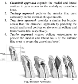
Various approaches are described for harvesting the anterior iliac crest.
1. Clamshell approach expands the medial and lateral cortices to gain access to the underlying cancellous bone.
2. Tschopp approach pedicles the anterior iliac crest osteotomy on the external oblique muscle.
3. Trap door approach provides a similar but broader access than the clamshell approach by pedicling the medial and lateral cortices on the external oblique and tensor fascia lata, respectively.
4. Tessier approach creates oblique osteotomies to pedicle the medial and lateral walls of the anterior iliac crest to access the cancellous bone5.
Fig 3 Different Approaches
Hemostatic agents, such as bone wax, microfibrillar collagen, and gelatin sponge, may be used to limit oozing from the cancellous bone. A drain may be used to limit postoperative fluid collection. Closure should be achieved to reapproximate the periosteal envelope over the iliac crest followed by subcutaneous and skin closure.The patient is allowed to ambulate on postoperative day one but may require assistance with a walking device in the immediate postoperative period5.
Costochondral Rib Bone Grafting
The use of rib for closing the alveolar cleft has shown good results and is the graft of choice under Rosenstein’s primary bone grafting protocol. Rib grafting has certain donor site morbidities, however, including visible scarring and pain. Others have also reported difficulty in orthodontic tooth movement with rib grafts.
Surgical Technique
The incision is taken approx. 2.5 cm below the nipples
The most commonly harvested rib as graft is 8th or 9th rib.
The periosteum is reflected superiorly and inferiorly, and the inner aspect is reflected with the aid of a Doyen (pigtail) retractor.
Once the appropriate length is dissected, the medial cut is done followed by the posterior cut with the aid of a rib cutter.
If cartilage cap is needed - dissection is extended to the costochondral junction and leaving overlying periosteum/perichondrium in the area intact.
During harvesting, continuous palpation of the underlying rib is essential to avoid pleural injury. Pneumothorax was excluded by the absence of air
leakage after filling the wound with saline solution while the anesthetist was applying positive pressure into the lungs5.
The wound was then infiltrated with 5–10 ml of 2% xylocaine and closed in layers.
Use of recombinant human Bone Morphogenetic Protein (rhBMP-2) in reconstruction of maxillary alveolar cleft
By the late 1980s, the active factor responsible for the induction of bone was identified: Bone Morphogenetic Protein (BMP). BMP replicates the embryonic induction of bone formation. It can induce pluripotent mesenchymal stem cells to differentiate into bone-forming osteoblasts.
BMP has been demonstrated to have osteoinductive properties. With recent approval of FDA, use of rhBMP2 has been indicated in for use in certain oral and maxillofacial bone grafting procedures, including sinus augmentation and localized alveolar ridge augmentation, maxillary alveolar clefts.
There is an obvious advantage of rhBMP2 use over iliac crest bone in terms of surgical, post surgical recovery parameters and the efficiency of radiographic obliteration of the defect. It avoids the unnecessary surgery, loss of blood and postoperative morbidity of iliac crest harvesting. Furthermore the presence of scar is objectionable in case of females.
The suggested human therapeutic doses (0.88 mg/mL of sterile water for rhBMP-7 and 1.50 mg/mL of sterile water for rhBMP-2) were derived from nonhuman primate studies and verified in clinical orthopedic studies6.
Fig 4 Mechanism of action of BMPs in bone repair.
Guided Tissue Regeneration, Barrier Membranes and Reconstruction of the Cleft Maxillary Alveolus
In the mid 1980s, the Guided Tissue Regeneration (GTR) principle was introduced, according to which, regeneration of a certain type of tissue is achieved when cells with the capacity to regenerate the particular type of lost tissue are allowed to populate the defect during healing (Nyman et al. 1982; Gottlow et al. 1984).
The use of barrier membranes for bone regeneration is especially beneficial in the case of severely affected soft tissue. One useful indication may be the formation of an effective shell for bone grafts in maxilla cleft defect reconstruction.
The application is contraindicated in the case of a wound infection or with the periodontal repair in smokers.
Depending on their reaction to a biological environment, the membrane barriers are either resorbable or non-resorbable. The resorbable barriers are used when their subsequent removal is not advisable, as in the case of cleft alveolar defect reconstruction. These membranes disintegrate either by hydrolysis or by enzymatic action depending on their composition. Their compounds are either of a natural (collagen) or synthetic (polymers, lactic acid, polyglycolic acid) origin.
Commonly used collagen membranes are: Biomend,OSSIX,Neomem.7
Procedure of Radiographic Assessment Of Bone10
Measurement of Cleft Width
Cleft width at the narrowest point, determined by inspection, was measured on a pre-surgical maxillary occlusal radiograph which was confirmed using study models.
To determine radiographic elongation or shortening, the mesio-distal width of the tooth adjacent to the cleft was measured and compared with the mesio-distal width of the same tooth measured on the orthodontic study model. The difference was used as the radiographic correction factor8.
Fig 5 Presurgical radiograph scan showing the cleft and tracing of the defect
Assessment of Canine Position
Six points on the pre-surgical IOPAR were used to measure the amount of permanent cuspid crown emerged into the cleft beyond the adjacent alveolar bone and the total crown length of this tooth.
To minimize the effect of radiographic foreshortening and elongation, the eruption measure was expressed as a ratio of the amount of crown emerged, divided by the total crown length.
Fig. 6 a Landmark points marked on the pre-surgical radiographs to identify the anatomic crown of the cuspid (points 1, 2 and 3) and the amount of cuspid crown emerged through the alveolus (points 4, 5 and 6) b A length of proximal segment anatomic root, B most coronal bone attachment along distal surface of proximal root, C location of alveolar cleft on distal surface of proximal root, D notching of graft, E length of distal root, F (similar to B) on distal root, G (similar to C) on distal root8.
Presurgical orthodontic care:-
Two major orthodontic considerations intimately integrate with the timing of the alveolar cleft grafting:
The correction of cross-bites and
The alignment of the anterior teeth.
If posterior cross-bites exist secondary to narrowed transverse dimension of the maxilla, maxillary expansion may be performed before grafting. If the graft is performed before the expansion of the maxilla, a 3-month period must elapse before this expansion.
The Quadhelix and Hyrax appliances that are usually used for maxillary expansion should be left in place for at least 3 months postoperatively to help prevent relapse9.
Surgical Procedure For Reconstruction Of The Alveolar-Anterior Maxillary Bone Defect In Unilateral And Bilateral Cleft Cases10.
Three basic surgical principles must be satisfied for the successful treatment of the alveolar cleft grafting:closure of oronasal fistula, adequate volume of graft material, and water tight and tension-free closure10.
Irrigation of the oral cavity with chlorhexidine solution is advised to decrease the chance of immediate postoperative infection.
Injection with local anesthesia with epinephrine not only aids with postoperative pain control and intraoperative hemostasis but also allows the surgeon to identify the margins of the bony cleft and the oronasal communication.
If the graft material allows for a two-team approach, it is more convenient to choose the donor site opposite the side of the cleft to decrease the amount of interference between the two teams and possible cross-contamination of the donor site field.
Surgical technique: unilateral cleft
About 10 minutes before initiating surgery, infiltration of 2% lidocaine with 1/100,000 epinephrine is performed palatally and throughout the vestibular sulcus.
Attention is first given to developing soft tissue flaps to reconstruct the nasal floor, palate, and labial mucosa and attached gingiva.
The initial incisions are planned to preserve adequate attached gingiva around the erupted teeth and permit the advancement of attached gingiva into the area of future tooth eruption (see Fig. 7).
In the region of the molar teeth, this incision is carried superiorly posteriorly into the free mucosa and sulcus approximately 1.0 to 1.5 cm to permit relaxation for advancement of this flap. This is completed bilaterally.
Fig 7
Incisions are next made along the outer margins of the bony cleft and carried superiorly into the depth of the vestibule.
At their superior extent in the depth of the vestibule, each of the incisions becomes horizontal and extends laterally approximately 1.0 to 2.0 cm from either side of the cleft (see Fig. 7).
Beginning inferior-medially, the mucoperiosteal tissues are undermined subperiosteally off the outer aspect of the alveolus and anterior maxilla along the entire length of both cleft margins.
Medially, this dissection is carried until the anterior nasal spine and the floor of the contralateral (noncleft side) nasal floor are identified.
Laterally, the dissection is carried superiorly on the maxilla to the level of the infraorbital neurovascular bundle (see Fig. 7).
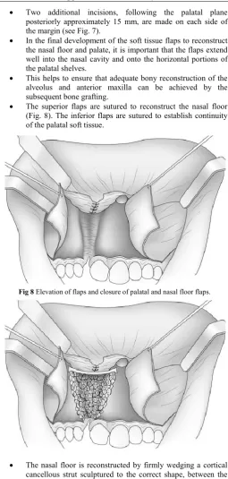
Two additional incisions, following the palatal plane posteriorly approximately 15 mm, are made on each side of the margin (see Fig. 7).
In the final development of the soft tissue flaps to reconstruct the nasal floor and palate, it is important that the flaps extend well into the nasal cavity and onto the horizontal portions of the palatal shelves.
This helps to ensure that adequate bony reconstruction of the alveolus and anterior maxilla can be achieved by the subsequent bone grafting.
The superior flaps are sutured to reconstruct the nasal floor (Fig. 8). The inferior flaps are sutured to establish continuity of the palatal soft tissue.
Fig 8 Elevation of flaps and closure of palatal and nasal floor flaps.
The nasal floor is reconstructed by firmly wedging a cortical cancellous strut sculptured to the correct shape, between the alveolar margins of the cleft, slightly superior to the contralateral nasal floor.
Horizontal mattress, 4-0, slow-absorbing sutures are preferred to close the vertical limb of these incisions, and a running, continuous, slow-absorbing suture is used to close the horizontal limbs of these incisions (Fig. 9).
Surgical technique: bilateral cleft
The operation for a bilateral cleft deformity is similar to that for a unilateral deformity. Because it is performed bilaterally and simultaneously, however, the differences described in the paragraphs that follow must be noted.
First, there can be only minimal elevation of the mucoperiosteum on the labial surface of the premaxilla because this soft tissue is the primary vascular supply of this segment. Hence, the lateral flaps need to be adequate (well relieved) for advancement and closure of the entire cleft area.
Second, significant lip and nose changes accompany bilateral alveolar-anterior maxillary reconstruction. It is recommended that extensive simultaneous lip-nose revision not be done simultaneously for the patient with a bilateral deformity; a delayed lip-nose revision is suggested.
Fig 9 Labial incisions for bilateral cleft alveolus-anterior maxilla bone
reconstruction.
Palatal incisions for bilateral cleft alveolus-anterior maxilla bone reconstruction.
Bone reconstruction with cortical strut superiorly and cancellous bone
Nasoalveolar molding (NAM) and gingivoperiosteoplasty (GPP) versus traditional secondary bone grafting10
Gingivoperiosteoplasty is the primary repair of the gingivoperiosteum at the site of an alveolar cleft, with the intention of forming an osseous union. Narrowing of the alveolar cleft and approximating the lateral segments through presurgical infant orthopedics followed by GPP at the time of lip repair may eliminate the need for this bone graft.
Its use is one of the most widely debated areas in the treatment of patients with cleft lip and palate; its advocates are few, but are well-respected.However, the degree of ossification after gingivoperiosteoplasty varies between 50% and 100%, and a third step ofbone grafting may be required. Some authors also argue that all alveolar clefts will remain deficient in osseous contourand bulk, which compromises the eruption and maintenanceof permanent dentition, so necessitating an eventual bonegraft.
One criticism of GPP is the potential for impairment of maxillary growth. However, despite the wide mucoperiosteal dissection performed by Skoog (1967), there was no impairment of maxillary growth in the anterior-posterior dimension (Ross,1987). Technique of GPP necessitates dissection only in the margins of the alveolar cleft because presurgical NAM is highly effective in aligning the cleft alveolar segments. Limited mucoperiosteal dissection will not impede maxillary growth.
A second advantage of NAM is its positive effect on the cleft nasal deformity. NAM acts to elevate the depressed nasal dome, stretches the nasal lining, and elongates the columella.
Presurgical infant orthopedics is a standard feature of our protocol for the management of both unilateral and bilateral clefts of the alveolus and palate, and it allows the close approximation and proper alignment of the alveolar segments prior to surgical repair.
Hence, Bone grafting in the mixed dentition was eliminated by the use of a combined approach of presurgical infant orthopedics and GPP10.
CONCLUSION
Although many controversies exist regarding the timing, choice of graft material, or even flap design, this procedure has provided a predictable means of resolving a unique and debilitating problem for the cleft patient.
Considering that these patients are exposed to numerous surgical procedures, it is the surgeons’ obligation to plan operations carefully and provide individualized treatment that allows for the best results for individual patients.
References
1. Richard P Juniper, William P Smith; Cleft Lip & Palate. Developmental Anomalies Of The Face, Palate, Jaws & Teeth; Bailey & Love Short Practice Of Surgery; Chapter 37.
2. Kazemi et al. Secondary grafting in the alveolar cleft patient; Oral Maxillofacial SurgClin N Am 14 (2002) 477-490.
3. Bruce N. Epker. Alveolar-anterior Maxillary Cleft Repair; Atlas Oral Maxillofacial SurgClin N Am 17 (2009) 167-173.
4. Y.L. Jia et al. Long-term outcome of secondary alveolar bone grafting in patients with various types of cleft;
British Journal of Oral and Maxillofacial Surgery 44 (2006) 308-312.
5. Kenneth J. Zouhary; Bone Graft Harvesting From Distant Sites: Concepts and Techniques; Oral Maxillofacial Surg Clin N Am 22 (2010) 301-316 6. Sarah D. Davies. Bone Morphogenetic Proteins in
Craniomaxillofacial Surgery; Oral Maxillofacial Surg Clin N Am 22 (2010) 17-31.
7. Retzepi M, Donos N. Guided Bone Regeneration: biological principle and therapeutic applications. Clin. Oral Impl. Res. 21, 2010; 567-576.
8. Varsha H. Upadyaetal. Radiographic Assessment of Influence of Cleft Width and Canine Position on Alveolar Bone Graft Success. J. Maxillofac. Oral Surg.
(Jan-Mar 2013) 12(1):68-72.
9. Santiago Et Al., Reduced Need For Alveolar Bone Grafting By Presurgical Orthopedics And Primary Gingivoperiosteoplasty Cleft Palate-Craniofacial Journal, January 1998, Vol. 35 No. 1.
10. Tracy M. Pfeifer .Nasoalveolar Molding And Gingivoperiosteoplasty Versus Alveolar Bone Graft:
Cleft Palate-Craniofacial Journal, January 2002, Vol. 39 No. 1.
11. Y.L. Jia et al. Long-term outcome of secondary alveolar bone grafting in patients with various types of cleft;British Journal of Oral and Maxillofacial Surgery
44 (2006) 308-312
12. Bajaj Et Al; Management Of Alveolar Clefts; The
Journal Of Craniofacial Surgery .Volume 14, Number 6 November 2003.
How to cite this article:
Payak A.P and Bhadouria.P (2018) 'An Complete Overview of Alveolar Cleft Bone Defects And Its Management ',
International Journal of Current Advanced Research, 07(2), pp. 9758-9764. DOI: http://dx.doi.org/10.24327/ijcar.2018.9764.1627