245
RESEARCH ARTICLE
CODEN (USA): IJPSPP
ISSN: 0975-248X
Development and Evaluation of Lapatinib Solid Lipid Nanoparticles
in the Management of Breast Cancer
Pamu Sandhya
*Department of Pharmaceutics, Shadan Women’s College of Pharmacy, Khairatabad, Hyderabad-500004, Telangana, India
Copyright © 2019 Pamu Sandhya. This is an open access article distributed under the terms of the Creative Commons Attribution-NonCommercial-ShareAlike 4.0 International License which allows others to remix, tweak, and build upon the work non-commercially, as long as the author is credited and the new creations are licensed under the identical terms.
ABSTRACT
The current research was aimed at formulation of Lapatinib loaded solid lipid nanoparticles (SLNs) followed by evaluation for effective treatment of breast cancer. The formulations prepared by homogenization and ultrasonication and evaluated for zeta potential, particle size, polydispersity index, entrapment efficiency and in-vitro dissolution studies. Entrapment efficiency studies indicated proportional relation between concentration of lipid and the amount of drug entrapped. The physicochemical parameter evaluation data indicated 94.27% entrapment efficiency, 130 nm particle size and -19.9 zeta potential for stable formulation. The in vitro drug dissolution studies indicated that Lapatinib loaded SLNs (F6) formulated with Dynasan 116 and Egg Lecithin was suitable for anti-cancer therapy with higher drug dissolution rate.
Keywords: Lapatinib, SLNs, Solubility, Breast cancer, Dynasan, Egg lecithin.
DOI: 10.25004/IJPSDR.2019.110515 Int. J. Pharm. Sci. Drug Res. 2019; 11(5): 245-249
*Corresponding author: Dr. Pamu Sandhya
Address: Department of Pharmaceutics, Shadan Women’s College of Pharmacy, Khairatabad, Hyderabad-500 004, Telangana, India Tel.: +91-9849395823
E-mail: [email protected]
Relevant conflicts of interest/financial disclosures: The authors declare that the research was conducted in the absence of any commercial or financial relationships that could be construed as a potential conflict of interest.
Received: 04 August, 2019; Revised: 05 September, 2019; Accepted: 10 September, 2019; Published: 25 September, 2019
INTRODUCTION
Despite of administration by the oral route remains most popular method of drug delivery, significant problems arise due to varying physical, chemical or biological characteristics of drug. Improper solubility, instability in gastrointestinal tract, poor membrane permeability or first pass effect is some of the major hurdles that lead to non-acceptance of drug entities with potentiality. [1] The drug delivery systems based on lipids are means of by-passing few physical and chemical hurdles related to drugs that exhibit poor absorption profile from GIT. These potential systems
Int. J. Pharm. Sci. Drug Res. September-October, 2019, Vol 11, Issue 5 (245-249) formation of chylomicrons, which guides the carrier
and drug entrapped within to follow transcellular pathway of absorption. [4] SLN enhances the bioavailability of lipophilic drugs by increasing the oral absorption of these drugs. [5]
Lapatinib acts as tyrosine kinase inhibitor that checks the growth of EGFR/ErbB1 and HER2/ErbB2 epidermal growth factors. [6] Lapatinib in combination with capecitabine was first approved by USFDA in 2007 for treatment of breast cancer in metastatic and advanced patients. [7-9]
METERIALS AND METHODS Materials
Lapatinib received as gift sample from Aurobindo Pharma Ltd., Hyderabad. The triglycerides Dynasan 112 and 116 were gifted by Sasol, Germany. Poloxamer-188 procured from Fizmerk Chemicals, UP., INDIA. Egg Lecithin (Lipoid E80) was obtained from Lipoid, Germany. The reagents and solvents were of analytical reagent (AR) grade.
Preparation of Lapatinib solid lipid nanoparticles
Lapatinib (drug 10 mg), solid lipid (50/100 mg) and egg lecithin (100 mg) were mixed with 10 mL of methanol and chloroform (1:1) followed by complete removal of organic solvents by employing a Rota evaporator. The contents heated to a temperature of 5°C higher than melting point of the lipid to melt model drug-embedded lipid layer. Poloxamer 188 dissolved in water was heated to similar temperature of oil phase. The hot aqueous phase mixed with the oil phase, followed by homogenization for 4min at a rate of 12,000 rpm. Sonication (12T-probe) was then performed for 20 minutes to obtained hot emulsion of oil-in-water. Lapatinib loaded solid lipid nanoparticles obtained by subsequently cooling the nanoemulsion to room temperatures.
Measurement of entrapment efficiency
Entrapment efficiency determined by the chromatographic estimation of free drug in aqueous phase separated from oil phase by ultra-filtration technique. The equipment fitted with centrisart tubes (Sartorius, USA) that contain a sample recovery chamber whose base is fitted with a filtration membrane (M. Wt. 20,000 Da). [10]
Estimation of total drug content
An equimolar mixture of carbinol and chloroform used to dissolve 100µL of the SLN formulation followed by dilutions mobile phase which were then estimated for Lapatinib present by HPLC.
In vitro drug dissolution studies
Dialysis bag method was employed for in-vitro drug dissolution studies. The membrane used for dialysis (pore size 2.4 nm; molecular weight 12,000-14,000 Da) was soaked in distilled water overnight prior to use and 0.1N Hydrochloric acid with 2% polysorbate 80 was used as dissolution medium. About 1 mL sample was withdrawn from receiver compartment at predefined intervals of 00.25, 0.5, 1, 2, 3, 4, 6, 8, 10, 12
and 24 hour time points simultaneously replacing it with equal quantity of release medium. These samples analyzed for drug concentration using UV-Visible Spectrophotometer at 260 nm after proper dilutions. [11]
Characterization
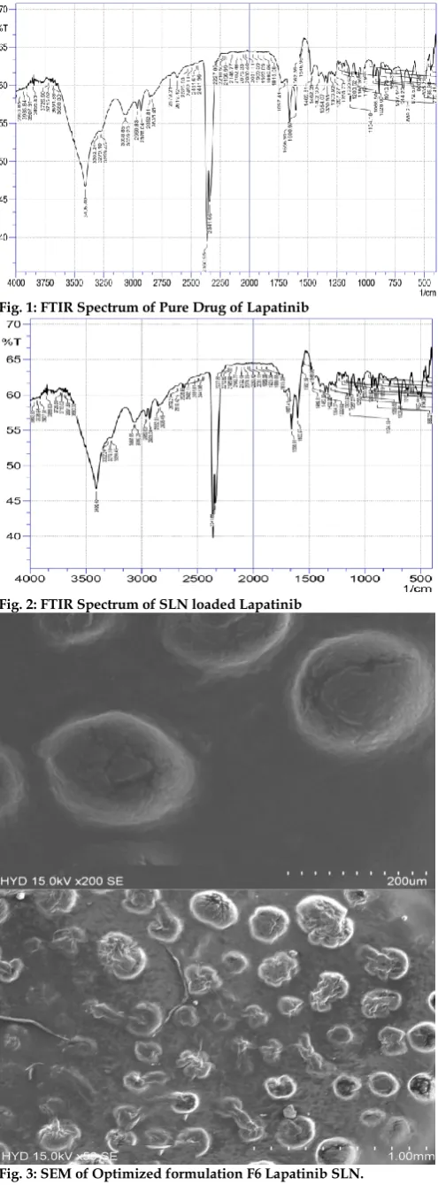
Fourier Transform infrared Spectroscopy FTIR
FTIR studies performed using Shimadzu spectrometer (Japan). Samples were prepared as KBr pellets
Differential Scanning Calorimetry (DSC)
The DSC chromatogram was recorded by placing accurately weighed 10 mg sample on aluminum plate and increasing the temperature of pan at rate 10ºC/min
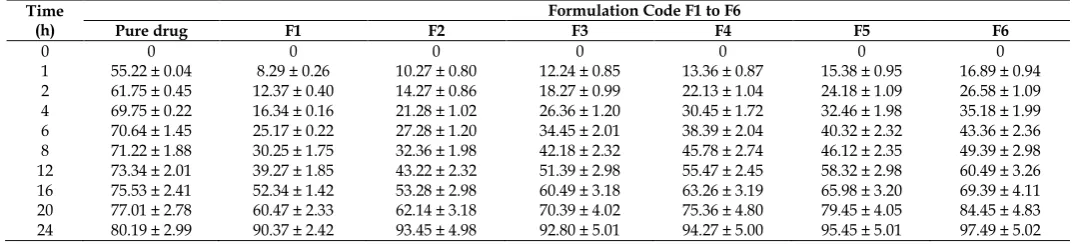
Surface Morphology
The formulation was vacuum dried to thin film after being transferred to a spherical aluminum plate followed by observation under scanning electron microscope (Quantum 200E Instrument). [12]
Determination of Zeta Potential, Particle Size and PDI of SLNs
Malvern Zeta seizer was used to measure zeta potential, size of the particle and polydispersity index (PDI) of Lapatinib loaded SLN’s (Nano ZS90). About 100µL of prepared SLN’s was mixed with 5mL water to obtain measurement of 50-200 Kilo Counts per Second (KCPS). [13]
Stability Studies
The stability Lapatinib SLNs checked at room temperature and 4°C for 60 days followed by determination of particle size, polydispersity index and zeta potential.
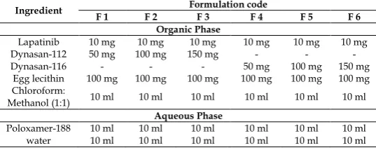
Table 1: Composition of Lapatinib SLNs
Ingredient Formulation code
F 1 F 2 F 3 F 4 F 5 F 6
Organic Phase
Lapatinib 10 mg 10 mg 10 mg 10 mg 10 mg 10 mg
Dynasan-112 50 mg 100 mg 150 mg - - -
Dynasan-116 - - - 50 mg 100 mg 150 mg
Egg lecithin 100 mg 100 mg 100 mg 100 mg 100 mg 100 mg
Chloroform:
Methanol (1:1) 10 ml 10 ml 10 ml 10 ml 10 ml 10 ml
Aqueous Phase
Poloxamer-188 10 ml 10 ml 10 ml 10 ml 10 ml 10 ml
water 10 ml 10 ml 10 ml 10 ml 10 ml 10 ml
RESULTS AND DISCUSSION Optimization of Process Parameters Homogenization time
A homogenization time of 5 minutes yielded optimal particle size and PDI. No considerable effect was observed when homogenization time was further increased.
Ultrsonication time
An Ultrsonication time of 20 min yielded optimal particle size and PDI.
Lipid concentration
Lipid concentration was optimized to 100 mg in increasing trend in particle size that slowed drug release was observed when lipid concentration was increased beyond 200 mg.
Surfactant concentration
Int. J. Pharm. Sci. Drug Res. September-October, 2019, Vol 11, Issue 5 (245-249) Fig. 1: FTIR Spectrum of Pure Drug of Lapatinib
Fig. 2: FTIR Spectrum of SLN loaded Lapatinib
Fig. 3: SEM of Optimized formulation F6 Lapatinib SLN.
Effect of lipid concentration on entrapment efficiency (EE)
Entrapment efficiency (EE) was one of the important parameter and to attain optimum efficiency the factors
like type of lipid, its concentration and type of surfactant were considered. The EE of Solid lipid nanoparticles loaded Dynasan-112 and Dynasan-116 indicated an increase in EE with increase in lipid ratio. Results are tabulated and represented in Table 2. All formulations exhibited good entrapment efficiency that ranged from 88.39% to 94.27% with F6 formulation showing best entrapment efficiency.
Table 2: Entrapment efficiency and total drug content of SLNs (n=3) Formulation code Drug content (mg) ± SD Entrapment efficiency (%) ± SD
F1 9.537 ± 0.022 88.39 ± 0.115
F2 9.149 ± 0.042 92.35 ± 0.248
F3 9.459 ± 0.016 90.27 ± 0.311
F4 9.349 ± 0.020 91.27 ± 0.049
F5 9.022 ± 0.006 92.34 ± 0.211
F6 9.891 ± 0.048 94.27 ± 0.222
Estimation of drug content
The maximum drug content of 09.891 mg was observed in formulation F6. The drug content of all prepared formulations is within 9.022-9.891 mg (Table 2).
The drug release from SLNs and suspension by dialysis method
The in vitro drug dissolution studies of Lapatinib SLN
formulations were determined by Dialysis bag diffusion technique. Lapatinib-loaded SLN dispersions containing equivalent amount of drug was sealed in dialysis bag. The dialysis bag suspended in 250 mL of 0.1N Hydrochloric acid with 2% polysorbate 80 and stirred at 50 rpm at 37°C ± 0.5°C. The cumulative percent release was more initially during the first hour for pure drug and gradually the drug release was low when compared with other formulations and cumulative release of 97.49 ± 5.02% was noted for F6 formulation which was highest of all formulations and pure drug.
Characterization of prepared Lapatinib solid lipid nanoparticles
The presence of characteristic absorption bands of Lapatinib and the optimized SLNs containing Lapatinib suggest no interaction between the drug and excipients (Figure 1 and 2).
Drug-excipients compatibility study by DSC
DSC chromatograms of drug, excipients and drug-excipients mixtures (Figure 3) show thermograms of Lapatinib, Dynasan-112 and Dynasan-116 and the DSC thermograms of Lapatinib exhibits a sharp endothermic peak at 178.60°C
Scanning Electron Microscopy
The SEM micrograph indicates rod and spherical shape particles with smooth surface (Figure 4).
Zeta potential and Particle size of prepared SLNs
Int. J. Pharm. Sci. Drug Res. September-October, 2019, Vol 11, Issue 5 (245-249) Table 3: In vitro dissolution studies of Lapatinib loaded SLNs (F1 to F6)
Time (h)
Formulation Code F1 to F6
Pure drug F1 F2 F3 F4 F5 F6
0 0 0 0 0 0 0 0
1 55.22 ± 0.04 8.29 ± 0.26 10.27 ± 0.80 12.24 ± 0.85 13.36 ± 0.87 15.38 ± 0.95 16.89 ± 0.94
2 61.75 ± 0.45 12.37 ± 0.40 14.27 ± 0.86 18.27 ± 0.99 22.13 ± 1.04 24.18 ± 1.09 26.58 ± 1.09
4 69.75 ± 0.22 16.34 ± 0.16 21.28 ± 1.02 26.36 ± 1.20 30.45 ± 1.72 32.46 ± 1.98 35.18 ± 1.99
6 70.64 ± 1.45 25.17 ± 0.22 27.28 ± 1.20 34.45 ± 2.01 38.39 ± 2.04 40.32 ± 2.32 43.36 ± 2.36
8 71.22 ± 1.88 30.25 ± 1.75 32.36 ± 1.98 42.18 ± 2.32 45.78 ± 2.74 46.12 ± 2.35 49.39 ± 2.98
12 73.34 ± 2.01 39.27 ± 1.85 43.22 ± 2.32 51.39 ± 2.98 55.47 ± 2.45 58.32 ± 2.98 60.49 ± 3.26
16 75.53 ± 2.41 52.34 ± 1.42 53.28 ± 2.98 60.49 ± 3.18 63.26 ± 3.19 65.98 ± 3.20 69.39 ± 4.11
20 77.01 ± 2.78 60.47 ± 2.33 62.14 ± 3.18 70.39 ± 4.02 75.36 ± 4.80 79.45 ± 4.05 84.45 ± 4.83
24 80.19 ± 2.99 90.37 ± 2.42 93.45 ± 4.98 92.80 ± 5.01 94.27 ± 5.00 95.45 ± 5.01 97.49 ± 5.02
Table 4: Stability studies of the optimized formulation (F6) at both 25°C and 4°C for 60 days
Day 25°C 4°C
Size (nm) PDI Zeta potential (mV) Size (nm) PDI Zeta potential (mV)
1 88.69 ± 1.45 0.517 ± 0.045 -22.9 ± 1.10 88.69 ± 1.58 0.517 ± 0.017 -22.9 ± 1.10
30 102.24 ± 5.27 0.521 ± 0.152 -21.24 ± 2.06 99.27 ± 2.84 0.519 ± 0.047 -21.1 ± 2.17
60 124.14 ± 8.29 0.533 ± 0.076 -20.24 ± 2.08 112.47 ± 3.47 0.521 ± 0.023 -20.6 ± 2.89
Fig. 4: Particle size analysis of optimized formulation (F6) of Lapatinib SLN
Fig. 5: Zeta potential of the Lapatinib SLN optimized formulation F6
Fig. 6: Cumulative % drug release from Lapatinib SLNs
The Poly-dispersity index of SLNs was in between 0.398 to 0.731 and it was reduced to narrow range due to increasing concentration of surfactant.
The zeta potential of SLNs varies from -19.9 to -26.3 mV. The results indicate that formulations containing Dynasan-112 exhibited smaller particle sizes and larger PDI, zeta potential values. The formulations comprising Dynasan-116 (F6) exhibited better particle size and zeta potential values in comparison with other formulations.
Stability studies
Stability studies conducted for 60 days indicated no significant variations in the particle size, PDI, zeta potential, EE and drug release data of the optimized formulation (F6).
The SLNs were successfully formulated for Lapatinib by the process Homogenization and Ultrsonication. These are economically manufactured from Dynasan-112, Dynasan-116 and Egg Lecithin etc. The prepared formulations characterized for particle size, PDI, Zeta potential, DSC, scanning electron microscopy, entrapment efficiency and in-vitro drug dissolution studies. Results indicated that increase in the concentration of Lipid content lead to higher entrapment efficiency. A controlled drug release was observed in In-vitro drug dissolution pattern of SLN. Thus, results conclude that the Lapatinib loaded SLNs are effective drug carrier with increased solubility and bioavailability.
REFERENCES
1. Bargoni A, Cavalli R, Zara GP. Transmucosal transport of tobramycin incorporated in solid lipid nanoparticles (SLN) after duodenal administration to rats. Part II – Tissue distribution. Pharm Res. 2001; 43: 497–502.
2. Freitas C, Müller RH. Effect of light and temperature on zeta potential and physical stability in solid lipid nanoparticles (SLN) dispersions. Int. J. Pharm. 1998; 168:221–229.
3. Cavalli R, Caputo O, Gasco MR. Solid lipospheres of doxorubicin and idarubicin. Int J Pharm. 1993; 89:9-12. 4. Stuchlik M, Zak S. Lipid-based vehicle for oral drug delivery.
Int. J. Pharm. Sci. Drug Res. September-October, 2019, Vol 11, Issue 5 (245-249) 5. Das S, Chaudhury A. Recent advances in lipid nanoparticle
formulations with solid matrix for oral drug delivery. AAPS Pharm SciTech. 2011; 12(1):62–76.
6. Rusnak D, Affleck W, Cockerill K, Stubberfield SG, Harris C, Page R, Smith M, Guntrip KJ, Carter SB, Shaw MC. The characterization of novel, dual ErbB-2/EGFR, tyrosine kinase inhibitors: Potential therapy for cancer. Cancer Res. 2001; 61:7196–7203.
7. Konecny GE Pegram MD, Venkatesan N, Finn R, Yang G, Rahmeh M, Untch M, Rusnak DW, Spehar G, Mullin RJ. Activity of the dual kinase inhibitor lapatinib (GW572016) against HER-2-overexpressing and trastuzumab-treated breast cancer cells. Cancer Res. 2006; 66:1630–1639.
8. Higa GM, Abraham J. Lapatinib in the treatment of breast cancer. Expert Review of Anticancer Therapy (Future Drugs). 2007; 7(9): 1183–92.
9. Glaxo Smith Kline (GSK). Glaxo Smith Kline receives marketing authorization in the EU for Tyverb (lapatinib), the
first oral targeted therapy for ErbB2-positive breast cancer. Glaxo Smith Kline (GSK) Press release. 2008; 06-12.
10. Venkateshwarlu V, Manjunath K. Preparation,
characterization and in vitro release kinetics of clozapine solid lipid nanoparticles. J Control Rel. 2004; 95:627-38. 11. Dudhipala N, Veerabrahma K. Improved anti-hyperlipidemic
activity of Rosuvastatin Calcium via lipid nanoparticles:
Pharmacokinetic and pharmacodynamic evaluation.
European Journal of Pharmaceutics and Biopharmaceutics. 2017; 110:47–57.
12. Singh S, Dobhal AK, Jain A, Pandit JK, Chakraborty S. Formulation and evaluation of solid lipid nanoparticles of a
water soluble drug: Zidovudine. Chemical and
Pharmaceutical Bulletin. 2010; 58(5):650–655.
13. Fundaro A, Cavalli R, Bargoni A. Non-stealth and stealth solid lipid nanoparticles (SLN) carrying doxorubicin: pharmacokinetics and tissue distribution after IV administration to rats. Pharm Res. 2000; 42: 337–43.
HOW TO CITE THIS ARTICLE: Sandhya P. Development and Evaluation of Lapatinib Solid Lipid