Human Identification Method: SCLERA
RECOGNITION
1
Vanita Patil, 2 Dr A. M. Patil
1, 2
Dept. of E&TC, J T Mahajan College of Engineering. Faizpur, Maharashtra
Abstract - The structure of blood vessels in the sclera- the white part of the human eye, is unique for every individual, hence it is best suited for human identification. However, this is a challenging research because it has a high insult rate (the number of occasions the valid user is rejected). In this survey firstly a brief introduction is presented about the sclera based biometric authentication. In addition, a literature survey is presented. We have proposed simplified method for sclera segmentation, a new method for sclera pattern enhancement based on histogram equalization and line descriptor based feature extraction and pattern matching with the help of matching score between the two segment descriptors. We attempt to increase the awareness about this topic, as much of the research is not done in this area.
Keywords -Sclera Recognition, Histogram Equalization, Image Processing, Line Descriptor, Sclera Segmentation
1. Introduction
iometrics is relegated to authentication of human being based on their physiological and behavioral characteristics. To detect person as accurately as possible number of techniques were proposed but it was found that no biometric method is perfect or accepted globally. To achieve higher recognition accuracy, to increase population coverage further research is required. There are various techniques in biometrics to identify an individual, among these sclera recognition is found out to be a best technique to complement previous trades because of sclera part, Because Sclera areas are highly secured part of an eye, which is impossible to stole. Identification of a human being by the vessel pattern of the sclera is possible because the vessel pattern of human beings is very different, and it is unique for every individual. Twins also have different vessel and this makes it suitable for human identification. Secondly, the vessel pattern of the person throughout lifetime is stable. Even the vessel patterns of left and right eye of a person differ from each other. Therefore, this system is best and reliable approach human identification. This paper organized as follows. Section II covers the background of sclera a recognition, after that segmentation process is clearly specified after that that we have concluded.
2. Background
Many Different methods are used such as fingerprint, iris, voice, face. Every method has its pros and cons. Almost all biometrics uses following objectives: Accuracy,
reliability, stability, identification, ID capability in distance, user cooperation and scalability to large population. For example, face detection is very natural way of identifying a person by humans, but human beings face structure changes according to his age increase and these changes affects the recognition correctness. Human being finger print structure is not change and its identification accuracy is more but it has one disadvantage we cannot us ed it as an ID for a certain distance. In addition, there are many reasons why people object the some methods for example culture, hygiene, religion, personal preference. Fingerprint recognition may cause hygiene and medical issues.
In real life applications, some biometrics techniques are more preferable than others in certain situations are. For example, the accuracy provided by fingerprint and iris recognition is higher than face recognition. Overall no biometric is said to be perfect or can be accepted globally. Researchers are trying to find new biometrics, which provides more options for human ID. In this paper, we have proposed a human ID system Sclera recognition, which gives us more accuracy than other methods. Sclera is the white part of the eye. It completely surrounds the eye and made up of for layering of tissues-the epsclera, stroma, lamina fusca, and endothelium, Structure of blood vessels are quite visible and stable over time. It is different for each person. Sclera recognition system consists of four modules –sclera segmentation, sclera vessel feature extraction, sclera vessel feature matching decision.
3. Sclera Segmentation
Sclera segmentation is the first method in the process of sclera recognition system. It has many steps involved in it. A. Pre-processing In image processing, as we know all the filters are applied to the grayscale image. If the input image to the system is colored image then it is converted into a grayscale image. We have incorporated one instruction in Matlab Software, which gives us exact output as grayscale image, which we want to process further in this paper
.
3.1 Color Plane Conversion
Any colored image has three dimensions that is, red, green and blue plane. In MATLAB a binary and gray-scale image is represented by one 2-dimensional array. A color image is represented by a 3-dimensional array (one 2-dimensional array for each of the color planes or color channels red, green and blue). The origin of the image is in the upper left and the size of the image is defined by the parameter width (number of columns of the array) and height (number of rows of the array). Note that the x- and y-coordinates are chosen such that the z-axis points to the front. If any image from the dataset has green or brown colored iris then it is necessary to get those color intensities. Hence the colored image is converted into the three planes using following formulae where 1,2,3 resembles to red, green and blue plane respectively [6].
: , : ,1; : , : ,2;
: , : ,3;
The glare area detection is the first step to do in the system. Glare area is a white colored bright area of an eye image. The glare area detection is carried out by brighten the part of eye except the skin and pupil part. To brighten the required area we have used contrast stretching mechanism as well as intensity enhancement technique.
3.2 Iris Boundary Detection
Fig 4. Iris boundary detection. (a) Finding the start point. (b) Searching along the radial direction
The algorithm searches along the radial direction at a predefined set of angles to estimate the pupil boundaries and then iteratively maps the highest edge value along the angular direction for π/2 radians for each of these starting angles. Starting at the estimated center of the pupil, the algorithm searches along a radial direction for the highest edge value within some radial length range.
, , | ,, arctan $%&%'(&(') Ɵ+---1 two nearest neighbors along the radial dimension
, , , | , , - 0 / 01Ɵ 0 / Ɵ 23 1 4 2/ 156--2
3.3 Sclera Area Detection
area detection step. The left and right ROIs are selected based on the iris center and boundaries.
Fig. 4. Sclera area detection using Otsu‘s method of thresholding
3.4 Iris and Eyelid Detection and Refinement
The upper eyelid, lower eyelid, and iris boundaries are then refined using the Fourier active contour method [18].
3.5 Sclera Feature Extraction
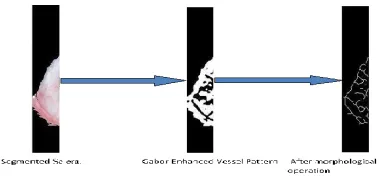
The feature extraction of sclera has few steps: Sclera Vessel pattern Enhancement, and Vascular Pattern Extraction. Fig. 8 shows the sclera feature extraction process.
3.6 Sclera Vessel Pattern Enhancement
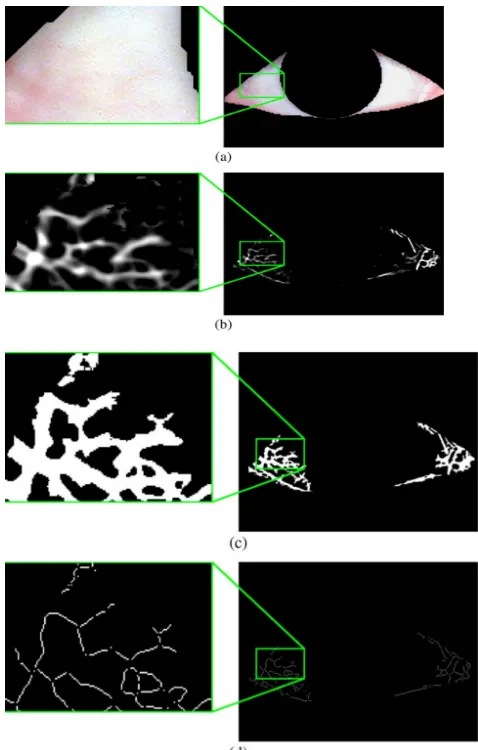
The vessel structure in the sclera region is very difficult to see; the sclera vascular patterns are often blurry and/or have very low contrast. To overcome this, it is important to enhance the vascular patterns. In [19], Daugman shows that the family of Gabor filters is good approximations of the vision processes of the primary visual cortex. Because the vascular patterns could have multiple orientations.
The image is first filtered with Gabor filters with different orientations and scales-
78 9, :, :, 7 9,: ∗ 9, :, :, -(3)
All the filtered images are fused together to generate the
vessel-boosted image< , .
8 , =∑C∈Ɵ ∑ 78 , , , 78 , , , A∈A @(4)
Fig. 5. Vessel patterns—before and after Gabor enhancement. (a) Segmented sclera region. (b) After Gabor enhancement (vessel-boosted
image). (c) After thresholding (binary vessel image). (d) After morphological operations.
3.7 Vascular Pattern Extraction
In this study, binary morphological operations are used to thin the detected vein structure down to a single-pixel wide skeleton and remove the branch points. The line segments are then used to create a template for the vein structure. The segments are described by three quantities the segments ‘angle to some reference angle at the pupil‘s center, the segments distance to the pupil center, and the dominant angular orientation of the line segment. Fig.7 shows a visual description of the line descriptor.
A descriptor is
Ɵ ФE
Ɵ tan&F$ %G&%H
(G&(H),
(5)
= I 3 @/ I 3 @
(6)
Ɵ tan&F$ J%
J(8GHKL )
---7
Here
8
GHKLis the polynomial approximation of the line segment
I, I
is the center point of the line segment,,
is the center of the detected iris, and is the line descriptor.Fig. 6. Sclera Feature Extraction 3.8 Sclera Feature Matching
The matching process consists of two steps: i) Sclera Template Registration, and ii) Sclera Template Matching.
3.9 Sclera Template Registration
Using these values, it calculates a fitness value for the registration using these parameters. The two descriptors and
M
.(H ,
Ɵ Ф6and
%H ,
ƟM M
ФM6
(8)
Fig. 7. Weighting Image
First, an offset vector is created using the shift offset and randomly determined scale and angular offset values
N' ,
0 0
06
(9)
Where ' (H01Ɵ(H3
%O01Ɵ%O
and ' (HƟ(H3 %OƟ%O
The fitness of two descriptors is the minimal summed pair-wise distance between the two descriptors given some offset vector. Where,
PQ (, %R S'P~ (, %, N'
Where,
P~Q(, %, N'R ∑(H∈ULAVP 8Q (H, N'R, %
---10
Here,
8
(H,
N
0
is the function that applies the registration given the offset vector to a sclera line descriptor.The minimum pairwise distance is calculated using
P (H%=MOWX (H,%OY (11)
The distance between two points calculated using XQ(H,%OR = ' @/ '@(12)
Where
the first descriptor used for registration,M
is the second descriptor, is the set of offset parameter valuesthat modifies the descriptor with the given offset values, s is the scaling factor, and is the rotation value.
Sclera Template matching
Fig. 8. Sclera Template Matching
After the templates are registered, each line segment in the test template is compared with the line segments in the target template for matches are, segments and , is the Euclidean distance between the segment descriptor center points , is the matching distance threshold, and match is the matching angle threshold
4. Conclusions
30 distance values are calculated. Based upon minimum distance value the authentication has been done. Testing is done by measuring min distance value between template matching
Fig.9 SCLERA Matching score
Acknowledgments
I would like to express profound gratitude to my guide andcoordinator M.E. Program Prof. K. S. Bhagat for his invaluable support, encouragement, supervision and useful suggestions throughout this project work. His moral support and continuous guidance enabled me to complete my work successfully.
I am very grateful for the valuable cooperation and constant encouragement from Prof. K. S. Bhagat, Prof. A. M. Patil and my honorable Head of Department Dr. A. M. Patil. His regular suggestions made my work easy and proficient.
I wish to express my appreciation to Principal Dr. Nandani Chaudhari who helps me to overcome my dough in doing this project work.
Last but not the least, I am thankful and indebted to my family and all those who helped me directly or indirectly in completion of this project report.
References
[1] R. Derakhshani, A. Ross, and S. Crihalmeanu, -A new biometric modality based on conjunctival vasculature,ǁ
in Proc. ANNIE, 2006, pp. 1–8.
[2] R. Derakhshani and A. Ross, ―A Texture-Based Neural Network Classifier for Biometric Identification Using Ocular Surface Vasculature, in Proc. IJCNN, 2007, pp. 2982–2987.
[3] Mohammad Hossein Khosravi and Reza Safabakhsh, Human eye sclera detection and tracking using a modified time-adaptive self-organizing map, ‘‘Science
Direct Journal on Pattern Recognition, Volume 41, Issue 8, pp. 2571–2593, August 2008.
[4] J. R. Parker and A. Q. Duong, Gaze Tracking: A Sclera Recognition Approach,‘‘ in Proceedings of the 2009 IEEE International Conference on Systems, Man, and Cybernetics San Antonio, TX, USA - October 2009, pp.3836-3841 .
[5] Z. Luo and T. Lin, Detection of non-iris region in the iris recognition, in Proc. of ISCSCT, 2008, pp. 45–48.
[6] N. L. Thomas, Y. Du, and Z. Zhou, A new approach for sclera vein recognition, Proc. SPIE, vol. 7708, p. 770 805, 2010.
[7] Zhi Zhou, Eliza Yingzi Du, N. Luke Thomas, and Edward J. Delp, Multi angle Sclera Recognition Systemǁ, in Proc. of 2011 IEEE Workshop on Computational Intelligence in Biometrics and Identity Management (CIBIM), 11-15 April 2011, pp. 103-108.
[8] Tatiana Tambouratzis and Michael Masouris, GA-Based Iris/Sclera Boundary Detection for Biometric Iris Identification., Springerlink Lecture Notes in Computer Science Volume 4432, pp 457-466, 2008.
[9] Fernando Alonso-Fernandez and Josef Bigun, Iris Boundaries Segmentation Using the Generalized Structure Tensor. A Study on the Effects of Image Degradatio, in Proc. of IEEE Fifth International Conference on Biometrics: Theory, Applications and Systems (BTAS), 23-27 Sept. 2012, pp. 426 – 431.
[10] Tae-Hong Min and Rae-Hong Park, COMPARISON OF EYELID AND EYELASH DETECTION
ALGORITHMS FORPERFORMANCE
IMPROVEMENT OF IRIS RECOGNITIONǁ, in proc. of 15th IEEE International Conference on Image Processing, 12-15 Oct. 2008, pp. 257 – 260.
[11] M. Abdullah-Al-Wadud and Oksam Chae, Skin Segmentation Using Color Distance Map and Water-flow Propertyǁ, in proc. of Fourth International Conference on Information Assurance and Security, 8-10 Sept. 2008, pp. 83 -88
[12] M. Abdullah-Al-Wadud and Oksam Chae, Region-of-Interest Selection for Skin Detection Based Applicationsǁ, in proc. of International Conference on Convergence Information Technology, 21-23 Nov. 2007, pp. 1999 – 2004.
[13] Zhi Zhou, Yingzi Du1, N. Luke Thomas1, and Edward J. Delp, Multimodal Eye Recognitio, SPIE journal on Mobile Multimedia/Image Processing, 2010, Volume 7708, article id. 770806, pp. 1-10.
[14] Zhi Zhou, Eliza Yingzi Du, Craig Belcher, N. Luke Thomas, and Edward J. Delp, Quality Fusion Based Multimodal Eye Recognitionǁ, in proc. of IEEE International Conference on Systems, Man, and Cybernetics, October 14-17 2012, Seoul, Korea, pp. 1297-1302.
[15] Zhi Zhou, Eliza Yingzi Du, N. Luke Thomas, and Edward J. Delp, A New Human Identification Method: Sclera Recognitionǁ, IEEE TRANSACTIONS on Systems, MAN, and Cybernetics Part A: Systems and Humans, Vol. 42, No. 3, pp. 571-583, May 2012.
1 2 3 4 5 6 7 8 9 10
9 9 .2 5 9 9 8 9 8
8 96
8
9
8
7
8
7 92
9
1
1 2 3 4 5 6 7 8 9 1 0
M A T C H IN G S C O R E IMAGE NUMBER
S C L E R A A M TC H I N G
S C O R E
[16] M. A. Fischler and R. C. Bolles, Random sample consensus: A paradigm for model fitting with applications to image analysis and automated cartography, Commun. ACM, vol. 24, no. 6, pp. 381– 395, Jun. 1981. [17] N. Otsu, A threshold selection method from gray-level histograms, Automatica, vol. 11, pp. 285–296, 1975.
[18] J. Daugman, New methods in iris recognition, IEEE Trans. Syst., Man, Cyber. B, Cybern. vol. 37, no. 5, pp. 1167–1175, Oct. 2007.
[19] J. Daugman, Two-dimensional spectral analysis of cortical receptive field profiles, Vis. Res., vol. 20, no. 10, pp. 847–856, 1980.
[20] H. Proença and L. A. Alexandre, UBIRIS: A noisy iris image database, in Proc. 13th Int. Conf. Image Anal. Process. vol. 3617, LNCS, 2005, pp. 970– 977. (http://iris.di.ubi.pt/).
Authors –
Ms. Vanita Patil had completed her BE in E&TC and pursuing M.E. from the J.T. Mahajan college of Enginerering Faizpur. Maharashtra.