Open Access
Research article
Decision making in patients with acute abdominal pain at a
university and at a rural hospital: does the value of abdominal
sonography differ?
Aristomenis K Exadaktylos*
1, Charlotte Sadowski-Cron
1, Paul Mäder
2,
Monika Weissmann
1, Hans Peter Dinkel
3, Marco Negri
2and
Heinz Zimmermann
1Address: 1Department of Emergency Medicine, University of Berne, Inselspital, Berne, Switzerland, 2Rural Hospital Frutigen, Switzerland and 3Department of Radiology University of Berne, Inselspital, Berne, Switzerland
Email: Aristomenis K Exadaktylos* - aristomenis@exadaktylos.ch; Charlotte Sadowski-Cron - sadowskich@hotmail.com; Paul Mäder - paul.maeder@hin.ch; Monika Weissmann - notfallzentrum@insel.ch; Hans Peter Dinkel - notfallzentrum@insel.ch; Marco Negri - marco.negri@spitalfmi.ch; Heinz Zimmermann - heinz.zimmermann@insel.ch
* Corresponding author
Abstract
Introduction and objectives: Abdominal sonography is regarded as a quick and effective diagnostic tool for acute abdominal pain in emergency medicine. However, final diagnosis is usually based on a combination of various clinical examinations and radiography. The role of sonography in the decision making process at a hospital with advanced imaging capabilities versus a hospital with limited imaging capabilities but more experienced clinicians is unclear.
The aim of this pilot study was to assess the relative importance of sonography and its influence on the clinical management of acute abdominal pain, at two Swiss hospitals, a university hospital (UH) and a rural hospital (RH).
Methods: 161 patients were prospectively examined clinically. Blood tests and sonography were performed in all patients. Patients younger than 18 years and patients with trauma were excluded. In both hospitals, the diagnosis before and after ultrasonography was registered in a protocol. Certainty of the diagnosis was expressed on a scale from 0% to 100%.
The decision processes used to manage patients before and after they underwent sonography were compared. The diagnosis at discharge was compared to the diagnosis 2 – 6 weeks thereafter.
Results: Sensitivity, specificity and accuracy of sonography were high: 94%, 88% and 91%, respectively.
At the UH, management after sonography changed in only 14% of cases, compared to 27% at the RH. Additional tests were more frequently added at the UH (30%) than at the RH (18%), but had no influence on the decision making process-whether to operate or not. At the UH, the diagnosis was missed in one (1%) patient, but in three (5%) patients at the RH. No significant difference was found between the two hospitals in frequency of management changes due to sonography or in the correctness of the diagnosis.
Published: 8 October 2008
World Journal of Emergency Surgery 2008, 3:29 doi:10.1186/1749-7922-3-29
Received: 14 August 2008 Accepted: 8 October 2008
This article is available from: http://www.wjes.org/content/3/1/29
© 2008 Exadaktylos et al; licensee BioMed Central Ltd.
Conclusion: Knowing that sonography has high sensitivity, specificity and accuracy in the diagnosis of acute abdominal pain, one would assume it would be an important diagnostic tool, particularly at the RH, where tests/imaging studies are rare.
However, our pilot study indicates that sonography provides important diagnostic information in only a minority of patients with acute abdominal pain.
Sonography was more important at the rural hospital than at the university hospital. Further costly examinations are generally ordered for verification, but these additional tests change the final treatment plan in very few patients.
Introduction
Acute abdominal pain is a non-specific symptom of many diseases. An efficient initial diagnostic evaluation, includ-ing physical examination and blood tests, is performed in most cases [1,2]. Different authors have shown that sonography adds up to 40% more information to these clinical examinations [3-6] and leads to a change in man-agement in 20% of cases [7-9]. Thus, sonography is con-sidered an important diagnostic tool in emergency departments (ED) [9-11].
The accuracy of sonography has been found to be between 71–98% for acute appendicitis and billiary tract disease [4,8,11]. The sensitivity and specificity of sonography are high in the diagnosis of cholecystitis, ileus and diverticu-litis [3,5,12] but rather low in the diagnosis of appendici-tis [13]. The accuracy of CT is superior to that of sonography in the diagnosis of acute appendicitis [9,14-16], but a CT-scan is rarely available at rural hospitals (RH) in Switzerland. However, CT scans [17,18], diagnos-tic laparoscopy [18], clinical scores [4,8,19,20] and the use of diagnostic algorithms [4,21] have been shown to be helpful in the decision making process. In many unclear situations, prolonged observation is used too, but is a financial burden, as it contributes to increasing hospital costs [3,22,23]. It is obvious that the decision making process in patients with acute abdominal pain is still a major challenge, as in up to 45% of cases no specific diag-nosis is made [7,10,24]. The aim of this pilot study was therefore to further clarify to what extent sonography influences the decision making process in patients with acute abdominal pain at a university hospital (UH), com-pared with a rural hospital (RH), and also addressed the issue of how additional examinations influence surgical management.
To the best of our knowledge, this is the first study com-paring the impact of sonography at a university and rural hospital emergency unit under everyday situations.
Materials and methods
This investigation was a prospective study of a conven-ience sample of 161 consecutive patients with acute
abdominal pain who were treated in the emergency department of both hospitals, 106 at the University Hos-pital InselsHos-pital in Berne (UH) and 55 at a small rural hos-pital (RH), both in the district of the Bernese Alps, Switzerland. Patients younger than 18 years and trauma patients were excluded.
At both hospitals, only patients were included in the study when they had abdominal symptoms of unclear origin. Patients with clinically clear findings were not given an unnecessary sonography and the usual procedure was not changed. The UH has approximately 1000 beds, serving 1.5 million people in the region. Over 50,000 in-patients and 150,000 out-patients are treated annually, including 30,000 in the adult emergency department. Sonography, CT, MRI, gastroscopy and colonoscopy and laboratory tests are available for the diagnosis of the acute abdomen 24 hours a day. The RH has approximately 50 beds serving 20,000 inhabitants and several thousand tourists per year. Over 2,000 in-patients and 6,000 out-patients are treated annually. Sonography, X-Ray, gastroscopy and colonos-copy and laboratory examinations are available 24 hours a day. For advanced diagnostic procedures, patients have to be transported to the next district hospital, which is 25– 50 km away.
At both the UH and the RH, as illustrated in Fig. 1, the diagnosis before and after sonography was registered by the surgeon on call at stage I and III and by the registrar who performed the examination at stage II. The stages are defined below.
The probability of the diagnosis at each of these stages was expressed on a scale from 0% to 100% (open in scale), broken down into the following categories.
0 to 10%: unlikely
≥ 10% to ≤ 60%: low to intermediate probability
≥ 60% to ≤ 90%: probable
The diagnostic pathway Figure 1
The diagnostic pathway.
Stage
STAGE I
Sur geon on call
Examination
Performed by
Clinical Examination
Ъ Conservative treatment
Suspected diagnosis (Probability scale 0% -100%)
Ь Operative treatment
STAGE II
Sonography
US Examiner
STAGE III
Clinical examination
Sur geon on call
+
Sonography
-> Working diagnosis
Affirmation of the initial
Change of the initial
diagnosis and treatment
diagnosis and treatment
in Step I (Probability scale 0% -100%) in Step I (Probability scale 0% -100%)
STAGE IV
Affirmation of the
Affirmation of the
The surgeon on call at the UH performed and interpreted the clinical examinations and defined the management plan. At the RH, the chief physician of the emergency department was involved in all cases.
At the UH, an experienced resident or fellow in radiology and at the RH a senior hospital physician performed and interpreted the ultrasound examinations. All doctors per-forming the ultrasound examination had a formal and accredited training therein. The ultrasound studies were documented at both hospitals as still images and reviewed by senior radiologists.
An Acuson 128XPIO with a 3.5 MHz transducer was used at the UH, and an Acuson 74XER with a 5 MHZ transducer at the RH. Curvilinear transducers and linear arrays were used, but no endocavitary probe True positive and true negative examinations, were recorded, together with sen-sitivity, specificity and accuracy.
The diagnoses were classified as appendicitis, cholecysti-tis, gynaecological disease, nephrolithiasis or other dis-eases. The surgeon had to allocate his diagnosis to one of these five groups (see Table 1 and Table 2).
Stage I
The diagnostic pathway [Fig. 1] was as follows: After basic clinical examination (history taking, clinical examination
and blood test), the surgeon on call recorded the proba-bility of the diagnosis in the protocol and decided whether the patient should be treated by operation or conservatively.
Blood count, leukocytes, CRP, liver function tests, amy-lase, creatinine and urine dipstick were performed.
Stage II
[Fig. 1]: Sonography was performed and interpreted as mentioned above. No examiner had access to history, physical examination or lab results.
Stage III
[Fig. 1]: Knowing the result of the sonography, the sur-geon on call had to decide which steps to take next on the basis of clinical necessity. Options included surgery, con-servative treatment, discharge, or additional examina-tions, such as CT-scan, endoscopic retrograde cholangiopancreatography (ERCP), gastroscopy, and oth-ers. Women whose pain was suspected to have gynaeco-logical causes following the investigations mentioned above were seen at both hospitals by the gynaecologist on call (UH 6%/RH 9%).
Stage IV
[Fig. 1]: The final diagnosis was either obtained from the operating surgeon's report or in a telephone interview
Table 1: Frequencies of Diagnoses at Stages D 1–4 of the Decision-Making Process % (n)
UH (n = 106) RH (n = 55)
Stage I
Clinical diagnosis
Stage II
diagnosis by sonography
Stage III
Working diagnosis
Stage IV
final diagnosis after 2 weeks
Appendicitis
UH 47% (50) 18% (19) 23% (24) 22% (23)
RH 33% (18) 24% (13) 24% (13) 22% (12)
Cholecystitis
UH 22% (23) 10% (11) 10% (11) 10% (11)
RH 11% (6) 5% (3) 5% (3) 5% (3)
Gynaecologica disease
UH 11% (12) 9% (10) 11% (12) 9% (10)
RH 13% (7) 7% (4) 7% (4) 7% (4)
Nephrolithiasis
UH 12% (13) 8% (8) 11% (12) 11% (12)
RH 9% (5) 9% (5) 9% (5) 9% (5)
Other disease
UH 8% (8) 55% (58) 44% (47) 44% (47)**
RH 35% (19) 55% (30) 55% (30) 56% (31)
** The 47 (31) final diagnoses at the UH (RH) were:
Constipation 14 (0), gastroenteritis 13 (8), NSAP 5 (5), diverticulitis 4 (2), gastritis 3 (0), UTI 2 (0), spleen infarction 1 (0), musculoskeletal pain 1 (2), ovulation pain 1 (0), diverticulosis 1 (0), incarcerated hernia 1 (0), pancreatitis 1 (1), ileus 0 (2), coecal necrosis 0 (1), Meckel 's diverticulum 0 (1), haematoma of the abdominal wall 0 (1), bladder retention 0 (1), coecal volvolus 0 (1), colitis 0 (1), hepatocellular carcinoma 0 (1),
with the family physician 2 – 4 weeks after presentation to the ED.
The term non-specific abdominal pain (NSAP) was used when no diagnosis was found.
We defined and identified the 3 following outcomes (table 2/table 3):
1. The number of times the clinical management was changed due to the sonography (Stage I -> Stage III)
2. The number of times the diagnosis by sonography was correct (Stage II -> Stage IV)
3. The differences between UH and RH
Statistics
All information was entered in a computerised database by means of a standard PC-based spread sheet and statis-tics were calculated with the assistance of a biostatistician. Patient groups were compared using the Mann-Whitney U-test, since criteria for normal distribution were not
ful-filled. Categorical data were analysed by the Chi-square test, or the Fisher exact test if the expected samples size was smaller than five. A p-value smaller than 0.05 was considered significant.
Results
At the UH, there were 68 (72%) male patients and 48 (28%) female patients with a median age of 35 years (range 18–88 years). At the RH, there were 22 (40%) male patients and 33 (60%) female patients, with a median age of 40 years (range 18–87 years). The time that elapsed from the first until the final clinical evaluation ranged between 30 minutes and 2 hours at both hospitals.
The difference in male: female ratio at the two hospitals is most likely due to the fact that at the RH every patient is seen in the emergency department, whereas at the UH females with acute abdominal pain are also treated by the emergency department of gynaecology, which was not involved in this study.
Table 2: Allocation of the Individual Diagnoses at Each Stage n (%)
Diagnosis
UH (n = 106) RH (n = 55)
Stage I (Basic clinical examination)
Stage II (Sonography)
Stage III (Working diagnosis)
Stage IV
(Histo; Operating surgeon report; Clinical process)
1 2 3(*)
Appendicitis
UH 50 (47%) 19 (18%) 0 (0%) 31 (29%)° 24 (23%)A 23 (22%)
RH 18 (33%) 11 (20%) 2 (4%) 7 (13%)° 13(24%) 12 (22%)
Cholecystitis
UH 23 (22%) 11 (10%) 0 (0%) 12 (11%)1 11 (10%) 11 (10%)
RH 6 (11%) 2 (4%) 1 (2%) 4 (7%)1 3 (5%) 3 (5%)
Gyn. Disease
UH 12 (11%) 8 (8%) 2 (2%) 2 (2%) 2 10 (9%) 10 (9%)
RH 7(13%) 3 (5%) 1 (2%) 4 (7%) 2 4 (7%) 4 (7%)
Nephrolithiasis
UH 13 (12%) 8 (8%) 0 (0%) 5 (5%)3 12 (11%) B 12 (11%)
RH 5 (9%) 3 (5%) 2 (4%) 3 (5%)3 5 (9%) 5 (9%)
(*) 1: affirmed with sonography
2: newly diagnosed by sonography (initially an other diagnosis at Stage I)
3: excluded with sonography
° 10 × Constipation/15 × Gastroenteritis/2 × NSAP/4 × Diverticulitis (UH) 1 × Mesenterialinfarct/3 × Gastroenteritis/2 × NSAP/1 × Diverticulitis (RH)
16 × Gastroenteritis/1 × Musculoskeletal/4 × Constipation/1 × Spleen infarction (UH)
2 × NSAP/1 × Musculoskeletal/1 × Liver Cancer (RH)
22 × Urinary tract infection
1 × Adhesive ileus/1 × Coecumvolvulus/1 × Nephrolithiasis/1 × Appendicitis perf (RH)
31 × Pyelonephritis/3 × Condition after nephrolith loss, diagnosis with CT/1 × praevesical Concrement missed by sonography (UH)
1 × Gastritis/1 x NSAP/1 × Musculoskeletal (RH)
Although there were more female patients seen at the RH, the number of gynaecological diagnoses found was in the same range at the two hospitals (UH 12%/RH 7%).
The management changed in response to the sonography results in 14% (15/106) of patients at the UH but in 27% (15/55) of patients at the RH [Table 3]. Specificity and accuracy of sonography were higher at the RH than at the UH: 94% vs. 82%, respectively [Table 4]. At the UH, there
were10 false negative and 3 false positive diagnoses. How-ever, they did not influence the decision making process, perhaps because the surgeon ignored them or did not believe them; at the RH, there were 2 false positive and 1 false negative diagnoses.
Sonography was helpful in discarding the initial diagnosis in 29% of cases and in affirming it in 32%. In the
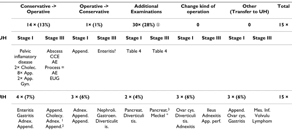
remain-Table 3: Change of Management Plan after Sonography
Conservative -> Operative
Operative -> Conservative
Additional Examinations
Change kind of operation
Other (Transfer to UH)
Total
14 × (13%) 1× (1%) 30× (28%) ① 0 0 15 ×
UH Stage I Stage III Stage I Stage III Stage I Stage III Stage I Stage III Stage I Stage III
Pelvic inflamatory
disease 2× Cholec.
8× App. 2× App. Gyn.
Abscess CCE
AE Process =
AE EUG
Append. Enteritis? Table 4 Table 4
RH 4 × (7%) 3 × (6%) 2 × (4%) 3 × (6%) 3 × (6%) 15 ×
Enteritis Gastritis Adnex. Append.
Append. Cholecy. Adnex. 1
Append.2
Adnex. Append. Append.
Nephroli. Gastroen. Diverticulit
is.
Pancreat. Diverticuli
tis.
Pancreat.3
Meckel °
Ovar cys. Diverticuli
tis. Adnexitis
Ileus Adnexitis App. perf.
Append. Ovar cys.
Gastritis
Mes. Inf. Volvulu Lymphom
① The additional examinations did not contribute to a change from initial management in any case. The additional examinations were ordered independent of the results of the sonography.
In the clinical process diagnosis of appendicitis and secondary operation. After basic clinical examination unlikely or low probability of appendicitis.
1 Basic clinical examination and sonography unclear. 2 Basic clinical examination and sonography unclear. 3 ERCP after sonography
° CT after sonography
+ 5× Change of the conservative therapy at RH after sonography: Nephrolithiasis -> Refluxoesophagitis
Hernia -> Nephrolithiasis
Cholecystolithiasis -> Musculoskeletal Nephrolithiasis -> Musculoskeletal Cholecystolithiasis -> Liver tumour
EUG = Extra uterine gravity; AE = Appendectomy; CCE = Cholecystectomy
Table 4: Sensitivity, Specificity and Accuracy of Sonography at the UH and RH
True Positive Sensitivity True Negative Specificity Total False positive False negative Total Accuracy
UH 50 93% 43 82% 93 3 10 13 87%
RH 29 95% 23 94% 52 2 1 3 95%
Total 79 94% 66 88% 145 5 11 16 91%
True positive and negative findings where the sonographic diagnosis corresponded to the intraoperative finding or to the final diagnosis. False positives or negatives were sonographic findings that were not identical to the diagnosis after 2 weeks.
ing 39% of cases, sonography did not make any contribu-tion to the diagnosis of acute abdominal pain.
In one third to one half of the patients who entered the emergency room with acute abdominal pain, appendicitis was suspected after clinical examination [Stage I, Table 1]. However, appendicitis could be confirmed by ultrasonic examination in only half of the cases [Stage II, Table 1 and 2]. For cholecystitis, nephrolithiasis and gynaecological disease, ultrasonic examination rarely changed the clinical diagnosis [Stage I -> Stage IV; Table 1 and 2].
Additional examinations were more often requested at the UH (30%; 32 patients) than at the RH (18%; 10 patients) and led to the final diagnosis at the UH in all but 1 (1%) patient (missed benign kidney tumor). The additional examinations at the UH were as follows: CT scan (20), intravenous pyelography (1), colon enema (1), endo-scopic retrograde cholangiopancreaticography (2), gynae-cological examination (6), diagnostic laparoscopy (0), gastroscopy (2). The additional examinations at the RH were as follows: CT scan (2), intravenous pyelography (1), colon enema (0), endoscopic retrograde cholangiopan-creaticography (2), gynaecological examination (1), diag-nostic laparoscopy (4), gastroscopy (0). At the RH, one diagnosis of appendicitis was missed by sonography and the patient was operated on 2 weeks later. One suspected appendicitis turned out to be gastroenteritis, and one Meckel's diverticulum was misdiagnosed as sigmoid diverticulitis. NSAP was diagnosed at discharge in 5% (5/ 106) of the patients at the UH and 9% (5/55) at the RH. The difference between the two hospitals was not signifi-cant (p > 0.05).
Discussion
Abdominal ultrasonography of patients with acute abdominal pain is very helpful for the confirmation or exclusion of clinically suspected appendicitis, billiary tract disease and aortic aneurysm and thus is an important diagnostic tool, albeit in a minority of patients [Table 3].
As a result of sonographic findings, the surgeon on call changed his initial decision as to whether to operate or to observe in 14% (15/106) of the patients at the university hospital, but in 27% (15/55) of the patients at the rural hospital [Table 3]. However, the difference was not statis-tically significant (p > 0.5), perhaps because of the sample size. Further, the accuracy of sonography was higher at the RH than at the UH [Table 4]. The surgeon on call might therefore have been aware of the limitation of the sonog-raphy at the UH and have chosen not to rely on the inves-tigation without additional examinations. However, there were 3 cases (5%) misdiagnosed at the RH [Table 4].
The differences in the accuracy of sonography between the RH and the UH in our study can be explained by the fact that sonography is operator dependent and requires ded-ication and experience: at the RH an experienced physi-cian performed all examinations, while at the UH the majority of ultrasounds were performed by ER residents or radiology fellows with different levels of experience. Thus sonography has great weight in decision making related to the diagnosis of acute abdominal pain at the RH. At the RH, the diagnostic possibilities are limited but the attending surgeon could, due to his great clinical expe-rience and the long time collaboration with the sonogra-pher, place more trust in the results of the sonography.
Our average accuracy rate of 91% in the diagnosis of acute abdominal pain is in accordance with most authors [12,15,16,25,26]. However, 10 false negative sonogra-phies at the UH illustrate the limits of sonographers. The examiners' varied levels of experience is evident and has implications for using diagnostic sonography as men-tioned above. Most authors claim that sonography increases costs without improving diagnostic perform-ance [9,27,28]. In fact, at the UH, 32 (30%) other exami-nations in addition to sonography were thought to be necessary to reach a final diagnosis. Nevertheless, our study found that these costly examinations had no influ-ence on the final diagnosis either. Further studies are urgently needed to clarify how much expensive additional examinations contribute to decision making in patients with acute abdominal pain.
The rate of non-specific abdominal pain (NSAP) at dis-charge was 5% (5/106) at the UH and 9% (5/55) at the RH, and no causes of abdominal pain were found in these patients 2 weeks later. These rates of NSAP are low com-pared to the frequencies of 25–40% reported from other centres [9,10,29].
It is often proposed that sonography should be used because of its reliability and simplicity. It has also been claimed that the correct diagnosis can be obtained after a short training period – even by non-radiologists [30]. Our results call such recommendations into doubt.
Limitations
Due to these and other differences between the two hos-pitals, no general recommendations can be made on the basis of the data from this study. Larger studies are neces-sary, which should consider the special conditions of the individual hospitals and include economic aspects.
Conclusion
Knowing that sonography has high sensitivity, specificity and accuracy in the diagnosis of acute abdominal pain, one would assume it would be an important diagnostic tool, particularly at the RH, where tests/imaging studies are rare.
However, our pilot study indicates that sonography pro-vides important diagnostic information in only a minor-ity of patients with acute abdominal pain.
Sonography was more important at the rural hospital than at the university hospital
In a time when medical expenses are rising steeply, further studies are urgently needed to investigate to what extent expensive additional examinations contribute to the deci-sion making plan in patients with acute abdominal pain.
This study can serve as a pilot for future well designed and methodologically stringent studies
Competing interests
The authors declare that they have no competing interests.
Authors' contributions
AKE wrote and revised paper. CSC organised study and planned study design. PM planned study design. MW recruited patients and collected patient data. HPD super-vised performed and interpreted sonographies at UH. MN supervised performed and interpreted sonographies at RH. HZ designed study.
Acknowledgements
We would like to thank Dr Rodney Yeates and Dr DS Evangelopulos MD PhD for support in the revision and editing of this manuscript.
References
1. Andersson RE, Hugander AP, Ghazi SH, Ravn H, Offenbartl SK, Nys-trom PO, Olaison GP: Diagnostic value of disease history, clin-ical presentation, and inflammatory parameters of appendicitis. World J Surg 1999, 23(2):133-40.
2. Davies AH, Mastorakou I, Cobb R, Rogers C, Lindsell D, Mortensen NJ: Ultrasonography in the acute abdomen. Br J Surg 1991,
78(10):1178-80.
3. Kratzer W, Pfeiffer M, Gebel M, Dietrich C, Adler G: The research situation in abdominal sonography in the gastroenterology departments of university hospitals in the Federal Republic of Germany. Z Gastroenterol 2000, 38(10):833-6.
4. Hoffmann J, Rasmussen OO: Aids in the diagnosis of acute appendicitis. Br J Surg 1989, 76(8):774-9.
5. Douglas CD, Macpherson NE, Davidson PM, Gani JS: Randomised controlled trial of ultrasonography in diagnosis of acute
appendicitis, incorporating the Alvarado score. BMJ 2000,
321(7266):919-22.
6. Guillemin F, Cao MM, Girard F, Tortuyaux JM, Chone L, Toussaint E, Bressler L, Regent D, Boissel P: Cost of medical imaging prac-tices in acute abdominal syndromes. Presse Med 2000,
29(15):829-34.
7. Spouge AR, Wilson SR, Wooley B: Abdominal sonography in asymptomatic executives: prevalence of pathologic findings, potential benefits, and problems. J Ultrasound Med 1996,
15(11):763-70.
8. Ohmann C, Yang Q, Franke C, Diagnostic scores for acute appendi-citis: Abdominal Pain Study Group. Eur J Surg 1995,
161(4):273-81.
9. Cassina P, Rothlin M, Largiader F: Efficacy of basic surgical diag-nosis in acute abdominal pain. Chirurg 1996, 67(3):254-60. 10. Dhillon S, Halligan S, Goh V: The therapeutic impact of
abdom-inalultrasounds in patients with acute abdominal symptoms.
Clin Radiol 2002, 57(4):268-271.
11. Allemann F, Cassina P: Ultrasound scans done by surgeons for patients with acute abdominal pain; a prospective study. Eur J Surg 1999, 165(10):966-970.
12. Ooms HW, Koumans RK, Ho Kang You PJ, Puylaert JB: Ultrasonog-raphy in the diagnosis of acute appendicitis. Br J Surg 1991,
78(3):315-8.
13. Balla JI, Elstein AS, Christensen C: Obstacles to acceptance of clinical decision analysis. BMJ 1989, 298(6673):579-82. 14. de Dombal FT, Dallos V, McAdam WA: Computer aided teaching
packages. BMJ 1991, 303(6808):995-6.
15. Pickuth D, Heywang-Kobrunner SH, Spielmann RP: Suspected acute appendicitis: is ultrasonography or computed tomog-raphy the preferred imaging technique? Eur J Surg 2000,
166(4):315-9.
16. Rao PM, Boland GW: Imaging of acute right lower abdominal quadrant pain. Clin Radiol 1998, 53(9):639-49.
17. Cobben LP, Van Otterloo AM, Puylaert JB: Spontaneously resolv-ing appendicitis: frequency and natural history in 60 patients.
Radiology 2000, 215(2):349-52.
18. Oudenhoven LF, Koumans RK, Puylaert JB: Right colonic diver-ticulitis: US and CT findings – new insights about frequency and natural history. Radiology 1998, 208(3):611-8.
19. Riesener KP, Klever P, Truong SN, Schumpelick V: [Role of sonog-raphy as primary diagnostic method in acute abdomen – a prospective study]. Langenbecks Arch Chir Suppl Kongressbd 1997,
114:1196-8.
20. Sutton GC: Computer-aided diagnosis: a review. Br J Surg 1989,
76(1):82-5.
21. Chen SC, Chen KM, Wang SM, Chang KJ: Abdominal sonography screening of clinically diagnosed or suspected appendicitis before surgery. World J Surg 1998, 22(5):449-52.
22. Lazzaron AR, Marques Ds, de Souza R: Re: Chen, SC, et al. Abdominal sonography screening of clinically diagnosed or suspected appendicitis before surgery. World J Surg 1998,
22:449. World J Surg 1999;23(4):425–6
23. Von Herbay A, Vogt C, Haussinger D: New methods in abdomi-nal ultrasound: do they have a clinical value? Panoramic imaging, harmonic imaging technologies and contrast medium enhanced ultrasound. Z Gastroenterol 2001,
39(4):295-304.
24. Dekker HM, Jagt EJ van der, van Leeuwen JT, Werf GT van der, Hun-ink MG: Role of abdominal sonography in excluding abdomi-nal malignancy in the initial workup of patients with abdominal complaints. AJR Am J Roentgenol 2001, 177(1):47-51. 25. Moberg AC, Ahlberg G, Leijonmarck CE, Montgomery A, Reiertsen
O, Rosseland AR, Stoerksson R: Diagnostic laparoscopy in 1043 patients with suspected acute appendicitis. Eur J Surg 1998,
164(11):833-41.
26. McGrath FP, Keeling F: The role of early sonography in the management of the acute abdomen. Clin Radiol 1991,
44(3):172-4.
27. Puylaert JB: Acute appendicitis: US evaluation using graded compression. Radiology 1986, 158(2):355-60.
28. Ford RD, Passinault WJ, Morse ME: Diagnostic ultrasound for suspected appendicitis: does the added cost produce a bet-ter outcome? Am Surg 1994, 60(11):895-8.
Publish with BioMed Central and every scientist can read your work free of charge "BioMed Central will be the most significant development for disseminating the results of biomedical researc h in our lifetime."
Sir Paul Nurse, Cancer Research UK
Your research papers will be:
available free of charge to the entire biomedical community
peer reviewed and published immediately upon acceptance
cited in PubMed and archived on PubMed Central
yours — you keep the copyright
Submit your manuscript here:
http://www.biomedcentral.com/info/publishing_adv.asp
BioMedcentral