Open Access
Research
Sparing effects of selenium and ascorbic acid on vitamin C and E in
guinea pig tissues
Jesse Bertinato
1, Nick Hidiroglou
1, Robert Peace
1, Kevin A Cockell
1,
Keith D Trick
1, Penny Jee
1, Alex Giroux
1, Réné Madère
1, Giuseppe Bonacci
1,
Monica Iskandar
1, Stephen Hayward
2, Nicholas Giles
2and Mary R L'Abbé*
1Address: 1Nutrition Research Division, Food Directorate, Health Products and Food Branch, Health Canada, Sir Frederick G. Banting Research
Centre, Ottawa, ON, Canada and 2Bureau of Biostatistics and Computer Applications, Food Directorate, Health Products and Food Branch, Health
Canada, Sir Frederick G. Banting Research Centre, Ottawa, ON, Canada
Email: Jesse Bertinato - [email protected]; Nick Hidiroglou - [email protected]; Robert Peace - [email protected]; Kevin A Cockell - [email protected]; Keith D Trick - [email protected]; Penny Jee - [email protected];
Alex Giroux - [email protected]; Réné Madère - [email protected]; Giuseppe Bonacci - [email protected]; Monica Iskandar - [email protected]; Stephen Hayward - [email protected]; Nicholas Giles - [email protected]; Mary R L'Abbé* - mary_l'[email protected]
* Corresponding author
Abstract
Background: Selenium (Se), vitamin C and vitamin E function as antioxidants within the body. In this study, we investigated the effects of reduced dietary Se and L-ascorbic acid (AA) on vitamin C and α-tocopherol (AT) status in guinea pig tissues.
Methods: Male Hartley guinea pigs were orally dosed with a marginal amount of AA and fed a diet deficient D/MC), marginal M/MC) or normal N/MC) in Se. An additional diet group (Se-N/NC) was fed normal Se and dosed with a normal amount of AA. Guinea pigs were killed after 5 or 12 weeks on the experimental diets at 24 and 48 hours post AA dosing.
Results: Liver Se-dependent glutathione peroxidase activity was decreased (P < 0.05) in guinea pigs fed Se or AA restricted diets. Plasma total glutathione concentrations were unaffected (P > 0.05) by reduction in dietary Se or AA. All tissues examined showed a decrease (P < 0.05) in AA content in Se-N/MC compared to Se-N/NC guinea pigs. Kidney, testis, muscle and spleen showed a decreasing trend (P < 0.05) in AA content with decreasing Se in the diet. Dehydroascorbic acid concentrations were decreased (P < 0.05) in several tissues with reduction in dietary Se (heart and spleen) or AA (liver, heart, kidney, muscle and spleen). At week 12, combined dietary restriction of Se and AA decreased AT concentrations in most tissues. In addition, restriction of Se (liver, heart and spleen) and AA (liver, kidney and spleen) separately also reduced AT in tissues.
Conclusion: Together, these data demonstrate sparing effects of Se and AA on vitamin C and AT in guinea pig tissues.
Published: 26 March 2007
Nutrition Journal 2007, 6:7 doi:10.1186/1475-2891-6-7
Received: 27 July 2006 Accepted: 26 March 2007
This article is available from: http://www.nutritionj.com/content/6/1/7
© 2007 Bertinato et al; licensee BioMed Central Ltd.
Background
Vitamin C is a water soluble antioxidant. In contrast to many mammals, humans (and guinea pigs) are unable to synthesise vitamin C due to the lack of the enzyme L-gulono-gamma-lactone oxidase [1] and therefore must rely on diet for maintaining adequate levels of the vita-min. In tissues, the active form of vitamin C, L-ascorbic acid (AA), can be regenerated by the reduction of its oxi-dised forms, dehydroascorbic acid (DHAA) and the ascor-bate free radical in a process mediated by glutathione (GSH) [2-6]. Notably, however, other systems have also been implicated in the regeneration of AA [7,8].
Selenium (Se) and vitamin E also function as important antioxidants within the body. Se is an essential trace ele-ment that functions in oxidant defence as a component of selenoproteins [9,10]. Vitamin E is a lipid soluble antioxi-dant present in cell membranes where it plays a vital role in protecting against lipid peroxidation [11-13]. Vitamin E refers to several structurally related compounds; how-ever, α-tocopherol (AT) is the predominant form found in animal tissues. Like vitamin C, vitamin E must be obtained from the diet.
The importance of maintaining adequate levels of Se, vita-min C and vitavita-min E is underscored by studies indicating that low antioxidant status may be associated with increased risk of developing various diseases [14-16]. Se has been shown to spare both AA [7,8] and AT [17]. Fur-ther, sparing effects of AA on AT have also been reported [18-20]. Given that Se, vitamin C and vitamin E activities are interconnected, it is important to understand how deficiency in one or two of these antioxidants influences the other(s). In this study we sought to explore the sparing effects of Se and AA on vitamin C and AT in guinea pig, an in vivo model that cannot synthesise vitamin C.
Methods
Animals and test diets
On arrival, male Hartley guinea pigs (~ 10 days old) (Elm Hill Breeding Labs, Inc., Chelmsford, MA) were subjected to a 2 week adaptation period. Following the adaptation period, guinea pigs (n = 22/diet group) had free access to one of 4 test diets (Table 1) and demineralised drinking water. Normal or marginal amounts of AA were given to each guinea pig in a 0.5 mL aqueous solution via oral dos-ing by gavage three times per week (i.e. Monday, Wednes-day and FriWednes-day). Amount of AA was calculated from the previous day's mean body weight for the diet group [2.4 (normal) or 0.3 (marginal) mg AA/100 g body weight]. Normal and marginal AA levels were chosen based on the AA requirement for growing guinea pigs and previous studies demonstrating suboptimal dietary AA levels [21-23]. Test diets were torula yeast-based diets deficient in Se and similar to diets previously used to induce Se
defi-ciency in guinea pigs [24]. Test diets were supplemented with 0 (deficient), 0.05 (marginal) or 0.20 (normal) mg Se/kg diet.
Guinea pigs were killed following an overnight fast by exsanguination while anesthetised with 3% isoflurane. Half the guinea pigs per diet group were killed after 5 weeks and the remainder after 12 weeks on the experi-mental diets at 24 and 48 hrs post AA dosing. Blood was withdrawn from the abdominal aorta and collected in heparinised tubes. Plasma was separated from cells by centrifugation (1000 × g, 20 min, 4°C). Skeletal muscle (from quadriceps) and soft tissues were extracted and immediately frozen in liquid nitrogen. Plasma and tissues were stored at -80°C until analysis. The Health Canada Animal Care Committee approved the experimental pro-tocol. Guinea pigs were treated in accordance with the guidelines of the Canadian Council on Animal Care.
Determination of vitamin C and α-tocopherol in tissues
AA and total vitamin C (following reduction of the sam-ple with homocysteine) were measured by reverse-phase HPLC with electrochemical detection as described [25]. DHAA was calculated as the difference between total vita-min C and AA. Excised tissues were immediately frozen in liquid nitrogen, a procedure that has been shown to pre-vent oxidation of vitamin C [26]. To further prepre-vent oxi-dation of vitamin C, plasma and tissue homogenates were preserved by treating with metaphosphoric acid to a final concentration of 0.85% w/v. AA and DHAA standards could be completely recovered when spiked into tissue homogenates indicating that with this method both AA and DHAA are stable in a tissue matrix [27]. Further, time course experiments revealed that AA and DHAA concen-trations remained constant over the course of 90 minutes indicating that AA was not being converted to DHAA. AA concentrations determined using this method were also closely correlated with AA concentrations determined using the 2, 4-dinitrophenylhydrazine method [28]. AT content was determined by reverse-phase HPLC with flu-orescence detection [29].
Enzyme and other assays
Se-dependent glutathione peroxidase (Se-GSHPx) activity was measured essentially as described [30] using a SPEC-TRAmax PLUS microplate spectrophotometer (Molecular Devices, Sunnyvale, CA). Liver extracts were prepared by homogenising in 0.2% Triton-X-100. Se-GSHPx activity is expressed as U/g protein, where one unit of activity catal-yses the oxidation of 1.0 mmol of reduced NADPH/ minute. Total plasma GSH was determined by HPLC
using a manual adaptation of the automated NaBH4
immunoassay using reagents from an OxyBlot Protein Oxidation Detection Kit (Intergen, NY, USA) as previously described [33]. Lipid peroxide concentrations in liver homogenates and plasma were determined using a com-mercially available kit (LPO-CC Lipid Peroxides, Kamiya Biomedical, Seattle, WA, USA). Protein concentration was determined by the bicinchoninic acid method [34].
Statistical analyses
Data were analysed by one-way ANOVA and differences between means were determined by Fisher's least signifi-cant difference test. For tissue vitamin C, data were ana-lysed using univariate ANOVA with diet as the main effect. Since the variability of tissue AA levels increased with an increase in mean, AA data were transformed using the square root transformation. Two contrasts were included in the analyses to test for an effect of Se or AA intake on tissue AA, DHAA and total vitamin C concentrations. The Se-D/MC, Se-M/MC and Se-N/MC diet groups were used to test for an effect of Se intake. These diet groups were also used to determine whether a decreasing or increasing trend was present as the amount of Se decreased in the diet. Trend here refers to an overall increasing or decreas-ing response to a decrease in dose. To test for an effect of AA intake, the Se-N/MC and Se-N/NC diet groups were compared. For vitamin C analyses, data from guinea pigs killed at week 5, 48 hrs post AA dosing and week 12, 24 hrs post AA dosing were combined in order to increase the power of the statistical comparisons. The ability to
com-bine these data is predicated on the similarity of the response (i.e. tissue vitamin C concentrations) of week 5, 48 hrs and week 12, 24 hrs guinea pigs to changes in die-tary Se or AA. Similarity of the response was determined by ANOVA. Data are shown as means ± SEM. Statistical significance was set at P < 0.05. Data were analysed using Statistica 7 (StatSoft, Tulsa, OK) and SAS (SAS Canada, Ottawa, Canada) software.
Results
To investigate the sparing effects of Se on vitamin C and AT when intake of AA is low, guinea pigs were orally dosed with a marginal amount of AA and fed a diet defi-cient (Se-D/MC), marginal (Se-M/MC) or normal (Se-N/ MC) in Se. An additional diet group (Se-N/NC) dosed with a normal amount of AA and fed a normal Se diet was included in the experimental protocol to allow investiga-tion of the sparing effects of AA on vitamin C and AT (comparison with diet group Se-N/MC). Approximately one third of the guinea pigs fed the Se-D/MC diet devel-oped paralysis of their hind limbs and showed poor mobility beginning as early as 4 weeks on the diet. Three Se-D/MC guinea pigs died or were euthanised prior to the week 5 necropsy and two prior to the week 12 necropsy.
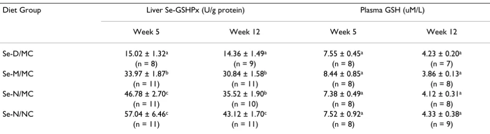
At week 5, liver Se-GSHPx activity showed a dose-depend-ent decrease (P < 0.05) with decreasing amounts of Se in the diet, confirming induction of graded levels of Se status in the guinea pigs (Table 2). At week 12, Se-D/MC guinea
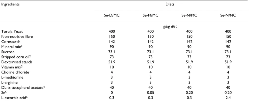
Table 1: Composition of experimental diets
Ingredients Diets
Se-D/MC Se-M/MC Se-N/MC Se-N/NC
g/kg diet
Torula Yeast 400 400 400 400
Non-nutritive fibre 150 150 150 150
Cornstarch 142 142 142 142
Mineral mix1 90 90 90 90
Sucrose 73.1 73.1 73.1 73.1
Stripped corn oil2 73 73 73 73
Dextrinised starch 51.9 51.9 51.9 51.9
Vitamin mix3 10 10 10 10
Choline chloride 4 4 4 4
L-methionine 3 3 3 3
L-arginine 3 3 3 3
DL-α-tocopherol acetate4 40 40 40 40
Se5 0 0.05 0.20 0.20
L-ascorbic acid6 0.3 0.3 0.3 2.4
1 Dyets no 200151 salt mix for Reid-Briggs guinea pig diet. 2 Containing 0.2 mg Tert-butylhydroquinone/g stripped corn oil.
3 Dyets no 300151 vitamin mix for Reid-Briggs guinea pig diet (Vitamin E omitted). 4 IU/kg diet; added via stripped corn oil.
pigs had lower Se-GSHPx activity compared to Se-M/MC or Se-N/MC guinea pigs. Se-N/MC guinea pigs had lower Se-GSHPx activity compared to Se-N/NC guinea pigs at week 12. Plasma total GSH concentrations were similar (P > 0.05) in guinea pigs fed the different test diets, consist-ent with a previous study showing no change in GSH lev-els with decreased Se and vitamin C status [7]. However, plasma GSH concentrations were lower (P < 0.05) in week 12 compared to week 5 guinea pigs for all diet groups.
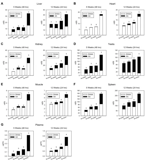
Tissue vitamin C and AT concentrations are only pre-sented for guinea pigs killed at week 5, 48 hrs post AA dos-ing and week 12, 24 hrs post dosdos-ing (see Discussion). Proportion of DHAA to AA in each tissue was similar between week 5 and 12 guinea pigs (Fig. 1). The only exception was plasma, where most of the vitamin C was present as AA at week 5 and as DHAA at week 12 (Fig. 1G). The ratio of DHAA to AA varied markedly between tissues. In testis, the majority of vitamin C was AA (Fig. 1D), whereas in heart and muscle most of the vitamin C was DHAA (Fig. 1B and 1E).
Because of the large variability in vitamin C concentra-tions between individual guinea pigs and the relatively small number of guinea pigs analysed per diet group, vita-min C data were combined for week 5, 48 hrs and week 12, 24 hrs guinea pigs to increase the power of the statis-tical comparisons (see Methods and Discussion). Effects of Se or AA intakes on tissue AA, DHAA and total vitamin C concentrations were determined by univariate ANOVA (Table 3). Differences between the Se-D/MC, Se-M/MC and Se-N/MC diet groups were determined to test for spar-ing effects of Se (Se Effect). Additional statistical analyses were performed to test whether there was a decreasing or increasing trend in vitamin C concentrations with decreas-ing Se in the diet [Se Effect (Trend)]. Differences between the Se-N/MC and Se-N/NC diet groups were determined to test for sparing effects of AA (AA Effect).
Se intake affected (P < 0.05) AA concentrations in kidney and muscle (Table 3, Se Effect). Trend analyses confirmed that the differences detected reflected a decrease in AA concentrations with decreasing Se in the diet [Table 3, Se Effect (Trend)]. Significant differences in AA concentra-tions were not detected (P > 0.05) in liver, heart, testis, spleen and plasma. However, testis and spleen showed a decreasing trend for AA with reduction in dietary Se. Total vitamin C concentrations were affected by Se intake in liver, heart, testis, muscle and spleen and showed a decreasing trend with decreasing Se. A Se effect and simi-lar decreasing trend was observed for DHAA in heart and spleen. In contrast, the Se effect on liver DHAA reflected an increasing trend with decreasing Se.
Se-N/MC guinea pigs had reduced (P < 0.05) AA concen-trations in all tissues compared to Se-N/NC guinea pigs [Table 3, AA Effect]. DHAA concentrations in Se-N/MC guinea pigs were also reduced (P < 0.0001) in liver, heart, kidney, muscle and spleen. All tissues from Se-N/MC guinea pigs showed decreased (P < 0.0001) concentra-tions of total vitamin C.
At week 5, there were no significant (P > 0.05) differences in AT concentrations between diet groups for any of the tissues analysed (Table 4). At week 12, Se-D/MC guinea pigs had lower (P < 0.05) AT concentrations in liver, heart and spleen compared to Se-M/MC guinea pigs (Table 4). AT was lower in liver, kidney and spleen of Se-N/MC com-pared to Se-N/NC guinea pigs. In plasma, while sole restriction of Se or vitamin C showed no effects on AT concentrations, combined restriction of Se and vitamin C decreased AT (compare Se-D/MC and Se-N/NC). Com-bined restriction of Se and vitamin C also decreased AT in liver, heart, kidney and spleen. Collectively, these data indicate that reductions in dietary Se and AA singly or in combination decrease AT concentrations in guinea pig tis-sues.
Table 2: Liver Se-GSHPx activity and plasma GSH concentration of guinea pigs after 5 and 12 weeks on the experimental diets1
Diet Group Liver Se-GSHPx (U/g protein) Plasma GSH (uM/L)
Week 5 Week 12 Week 5 Week 12
Se-D/MC 15.02 ± 1.32a 14.36 ± 1.49a 7.55 ± 0.45a 4.23 ± 0.20a
(n = 8) (n = 9) (n = 8) (n = 7)
Se-M/MC 33.97 ± 1.87b 30.84 ± 1.58b 8.44 ± 0.85a 3.86 ± 0.13a
(n = 11) (n = 11) (n = 8) (n = 8)
Se-N/MC 46.78 ± 2.70c 35.52 ± 1.90b 7.38 ± 0.49a 4.12 ± 0.31a
(n = 11) (n = 10) (n = 8) (n = 8)
Se-N/NC 57.04 ± 6.46c 43.12 ± 1.70c 7.52 ± 0.92a 4.33 ± 0.38a
(n = 11) (n = 11) (n = 8) (n = 9)
1 Data from guinea pigs killed at both 24 and 48 hrs post AA dosing.
Vitamin C concentrations in tissues of guinea pigs fed the experimental diets
Figure 1
Vitamin C concentrations in tissues of guinea pigs fed the experimental diets. Bars signify the amount of total vita-min C (DHAA + AA) in tissues of guinea pigs fed the Se-D/MC, Se-M/MC, Se-N/MC or Se-N/NC diets. The proportions of DHAA (white portion of bar) and AA (black portion of bar) are shown. Values are reported as the mean. Number of tissues analysed for each diet group are indicated above the bars. For each tissue, data are shown for guinea pigs killed after 5 and 12 weeks on the experimental diets at 48 and 24 hrs post AA dosing, respectively.
5 Weeks (48 hrs)
Se-D/M C Se-M/M C Se-N/M C Se-N/N C μ g/ g 0 10 20 30 40 DHAA AA
12 Weeks (24 hrs)
Se-D/M C Se-M/M C Se-N/M C Se-N/N C μ g/ g 0 10 20 30 40 50 DHAA AA Liver
A
5 5 6 5
4 5 6
5
5 Weeks (48 hrs)
Se-D/M C Se-M/M C Se-N/M C Se-N/N C μ g/ g 0 5 10 15 20 DHAA AA
12 Weeks (24 hrs)
Se-D/M C Se-M/M C Se-N/M C Se-N/N C μ g/ g 0 5 10 15 20 25 DHAA AA Heart
B
5 5 6 5 4 5 6 55 Weeks (48 hrs)
Se-D/M C Se-M/M C Se-N/M C Se-N/N C μ g/ g 0 10 20 30 40 DHAA AA
12 Weeks (24 hrs)
Se-D/M C Se-M/M C Se-N/M C Se-N/N C μ g/ g 0 10 20 30 40 50 DHAA AA Kidney
C
4 5 6 5 5 5 6 5 Se-D/M C Se-M/M C Se-N/M C Se-N/N C μ g/ g 0 20 40 60 80 100 120 140 160 180 DHAA AA Se-D/M C Se-M/M C Se-N/M C Se-N/N C μ g/ g 0 20 40 60 80 100 120 140 DHAA AA5 Weeks (48 hrs) 12 Weeks (24 hrs) Testis
D
5 5 6 5 4 5 6 5 Se-D/M C Se-M/M C Se-N/M C Se-N/N C μ g/ g 0 2 4 6 8 DHAA AA Se-D/M C Se-M/M C Se-N/M C Se-N/N C μ g/ g 0 1 2 3 4 5 6 7 DHAA AA5 Weeks (48 hrs) 12 Weeks (24 hrs) Muscle
E
4 5 6 5 5 5 6 5 Se-D/M C Se-M/M C Se-N/M C Se-N/N C μ g/ g 0 20 40 60 80 100 120 140 160 180 DHAA AA Se-D/M C Se-M/M C Se-N/M C Se-N/N C μ g/ g 0 50 100 150 200 250 DHAA AA5 Weeks (48 hrs) 12 Weeks (24 hrs) Spleen
F
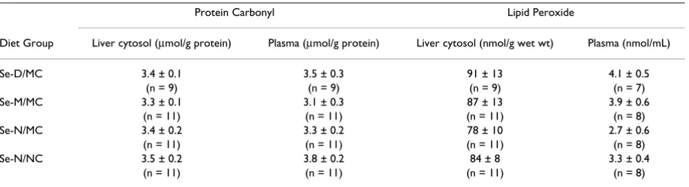
5 5 6 5 4 5 6 5 Se-D/M C Se-M/M C Se-N/M C Se-N/N C μ g/ m L 0.0 0.1 0.2 0.3 0.4 0.5 0.6 DHAA AA Se-D/M C Se-M/M C Se-N/M C Se-N/N C μ g/ m L 0.0 0.2 0.4 0.6 0.8 1.0 DHAA AAGiven that Se, vitamin C and AT function as antioxidants, it prompted us to examine tissues for oxidative damage. Liver cytosol and plasma protein carbonyl and lipid per-oxide concentrations were similar (P > 0.05) in guinea pigs fed the different experimental diets (Table 5).
Discussion
The primary objective of this study was to investigate the sparing effects of dietary Se and AA on tissue vitamin C and AT concentrations. Guinea pigs were chosen for these experiments as they are similar to humans in their inabil-ity to make vitamin C and therefore likely provide a more relevant model system compared to previously used cell culture systems [17,18,20] or animal models that have the ability to make vitamin C [7]. Further, we chose to inves-tigate the effects of Se under conditions of marginal AA intake, given that Se may play a more biologically signifi-cant role in sparing vitamin C and AT when intake of AA is low.
Only guinea pigs fed the Se-D/MC diet developed paraly-sis of their limbs. In some cases, the paralyparaly-sis was severe enough that the guinea pigs died or had to be euthanised. These results are consistent with previous studies demon-strating sensitivity of guinea pigs to disturbances in anti-oxidant status. Particularly, Se deficiency combined with vitamin E or C deficiency has been reported to cause skel-etal muscle damage [24,35]. Further, vitamin E combined with vitamin C deficiency has been shown to promote limb paralysis and death due to severe damage in the brainstem and spinal cord [36].
As part of the study design, guinea pigs were killed after 5 and 12 weeks on the experimental diets at 24 and 48 hrs following AA dosing. Although analyses of vitamin C data at each of the four separate time points revealed little
dif-ference between guinea pigs fed different levels of Se, a discernable decreasing trend for vitamin C concentrations in tissues with decreasing dietary Se was observed for week 5, 48 hrs and week 12, 24 hrs guinea pigs. In contrast, no trend was observed for week 5, 24 hrs and week 12, 48 hrs guinea pigs. The reason for the observed Se effect at differ-ent times post AA dosing for week 5 and 12 guinea pigs may be explained by differences in the metabolism of the dosed AA between younger (week 5) and older (week 12) guinea pigs. Notably, vitamin C concentrations were higher for guinea pigs killed at 24 compared to 48 hrs post dosing for both week 5 and 12 guinea pigs (data not shown) indicating that vitamin C concentrations rise in tissues following dosing and then fall over time as the vitamin is consumed. Increases in tissue vitamin C con-centrations at early times post dosing and low concentra-tions after an extended time post dosing may mask any effects of Se on vitamin C concentrations. Therefore, if the younger and older guinea pigs metabolised the dosed AA differently (e.g. differences in AA absorption or rate of AA consumption by tissues), it would not be surprising that the Se effects on vitamin C are observed at different times post dosing for week 5 and 12 guinea pigs. However, addi-tional studies are required to definitively show age related differences in AA metabolism in guinea pigs. Nonetheless, whatever the underlying mechanism for this difference, we clearly demonstrate here that dietary Se influences tis-sue vitamin C concentrations.
In vivo, AA is oxidised to DHAA. We show that Se or AA restriction decreases both the reduced (AA) and oxidised (DHAA) forms of vitamin C. Interestingly, liver was the only tissue that showed an increasing trend in DHAA with decreasing Se in the diet. Impaired regeneration of AA from DHAA with Se restriction may have resulted in
accu-Table 3: Univariate significance tests for the effects of Se and AA intakes on vitamin C concentrations in guinea pig tissues1
Tissue Se Effect2 AA Effect 3 Se Effect (Trend)4
AA DHAA Total AA DHAA Total AA DHAA Total
Liver NSD < 0.05 < 0.01 < 0.05 < 0.0001 < 0.0001 NSD < 0.005 < 0.005 Heart NSD < 0.05 < 0.05 < 0.01 < 0.0001 < 0.0001 NSD < 0.01 < 0.01
Kidney < 0.05 NSD NSD < 0.005 < 0.0001 < 0.0001 < 0.05 NSD NSD
Testis NSD NSD < 0.05 < 0.0001 NSD < 0.0001 < 0.05 NSD < 0.005
Muscle < 0.05 NSD < 0.05 < 0.001 < 0.0001 < 0.0001 < 0.05 NSD < 0.05 Spleen NSD < 0.05 < 0.01 < 0.0001 < 0.0001 < 0.0001 < 0.05 < 0.05 < 0.005
Plasma NSD NSD NSD < 0.00055 NSD5 < 0.00015 NSD NSD NSD
1 Type III P values are shown; NSD = no statistical difference.
2 Comparison between Se-D/MC, Se-M/MC and Se-N/MC guinea pigs killed at week 5 and 12, 48 and 24 hrs post AA dosing, respectively (n = 31). 3 Comparison between Se-N/MC and Se-N/NC guinea pigs killed at week 5 and 12, 48 and 24 hrs post AA dosing, respectively (n = 22).
4 Analyses to determine a decreasing or increasing trend with decreasing Se in the diet. Comparison of Se-D/MC, Se-M/MC and Se-N/MC guinea
pigs killed at week 5 and 12, 48 and 24 hrs post AA dosing, respectively (n = 31). All significant values indicate a decreasing trend with decreasing Se in the diet, except for liver DHAA which indicates an increasing trend.
mulation of DHAA in liver, perhaps due to slower elimi-nation of DHAA in liver compared to other tissues.
The observed sparing effects of Se on vitamin C may be explained by Se's role as a component of selenoproteins. It has been reported that the Se-dependent enzyme thiore-doxin reductase (TR) can regenerate AA from DHAA [7] and the ascorbyl free radical [8]. Although we were unsuc-cessful in developing an assay to measure TR activity in guinea pig tissues, it is possible that the low Se diets reduced TR activity which may have contributed to lower concentrations of vitamin C. Decreased antioxidant activ-ity due to decreased activactiv-ity of Se-dependent enzymes may also have contributed to the lower vitamin C and AT concentrations in tissues, since demand for their antioxi-dant activity may have been increased. The observed spar-ing effects of Se on AT may also be partly explained by a secondary effect of Se on AT given that vitamin C may play a role in the regeneration of vitamin E [37,38]. In this
regard, marginal AA intake reduced AT concentrations in liver, kidney and spleen.
A reduction in AT with decreased Se or AA intake was only observed in week 12 guinea pigs suggesting that longer-term Se or AA deficiency is more detrimental to tissue AT status than short-term deficiency. Previous studies with guinea pigs failed to observe reductions in AT in tissues with Se [24] or vitamin C [39] deficiency, including liver, which was depleted in AT in this study. However, in con-trast to these previous studies, this study was of longer duration and Se-deficient guinea pigs were also fed a mar-ginal AA diet.
AT concentrations were lower in tissues of Se-D/MC com-pared to Se-M/MC guinea pigs, but not Se-N/MC guinea pigs. Given the absence of significant differences between guinea pigs fed the Se-M/MC or Se-N/MC diets, these data are likely explained by the large variability in tissue AT
Table 4: Effects of Se and AA intakes on α-tocopherol concentrations in guinea pig tissues1
Animals Liver Heart Kidney Testis Muscle Spleen Plasma2
Week 53 μg AT/g
Se-D/MC (n = 5) 6.50 ± 1.57a 2.22 ± 0.67a 2.68 ± 0.88a 2.42 ± 0.65a 1.46 ± 0.36a 3.71 ± 0.86a 1.26 ± 0.28a
Se-M/MC (n = 5) 12.13 ± 3.42a 3.20 ± 0.86a 2.77 ± 0.81a 2.61 ± 0.84a 1.11 ± 0.42a 5.53 ± 1.22a 1.11 ± 0.18a
Se-N/MC (n = 6) 10.30 ± 2.88a 2.71 ± 1.12a 2.92 ± 1.11a 3.07 ± 0.92a 1.19 ± 0.49a 5.53 ± 1.23a 1.00 ± 0.31a
Se-N/NC (n = 5) 14.78 ± 4.11a 3.70 ± 1.41a 4.91 ± 1.15a 2.74 ± 0.63a 1.38 ± 0.34a 6.75 ± 1.63a 1.13 ± 0.33a4
Week 123
Se-D/MC (n = 4) 10.35 ± 3.58a 2.64 ± 0.77a 3.90 ± 0.92a 2.34 ± 0.59a 0.95 ± 0.32a 5.32 ± 1.58a 0.59 ± 0.27a
Se-M/MC (n = 5) 20.71 ± 1.54bc 6.79 ± 0.72b 5.46 ± 0.72a 2.98 ± 0.35a 1.68 ± 0.31a 8.75 ± 1.02bc 1.10 ± 0.15ab
Se-N/MC (n = 6) 15.10 ± 1.81ab 5.49 ± 0.67ab 5.12 ± 0.64a 2.49 ± 0.21a 1.53 ± 0.25a 7.37 ± 0.87ab 0.73 ± 0.11ab
Se-N/NC (n = 5) 23.87 ± 3.86c 6.37 ± 1.59b 7.98 ± 1.03b 3.63 ± 0.62a 1.66 ± 0.23a 10.76 ± 0.98c 1.48 ± 0.47b 1 AT concentrations in tissues from guinea pigs killed at week 5 and 12, 48 and 24 hrs post AA dosing, respectively.
2μg AT/mL.
3 For week 5 and week 12, values in a column without a common letter differ, P < 0.05. Values are means ± SEM. 4 n = 4.
5 n values are in parentheses.
Table 5: Protein carbonyl and lipid peroxide concentrations in liver cytosol and plasma of guinea pigs after 12 weeks on the experimental diets1
Protein Carbonyl Lipid Peroxide
Diet Group Liver cytosol (μmol/g protein) Plasma (μmol/g protein) Liver cytosol (nmol/g wet wt) Plasma (nmol/mL)
Se-D/MC 3.4 ± 0.1 3.5 ± 0.3 91 ± 13 4.1 ± 0.5
(n = 9) (n = 9) (n = 9) (n = 7)
Se-M/MC 3.3 ± 0.1 3.1 ± 0.3 87 ± 13 3.9 ± 0.6
(n = 11) (n = 11) (n = 11) (n = 8)
Se-N/MC 3.4 ± 0.2 3.3 ± 0.2 78 ± 10 2.7 ± 0.6
(n = 11) (n = 11) (n = 11) (n = 8)
Se-N/NC 3.5 ± 0.2 3.8 ± 0.2 84 ± 8 3.3 ± 0.4
(n = 11) (n = 11) (n = 11) (n = 8)
1 Data from guinea pigs killed at both 24 and 48 hrs post AA dosing.
concentrations between individual guinea pigs. However, these data suggest that marginal amounts of Se are suffi-cient to maintain tissue AT concentrations.
In most tissues, a large proportion of the total vitamin C was detected in the oxidised form. The large DHAA/AA ratios reported here are consistent with data from an ear-lier study by Hidiroglou et al [40] that reported compara-bly large DHAA/AA ratios in tissues of guinea pigs dosed with 1 mg AA/day. In addition, a study by Martensson et al [41] that used different methodology to measure vita-min C detected most of the total vitavita-min C in liver, lung, kidney and brain of control guinea pigs fed a standard guinea pig chow (Purina) diet as AA; however, when guinea pigs were fed an ascorbate-deficient diet for 21 days, 46 and 45% of the total vitamin C was detected as DHAA in liver and kidney, respectively. It should be noted that liver and kidney vitamin C concentrations reported in this study and that of Hidiroglou et al [40] are comparable to those of the ascorbate-deficient guinea pigs in the study by Martensson et al [41] showing large DHAA/AA ratios in tissues. The low tissue vitamin C concentrations reported in this study reflect the relatively low amounts of AA administered to the guinea pigs. Given these data, it is conceivable that reduced vitamin C intakes and conse-quently tissue vitamin C concentrations promote an increase in the DHAA/AA ratio in guinea pig tissues.
Se-GSHPx activity decreases with a reduction in Se status and is often used as a measure of Se nutriture in experi-mental animals, including guinea pigs [24,42,43]. Inter-estingly, guinea pigs dosed with marginal AA had lower Se-GSHPx activity compared to guinea pigs dosed with normal AA demonstrating a sparing effect of AA on Se-GSHPx activity. It remains to be determined whether the decrease in Se-GSHPx activity reflects a decrease in Se sta-tus or change in some other metabolic process that influ-ences Se-GSHPx activity.
Lastly, since decreased antioxidant status can lead to oxi-dation of cellular components, we examined liver and plasma for oxidative modifications of proteins and lipids. We failed to detect any differences in protein carbonyl and lipid peroxide concentrations in liver cytosols or plasma between guinea pigs fed the different diets. Although these data indicate the absence of severe oxidative modifi-cations to proteins and lipids in these tissues, we cannot rule out the presence of subtle changes that may be detected with more sensitive assays or differences in other markers of oxidative stress.
Conclusion
In this study, we performed a comprehensive analysis of the sparing effects of Se and AA on vitamin C and AT in guinea pigs, an animal model that is similar to humans
and cannot synthesise vitamin C. Dietary restriction of Se and AA decreased both the reduced and oxidised forms of vitamin C as well as AT in tissues. Given these findings and recent studies indicating inadequate Se intakes in cer-tain population groups [44-46], further studies evaluating the health implications and biological significance of reduced vitamin C and E status attributed to a low Se or AA diet are warranted.
Competing interests
The author(s) declare that they have no competing inter-ests.
Authors' contributions
JB analysed and interpreted the data and wrote the manu-script. NH and RM performed the vitamin C and AT anal-yses. RP and PJ measured the plasma GSH. KC and GB performed the protein carbonyl and lipid peroxide deter-minations. KT measured the liver Se-GSHPx activity. AG assisted in the animal phase of the experiment. MI, SH and NG performed the statistical analyses. ML conceived and coordinated the study and was involved in interpret-ing the data. All authors read and approved the final man-uscript.
Acknowledgements
The authors thank the Animal Resource Division of Health Canada for assistance in care of the guinea pigs. This is publication no. 609 of the Bureau of Nutritional Sciences.
References
1. Nishikimi M, Fukuyama R, Minoshima S, Shimizu N, Yagi K: Cloning and chromosomal mapping of the human nonfunctional gene for L-gulono-gamma-lactone oxidase, the enzyme for L-ascorbic acid biosynthesis missing in man. J Biol Chem 1994,
269:13685-13688.
2. Li X, Qu ZC, May JM: GSH is required to recycle ascorbic acid in cultured liver cell lines. Antioxid Redox Signal 2001,
3:1089-1097.
3. Li X, Cobb CE, Hill KE, Burk RF, May JM: Mitochondrial uptake and recycling of ascorbic acid. Arch Biochem Biophys 2001,
387:143-153.
4. Xu DP, Washburn MP, Sun GP, Wells WW: Purification and char-acterization of a glutathione dependent dehydroascorbate reductase from human erythrocytes. Biochem Biophys Res Com-mun 1996, 221:117-121.
5. Maellaro E, Del Bello B, Sugherini L, Santucci A, Comporti M, Casini AF: Purification and characterization of glutathione-depend-ent dehydroascorbate reductase from rat liver. Biochem J
1994, 301(Pt 2):471-476.
6. May JM, Qu Z, Li X: Requirement for GSH in recycling of ascor-bic acid in endothelial cells. Biochem Pharmacol 2001, 62:873-881. 7. May JM, Mendiratta S, Hill KE, Burk RF: Reduction of dehy-droascorbate to ascorbate by the selenoenzyme thioredoxin reductase. J Biol Chem 1997, 272:22607-22610.
8. May JM, Cobb CE, Mendiratta S, Hill KE, Burk RF: Reduction of the ascorbyl free radical to ascorbate by thioredoxin reductase.
J Biol Chem 1998, 273:23039-23045.
9. Burk RF, Hill KE, Motley AK: Selenoprotein metabolism and function: evidence for more than one function for selenopro-tein P. J Nutr 2003, 133:1517S-1520S.
10. Sun QA, Wu Y, Zappacosta F, Jeang KT, Lee BJ, Hatfield DL, Glady-shev VN: Redox regulation of cell signaling by selenocysteine in mammalian thioredoxin reductases. J Biol Chem 1999,
Publish with BioMed Central and every scientist can read your work free of charge "BioMed Central will be the most significant development for disseminating the results of biomedical researc h in our lifetime."
Sir Paul Nurse, Cancer Research UK
Your research papers will be:
available free of charge to the entire biomedical community
peer reviewed and published immediately upon acceptance
cited in PubMed and archived on PubMed Central
yours — you keep the copyright
Submit your manuscript here:
http://www.biomedcentral.com/info/publishing_adv.asp
BioMedcentral
11. Niki E: Antioxidants in relation to lipid peroxidation. Chem Phys Lipids 1987, 44:227-253.
12. Hafeman DG, Hoekstra WG: Lipid peroxidation in vivo during vitamin E and selenium deficiency in the rat as monitored by ethane evolution. J Nutr 1977, 107:666-672.
13. Awad JA, Morrow JD, Hill KE, Roberts LJ 2nd, Burk RF: Detection and localization of lipid peroxidation in selenium- and vita-min E-deficient rats using F2-isoprostanes. J Nutr 1994,
124:810-816.
14. Rimm EB, Stampfer MJ, Ascherio A, Giovannucci E, Colditz GA, Wil-lett WC: Vitamin E consumption and the risk of coronary heart disease in men. N Engl J Med 1993, 328:1450-1456. 15. Hansson LE, Nyren O, Bergstrom R, Wolk A, Lindgren A, Baron J,
Adami HO: Nutrients and gastric cancer risk. A population-based case-control study in Sweden. Int J Cancer 1994,
57:638-644.
16. Mark SD, Qiao YL, Dawsey SM, Wu YP, Katki H, Gunter EW, Frau-meni JF, Blot WJ, Dong ZW, Taylor PR: Prospective study of serum selenium levels and incident esophageal and gastric cancers. J Natl Cancer Inst 2000, 92:1753-1763.
17. Li X, Hill KE, Burk RF, May JM: Selenium spares ascorbate and alpha-tocopherol in cultured liver cell lines under oxidant stress. FEBS Lett 2001, 508:489-492.
18. Huang J, May JM: Ascorbic acid spares alpha-tocopherol and prevents lipid peroxidation in cultured H4IIE liver cells. Mol Cell Biochem 2003, 247:171-176.
19. Liu JF, Lee YW: Vitamin C supplementation restores the impaired vitamin E status of guinea pigs fed oxidized frying oil. J Nutr 1998, 128:116-122.
20. Li X, Huang J, May JM: Ascorbic acid spares alpha-tocopherol and decreases lipid peroxidation in neuronal cells. Biochem Biophys Res Commun 2003, 305:656-661.
21. Suzuki E, Kurata T, Arakawa N: Effect of erythorbic acid admin-istration on activities of drug metabolic enzyme and phos-phatases in guinea pigs administered an adequate amount of ascorbic acid. J Nutr Sci Vitaminol (Tokyo) 1989, 35:123-131. 22. Suzuki E, Kurata T, Koda M, Arakawa N: Effect of graded doses of
erythorbic acid on activities of drug metabolic enzyme and phosphatases in guinea pigs. J Nutr Sci Vitaminol (Tokyo) 1988,
34:439-447.
23. Subcommittee on Laboratory Animal Nutrition, Committee on Ani-mal Nutrition, Board on Agriculture, National Research Council:
Nutrient Requirements of Laboratory Animals, Fourth Revised Edition, 1995
Washington, D.C.: National Academy Press; 1995.
24. Hill KE, Motley AK, Li X, May JM, Burk RF: Combined selenium and vitamin E deficiency causes fatal myopathy in guinea pigs. J Nutr 2001, 131:1798-1802.
25. Behrens WA, Madère R: A procedure for the separation and quantitative analysis of ascorbic acid, dehydroascorbic acid, isoascorbic acid and dehydroisoascorbic acid in food and ani-mal tissues. J Liq Chromatogr 1994, 17:2445-2455.
26. Schell DA, Bode AM: Measurement of ascorbic acid and dehy-droascorbic acid in mammalian tissue utilizing HPLC and electrochemical detection. Biomed Chromatogr 1993, 7:267-272. 27. Behrens WA, Madère R: A highly sensitive high-performance liquid chromatography method for the estimation of ascor-bic and dehydroascorascor-bic acid in tissues, biological fluids, and foods. Anal Biochem 1987, 165:102-107.
28. Behrens WA, Madère R: Improved automated method for determining vitamin C in plasma and tissues. Anal Biochem
1979, 92:510-516.
29. Thompson JN, Hatina G: Determination of tocopherols and tocotrienols in foods and tissues by high pressure liquid chro-matography. J Liq Chromatogr 1979, 2:327-344.
30. Hotz CS, Fitzpatrick DW, Trick KD, L'Abbé MR: Dietary iodine and selenium interact to affect thyroid hormone metabo-lism of rats. J Nutr 1997, 127:1214-1218.
31. Fiskerstrand T, Refsum H, Kvalheim G, Ueland PM: Homocysteine and other thiols in plasma and urine: automated determina-tion and sample stability. Clin Chem 1993, 39:263-271. 32. Pastore A, Massoud R, Motti C, Lo Russo A, Fucci G, Cortese C,
Fed-erici G: Fully automated assay for total homocysteine, cysteine, cysteinylglycine, glutathione, cysteamine, and 2-mercaptopropionylglycine in plasma and urine. Clin Chem
1998, 44:825-832.
33. Cockell KA, Wotherspoon AT, Belonje B, Fritz ME, Madère R, Hidi-roglou N, Plouffe LJ, Ratnayake WM, Kubow S: Limited effects of combined dietary copper deficiency/iron overload on oxida-tive stress parameters in rat liver and plasma. J Nutr Biochem
2005, 16:750-756.
34. Stoscheck CM: Quantitation of protein. Methods Enzymol 1990,
182:50-68.
35. Burk RF, Hill KE, Motley AK, Li X, May JM: Muscle necrosis in guinea pigs fed a diet deficient in selenium and vitamin C [abstract]. FASEB J 2003, 17:A1138.
36. Burk FR, Christensen JM, Maguire MJ, Austin LM, Whetsell WO, May JM Jr, Hill KE, Ebner FF: A combined deficiency of vitamins E and C causes severe central nervous system damage in guinea pigs. J Nutr 2006, 136:1576-1581.
37. Packer JE, Slater TF, Willson RL: Direct observation of a free rad-ical interaction between vitamin E and vitamin C. Nature
1979, 278:737-738.
38. Buettner GR: The pecking order of free radicals and antioxi-dants: lipid peroxidation, alpha-tocopherol, and ascorbate.
Arch Biochem Biophys 1993, 300:535-543.
39. Hill KE, Montine TJ, Motley AK, Li X, May JM, Burk RF: Combined deficiency of vitamins E and C causes paralysis and death in guinea pigs. Am J Clin Nutr 2003, 77:1484-1488.
40. Hidiroglou N, Madère R, L'Abbé MR: Influence of oral dosing with D-isoascorbic acid on L-ascorbic acid content in guinea pig tissues. J Nutr Biochem 1997, 8:13-18.
41. Martensson J, Han J, Griffith OW, Meister A: Glutathione ester delays the onset of scurvy in ascorbate-deficient guinea pigs.
Proc Natl Acad Sci USA 1993, 90:317-321.
42. Cammack PM, Zwahlen BA, Christensen MJ: Selenium deficiency alters thyroid hormone metabolism in guinea pigs. J Nutr
1995, 125:302-308.
43. Burk RF, Lane JM, Lawrence RA, Gregory PE: Effect of selenium deficiency on liver and blood glutathione peroxidase activity in guinea pigs. J Nutr 1981, 111:690-693.
44. McLachlan SK, Thomson CD, Ferguson EL, McKenzie JE: Dietary and biochemical selenium status of urban 6- to 24-month-old South Island New Zealand children and their postpartum mothers. J Nutr 2004, 134:3290-3295.
45. de Jong N, Gibson RS, Thomson CD, Ferguson EL, McKenzie JE, Green TJ, Horwath CC: Selenium and zinc status are subopti-mal in a sample of older New Zealand women in a commu-nity-based study. J Nutr 2001, 131:2677-2684.