R E S E A R C H A R T I C L E
Open Access
Double-level lumbar spondylolysis and
spondylolisthesis: A retrospective study
Shengtao Zhang
1,2, Conglin Ye
1,2, Qi Lai
1,2, Xiaolong Yu
1, Xuqiang Liu
1, Tao Nie
1, Haibo Zhan
1,2, Min Dai
1*†and Bin Zhang
1*†Abstract
Background:Lumbar spondylolysis and isthmic spondylolisthesis are common conditions. However, double-level lumbar spondylolysis and spondylolisthesis are rare. We report 24 cases of it along with a review of literature and a briefly description of the clinical and radiological features and integrated management of patients with this condition.
Methods: Of 1700 inpatients diagnosed with lumbar spondylolisthesis at our hospital between January 2008 and September 2015, we selected those with a diagnosis of double-level spondylolisthesis who underwent surgery. We analyzed the data regarding age, sex, and heavy physical labour. Japanese Orthopaedic Association (JOA) and Visual Analog Scale (VAS) scores were used to evaluate preoperative and postoperative neurological function and back pain. All patients underwent decompression, reduction, and posterior lumbar interbody fusion (PLIF) with autogenous bone chips from posterior decompression or with a cage. After the operation, we were followed up for more than 2 years to observe the effect of the operation. In the meantime, the height of the intervertebral discs was measured at follow-up, and all data are analyzed in SPSS stastic.
Results:Double-level spondylolisthesis occurred at the L2/L3 and L3/L4 levels in one patient, L3/4 and L4/L5 levels in 11 patients, and L4/L5 and L5/S1 levels in 12 patients. Nine patients also had spondylolysis. Twenty patients underwent posterior lumbar interbody fusion and internal fixation with autologous bone chip, and 4 of them underwent cage and autogenous bone graft fixation. Postoperatively, the major symptoms (neurological dysfunction and low-back pain) improved significantly. Comparison of JOA and VAS scores indicated effective recovery of neurological function (p< 0.05). Postoperative follow-up demonstrated satisfactory interbody fusion and pars interarticularis healing.
Conclusions: Double-level lumbar spondylolysis and spondylolisthesis occurred more often in women. Most common site of double lumbar spondylolisthesis was L3–L5. The treatment principle was the same as that for single-level spondylolisthesis, but the reset order is questionable. Both, posterior lumbar interbody fusion (PLIF) with autogenous bone chips from posterior decompression or with cage can relieve discomfort in most patients. In our follow-up, we found that there was a high degree of loss in disk height when autogenous bone was used. Therefore, we suggest the use of a cage.
Keywords: Double-level, Spondylolysis, Spondylolisthesis
* Correspondence:1135547344@qq.com;acker11@126.com
†Equal contributors 1
Department of Orthopedics, The First Affiliated Hospital of Nanchang University, Artificial Joints Engineering and Technology Research Center of Jiangxi Province, No. 17 Yong Wai Zheng Street, Nanchang, Jiangxi 330006, China
Full list of author information is available at the end of the article
Background
Spondylolisthesis, including degenerative spondylolisthesis (DS) and isthmic spondylolisthesis (IS), is a common de-generative spinal disease and is described as a condition in which a vertebral body, compared to the vertebral body be-neath it, shifts forward with an intact neural arch [1–3]. Lumbar spondylolisthesis is seen in 4–6% of the general population [4, 5]. It commonly occurs at the fourth and fifth lumbar vertebrae (L4 and L5) and accounts for more than 95% of the total cases of spondylolisthesis. For a single-segment spondylolisthesis without degenerative dis-ease in the adjacent level, posterior lumbar interbody fusion (PLIF) with pedicle screw fixation is an effective and safe surgical procedure as reported by several papers [6, 7]. However, double-level lumbar spondylolisthesis or lumbar spondylolysis is rare [8–12], and the postoperative results are not similar to those of single segments. We present the data of 24 cases we encountered along with a review of literature. We also briefly describe the clinical and radio-logical features and the integrated management of patients with this condition.
Methods
During January 2008 to September 2015, more than 1700 inpatients were diagnosed with lumbar spondylolisthesis at our hospital. Though conservative treatment is com-monly the first-line treatment, we adopted surgical inter-vention in patients whose neurological symptoms such as leg pain and numbness or lumbar back pain were not re-lieved or exacerbated resulting in an effect on their quality of life. Of these, we identified 24 patients with double-level spondylolisthesis who underwent surgery. Nineteen of them were engaged in heavy physical labour. The mean (± standard deviation, SD) age was 61 ± 8.76 (44–77) years with a mean (± SD) symptom duration of 6.11 ± 6.25 years and a mean (± SD) follow-up of 4.17 ± 1.25 (2–6.58) years. Double-level spondylolisthesis occurred at the L2–L3 level in one patient, L3–L4 level in 11 patients, and L4–L5 level in 12 patients. Nine patients had concomitant spondyloly-sis. Before the surgery, the patients underwent lateral, flexion, and extension lumbar radiographs; sagittal com-puted tomography (CT); and magnetic resonance imaging (MRI). Postoperatively, they underwent plain radiography at 1, 3, and 6 months and over 1 year to observe the post-operative effect and fusion rate of the bone graft.
Preoperative and postoperative assessments
We performed clinical and radiological assessments be-fore and after the surgery. Scores on the visual analog scale (VAS, 0–10 numerical rating scale) and Japanese Orthopaedic Association (JOA) were used to evaluate preoperative and postoperative symptoms and nerve in-volvement. Additionally, we measured the lumbar inter-vertebral disc height preoperatively and postoperatively
at 1 week, a year, or even longer, in order to observe the changes in the disc height.
Results
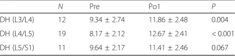
The mean follow-up duration was 4.17 ± 1.25 years. The results are valid because each patient was followed up for at least 2 years. The mean preoperative VAS was 8.88 ± 1.36, while it was 2.25 ± 1.26 postoperatively indi-cating that pain was relieved effectively (p< 0.05). Based on the JOA, the quality of life of the patients has been greatly improved. Bone healing was achieved in 24 patients and signs of root tension were negative in 23. No neurological deficits were noted; pain was not re-lieved in only one patient. Cage interbody fusion was used in three cases and autogenous bone in 21 patients. The changes in the mean difference of the intervertebral disc height at L3–L4, L4–L5, and L5–S1 were statisti-cally significant (P< 0.05) (Table1). The mean postoper-ative disc height at 1 year at L3–L4, L4–L5, and L5–S1 was lower than the preoperative values, and significant differences were noted at L4–L5 and L5–S1 (P< 0.05) (Table 2). It is proved that the use of autologous bone graft is difficult to maintain the height of the interverte-bral disc, hence it is't a good method of bone graft.
Case report
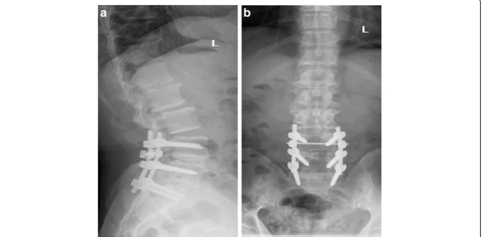
A 66-year-old man visited our outpatient clinic for severe back pain that he had developed spontaneously 3 years ago, in 2014. However, since it was not associated with significant disability, he was treated conservatively (anti-inflammatory painkillers, back exercises, etc.). Six months before hospitalization, he felt unbearable pain with numb-ness of the right lower limb and could only walk up to 100 m. On post-admission assessment, VAS was 10 points and JOA 12 points. X-ray, MRI, and CT showed double-level lumbar spondylolisthesis at L4–L5, with bilateral spondylolysis, and significant compression of the corre-sponding nerve (Figs.1and2). The muscle strength in the right lower limb was grade IV with normal muscle tension and negative straight leg raising test bilaterally. Therefore, we decided to treat him with surgery. We performed PLIF with autogenous bone graft, appropriate reduction, and sufficient decompression of the L4–L5 and L5–S1 verte-brae (Fig.3). Postoperatively, the back pain and numbness of the lower limbs were significantly reduced without re-striction of walking. Before discharge, VAS score was 2
Table 1Preoperative (Pre) and postoperative (Po1) lumbar disc height
N Pre Po1 P
DH (L3/L4) 12 9.34 ± 2.74 11.86 ± 2.48 0.004
DH (L4/L5) 19 8.17 ± 2.12 12.67 ± 2.41 < 0.001
and JOA was 23. At a follow-up after 1 year of self-care and only occasional soreness of the waist and back, VAS score was 1 and JOA score was 25, and imaging examin-ation showed that the lumbar vertebrae reached bony union.
Discussion
Spondylolisthesis is defined as the anterior or posterior migration, or slippage, of one vertebra in relation to the
next caudal vertebra. It mostly occurs in the lumbar spine and is considered to be due to with spondylolysis or degeneration [13]. Spondylolytic spondylolisthesis is distinguished by fracture of the pars interarticularis and is observed primarily during childhood or early adult life [14, 15]. Although the factors for the development of lumbar spondylolisthesis are still not clear, hereditary, traumatic, mechanical, and hormonal factors may play an important role in the process [16]. The occurrence of isthmus may be related to chronic strain, large spine pressure, and large shear force. It is known that women are approximately three times more likely to be affected by spondylolisthesis than men [14,17,18], which is con-sistent with our measurements (6 male/24 patients). Double-level lumbar spondylolysis is more serious spon-dylolisthesis, as some reports reported that female lum-bar spondylolisthesis more easily progress [4]. In this study, we also found that the greater degree of slipping of the L3–L4 and L4–L5 vertebrae promotes easy frac-ture of the isthmus. It is easy to understand that the greater the angle of slipping, the more unstable the ver-tebral body is, the easier it is to crack the isthmus. Ac-cording to VAS and JOA, surgery in most patients with symptoms is effective. Postoperative follow-up CT and X-ray showed good bone fusion at 1 year; therefore, both the surgical methods can be effective at improving the symptoms.
Although spondylolisthesis is usually asymptomatic, it can progress to spinal stenosis and result in neurogenic symptoms, such as leg pain, numbness, or weakness [19]. Conservative treatment is the first consideration, but if regular conservative treatment has not been allevi-ated and the quality of life is affected, surgery is an ef-fective solution. Treatment included a decompression alone, decompression and fusion, fusion including postero-lateral, interlaminar fusion, and interbody fusion. For two-level spondylolisthesis patients, spinal stability is poor and needs to be fixed and fused; at the same time, we adopted the most biomechanical inter-body fixation. So surgery in such cases is mainly for lumbar fixation, restoration of physiological curvature, and decompression of the compressed nerve root (Fig.4). Despite a variety of surgical methods as previously re-ported [20], there are five main approaches: posterior lumbar interbody fusion (PLIF), transforaminal lumbar interbody fusion (TLIF or MI-TLIF), oblique lumbar interbody fusion/anterior to psoas (OLIF/ATP), anterior lumbar interbody fusion (ALIF), and lateral lumbar interbody fusion (LLIF). The advantages of ALIF are that it retains all the posterior-stabilizing structures, reduces adjacent segment disease from denervation or injury to the adjacent facet joints and muscles, and avoids, both, epidural scarring and perineural fibrosis [21]. However, its risks include a potential for visceral injury (5%), Table 2Intervertebral disc height upon follow-up
N Po 1 Po 2 P
DH (L3/L4) 12 11.86 ± 2.48 10.71 ± 2.97 0.100
DH (L4/L5 19 12.67 ± 2.41 10.32 ± 1.74 < 0.001
DH (L5/S1) 11 11.41 ± 2.46 10.76 ± 1.74 0.035
Po 1: The intervertebral disc height was measured within a week of the operation
Po2: The intervertebral disc height was measured within a year of the operation
NPerfect number of follow-up patients
Fig. 1Magnetic resonance image shows II-degree spondylolisthesis at L4 and L5, and the corresponding segment of the
retrograde ejaculation and sympathetic dysfunction (3%) [22], and difficulty with revision due to the potential for scar tissue formation on the interface between the aorta and common iliac vein on the anterior border of the spine [23]. In our study, we adopt PLIF because it allows for adequate interbody height restoration and neural decompression while maintaining posterior support structures, [24]. After posterior pedicle screw fixation, most of the upper vertebral body force was transferred to the lower vertebral body through the nail bar system.
The bone conduction and internal pressure decreased significantly, which greatly reduced the incidence of pedicle screw loosening and fracture. Surgical decom-pression mainly includes the spinous process and the whole laminectomy, the medial part of the upper and inferior articular processes, nerve root canal scar tissue resection, and strive to complete decompression. The in-traoperative technique is mainly to accomplish moderate reduction, not necessarily completely reset, because ex-cessive reduction will also cause nerve root compression
Fig. 3D,EMRI, CT, X-ray show double-level spondylolisthesis at L4 and L5, in MRI and CT show the corresponding segment of the nerve com-pression. Preoperative lateral lumbar spine. Slipping of L4 and L5 resulting in II spondylolisthesis, and bilateral spondylolysis at L4 and L5
because of the soft tissue card. On the contrary, resetting can eliminate the spondylolisthesis and restore the normal physiological curvature of the lumbar spine, thereby improving the appearance of the lumbar spine and ensuring the normal physiological line of the lumbar spine. Spondylolisthesis can lead to narrowing of the intervertebral foramen, and resetting of the vertebral body can restore the height of the intervertebral
foramen, thus reducing the nerve root compression symptoms caused by the stenosis. In 23 patients, pain in the waist and back, and numbness and pain in the lower extremities disappeared or improved markedly after the surgery, and 1 case was not relieved. The success rate of interbody fusion was 87.5% (21/24), and there are no screw breakage and slippage phenomenon during the follow-up period.
Fig. 5a,bNo spondylolisthesis was seen after 1 year, and bone healing was achieved in the intervertebral bone graft
In earlier days, we used autologous bone fragments, and later on, cage was used. It has been reported that changes in disc height between the pre- and postopera-tive periods were significant with autologous bone and cage [25]. Based on the disc height measurements, we can see that whether interbody fusion with autogenous bone chips from posterior decompression or with cage, after pulling nail rod reduction, intraoperative distrac-tion, disc height can be restored, but resulting in postop-erative loss of disc height, statistics show that there are differences (P< 0.05). It may have several reasons: first, cage can provide an immediate stability, and second, during the process of bone fusion, bone chips undergo dissolution, absorption, cell growth, and trabecular bone absorption. Under the biological pressure, the height of intervertebral disc will decrease, and using cage fusion device may help maintain the height of intervertebral disc. In conclusion, the use of cage intervertebral bone graft is more helpful to maintain the height of the inter-vertebral disc (Fig.5).
The reduction order is worth discussing. According to the direction of slip, multi-segment degeneration slip-page is divided into three categories: the former spondy-lolisthesis, posterior spondylolisthesis, and mixed spondylolisthesis. Clinically, the most common are the former and mixed spondylolistheses. Due to different di-rections of vertebral spondylolistheses, reduction of multi-segment involvement is more difficult than that of a single segment. Intraoperatively, we found that due to the instability of adjacent vertebral bodies, the lower spondylolisthesis cannot provide mechanical fulcrum and the upper vertebral spondylolisthesis aggravated. In order to make the reduction simple, the“fulcrum” verte-bral body was reset first in the operation according to the type of slipping. We suggest that according to our experience, anterior spondylolisthesis should be first re-set, followed by fixation of the superior vertebral body and reattachment of the lower vertebral body. After the upper vertebral reduction, the lower vertebral body is repositioned through the upper metal rod. Mixed slip-page while fixing the upper and lower vertebral bodies, and then synchronize lifting intermediate vertebrae slip-page, may be reset from the bottom to the slide and fixed to the lower vertebral body, and then reset, the upper slide fixing vertebrae. While fixing difficulties, pedicle pulling system, pre-bent rods titanium leverage to reset the instrument.
There are certain limitations to our study. We adopt dynamic lateral flexion and extension X-ray films to evaluate the fusion state, which is widely used. However, CT is more advantageous in evaluating the imaging diagnosis of spinal fusion. Because of the low incidence rate, the number of single centre samples is small and the conclusions are limited.
Conclusion
In conclusion, we have reported 24 cases of double-level lumbar spondylolisthesis and have summarized the re-sults of a literature review. We found that both surgical techniques could significantly improve pain and disabil-ity in patients with double-level lumbar spondylolisthesis and achieve good mid-term prognosis. We compared autologous bones and cages and provided some thoughts on vertebral resuscitation. Further quantitative and de-tailed studies are required in this topic.
Abbreviations
ALIF:Anterior lumbar interbody fusion; CT: Computed tomography; JOA: Japanese Orthopaedic Association; PLIF: Posterior lumbar interbody fusion; Po: Postoperative; Pro: Preoperative; VAS: Visual Analog Scale
Acknowledgements
We greatly appreciate Editage who provided English language editing.
Funding
This project is supported by The Foundation of Health Department of Jiangxi Province (GJJ160127), Jiangxi Provincial Science and Technology Department (2015)24, and Gan-Po Talents Project 555 of Jiangxi Province.
Availability of data and materials
The authors confirm that all data underlying the findings are fully available from the corresponding author on reasonable request.
Authors’contributions
MD and BZ contributed to the experimental design; CY, LQ, and HZ contributed to the data collection; XY and XL contributed to the data analysis; and SZ completed the article writing. All authors read and approved the final manuscript.
Ethics approval and consent to participate
This research was approved by the ethics committee of the First Affiliated Hospital of Nanchang University. And agreed that participation was given to participants. Because of the retrospective nature of the study, informed consent was waived.
Consent for publication
Written informed consent for publication of their clinical details and/or clinical images was obtained from the patient/parent/guardian/relative of the patient.
Competing interests
This manuscript has not been published or presented elsewhere in part or in entirety and is not under consideration by another journal. All study participants provided informed consent, and the study design was approved by the appropriate ethics review board. We have read and understood your journal’s policies, and we believe that neither the manuscript nor the study violates any of these. The authors declare that they have no competing interests.
Publisher’s Note
Springer Nature remains neutral with regard to jurisdictional claims in published maps and institutional affiliations.
Author details
1
Received: 14 October 2017 Accepted: 16 January 2018
References
1. Smorgick Y, Park DK, Baker KC, Lurie JD, Tosteson TD, Zhao W, et al. Single-versus multilevel fusion for single-level degenerative spondylolisthesis and multilevel lumbar stenosis: four-year results of the spine patient outcomes research trial. Spine (Phila Pa 1976). 2013;38(10):797–805.https://doi.org/10. 1097/BRS.0b013e31827db30f. PubMed PMID: 23169068; PubMed Central PMCID: PMCPMC3757550
2. Weinstein JN, Lurie JD, Tosteson TD, Hanscom B, Tosteson AN, Blood EA, et al. Surgical versus nonsurgical treatment for lumbar degenerative spondylolisthesis. N Engl J Med. 2007;356(22):2257–70.https://doi.org/10. 1056/NEJMoa070302. PubMed PMID: 17538085; PubMed Central PMCID: PMCPMC2553804
3. Schuller S, Charles YP, Steib JP. Sagittal spinopelvic alignment and body mass index in patients with degenerative spondylolisthesis. Eur Spine J. 2011;20(5):713–9.https://doi.org/10.1007/s00586-010-1640-2. PubMed PMID: 21116661; PubMed Central PMCID: PMCPMC3082684
4. Ganju A. Isthmic spondylolisthesis. Neurosurg Focus. 2002;13(1):E1.https:// doi.org/10.3171/foc.2002.13.1.2. PubMed PMID: 15916408
5. Standaert CJ, Herring SA. Spondylolysis: a critical review. Br J Sports Med. 2000;34(6):415–22. PubMed PMID: 11131228; PubMed Central PMCID: PMCPMC1724260
6. Kuraishi S, Takahashi J, Mukaiyama K, Shimizu M, Ikegami S, Futatsugi T, et al. Comparison of clinical and radiological results of posterolateral fusion and posterior lumbar interbody fusion in the treatment of L4 degenerative lumbar spondylolisthesis. Asian Spine J. 2016;10(1):143–52.https://doi.org/ 10.4184/asj.2016.10.1.143. PubMed PMID: 26949470; PubMed Central PMCID: PMCPMC4764527
7. Liu X, Wang Y, Qiu G, Weng X, Yu B. A systematic review with meta-analysis of posterior interbody fusion versus posterolateral fusion in lumbar spondylolisthesis. Eur Spine J. 2014;23(1):43–56.https://doi.org/10.1007/ s00586-013-2880-8. PubMed PMID: 23812825; PubMed Central PMCID: PMCPMC3897839
8. Al-Khawashki H, Wasef Al-Sebai M. Combined dysplastic and isthmic spondylolisthesis: possible etiology. Spine (Phila Pa 1976). 2001;26(23):E542– 6. PubMed PMID: 11725254
9. Chung CH, Chiu HM, Wang SJ, Hsu SY, Wei YS. Direct repair of multiple levels lumbar spondylolysis by pedicle screw laminar hook and bone grafting: clinical, CT, and MRI-assessed study. J Spinal Disord Tech. 2007; 20(5):399–402.https://doi.org/10.1097/01.bsd.0000211253.67576.90. PubMed PMID: 17607107
10. Dunn AS, Baylis S, Ryan D. Chiropractic management of mechanical low back pain secondary to multiple-level lumbar spondylolysis with spondylolisthesis in a United States Marine Corps veteran: a case report. J Chiropr Med. 2009;8(3):125–30.https://doi.org/10.1016/j.jcm.2009.04.003. PubMed PMID: 19703668; PubMed Central PMCID: PMCPMC2732249 11. Ogawa H, Nishimoto H, Hosoe H, Suzuki N, Kanamori Y, Shimizu K.
Clinical outcome after segmental wire fixation and bone grafting for repair of the defects in multiple level lumbar spondylolysis. J Spinal Disord Tech. 2007;20(7):521–5.https://doi.org/10.1097/BSD. 0b013e3180335c1f. PubMed PMID: 17912129
12. Ravichandran G. Multiple lumbar spondylolyses. Spine (Phila Pa 1976). 1980; 5(6):552–7. PubMed PMID: 7466463
13. Wiltse LL, Newman PH, Macnab I. Classification of spondylolisis and spondylolisthesis. Clin Orthop Relat Res. 1976;117:23–9. PubMed PMID: 1277669
14. Fredrickson BE, Baker D, McHolick WJ, Yuan HA, Lubicky JP. The natural history of spondylolysis and spondylolisthesis. J Bone Joint Surg Am. 1984; 66(5):699–707. PubMed PMID: 6373773
15. Beutler WJ, Fredrickson BE, Murtland A, Sweeney CA, Grant WD, Baker D. The natural history of spondylolysis and spondylolisthesis: 45-year follow-up evaluation. Spine (Phila Pa 1976). 2003;28(10):1027–35.https://doi.org/10. 1097/01.BRS.0000061992.98108.A0. discussion 35. PubMed PMID: 12768144 16. Logroscino G, Mazza O, Aulisa G, Pitta L, Pola E, Aulisa L. Spondylolysis and spondylolisthesis in the pediatric and adolescent population. Childs Nerv Syst. 2001;17(11):644–55.https://doi.org/10.1007/s003810100495. PubMed PMID: 11734982
17. Kalichman L, Kim DH, Li L, Guermazi A, Berkin V, Hunter DJ. Spondylolysis and spondylolisthesis: prevalence and association with low back pain in the
adult community-based population. Spine (Phila Pa 1976). 2009;34(2):199– 205.https://doi.org/10.1097/BRS.0b013e31818edcfd. PubMed PMID: 19139672; PubMed Central PMCID: PMCPMC3793342
18. Amato M, Totty WG, Gilula LA. Spondylolysis of the lumbar spine: demonstration of defects and laminal fragmentation. Radiology. 1984;153(3):627–9. https://doi.org/10.1148/radiology.153.3.6494460. PubMed PMID: 6494460 19. Moller H, Sundin A, Hedlund R. Symptoms, signs, and functional disability in
adult spondylolisthesis. Spine (Phila Pa 1976). 2000;25(6):683–9. discussion 90. PubMed PMID: 10752099
20. Mobbs RJ, Phan K, Malham G, Seex K, Rao PJ. Lumbar interbody fusion: techniques, indications and comparison of interbody fusion options including PLIF, TLIF, MI-TLIF, OLIF/ATP, LLIF and ALIF. J Spine Surg. 2015;1(1): 2–18.https://doi.org/10.3978/j.issn.2414-469X.2015.10.05. PubMed PMID: 27683674; PubMed Central PMCID: PMCPMC5039869
21. Burkus JK, Gornet MF, Dickman CA, Zdeblick TA. Anterior lumbar interbody fusion using rhBMP-2 with tapered interbody cages. J Spinal Disord Tech. 2002;15(5):337–49. PubMed PMID: 12394656
22. Rao PJ, Loganathan A, Yeung V, Mobbs RJ. Outcomes of anterior lumbar interbody fusion surgery based on indication: a prospective study. Neurosurgery. 2015;76(1):7–23.https://doi.org/10.1227/NEU. 0000000000000561. discussion -4. PubMed PMID: 25255259 23. Kaiser MG, Haid RW Jr, Subach BR, Miller JS, Smith CD, Rodts GE Jr.
Comparison of the mini-open versus laparoscopic approach for anterior lumbar interbody fusion: a retrospective review. Neurosurgery. 2002;51(1): 97–103. discussion -5. PubMed PMID: 12182440
24. Lestini WF, Fulghum JS, Whitehurst LA. Lumbar spinal fusion: advantages of posterior lumbar interbody fusion. Surg Technol Int. 1994;3:577–90. PubMed PMID: 21319127
25. Song D, Chen Z, Song D, Li Z. Comparison of posterior lumbar interbody fusion (PLIF) with autogenous bone chips and PLIF with cage for treatment of double-level isthmic spondylolisthesis. Clin Neurol Neurosurg. 2015;138:111–6.https://doi.org/10.1016/j.clineuro.2015.08.012. PubMed PMID: 26318362
• We accept pre-submission inquiries
• Our selector tool helps you to find the most relevant journal
• We provide round the clock customer support
• Convenient online submission
• Thorough peer review
• Inclusion in PubMed and all major indexing services
• Maximum visibility for your research
Submit your manuscript at www.biomedcentral.com/submit