Lincoln
University
Digital
Thesis
Copyright
Statement
The
digital
copy
of
this
thesis
is
protected
by
the
Copyright
Act
1994
(New
Zealand).
This
thesis
may
be
consulted
by
you,
provided
you
comply
with
the
provisions
of
the
Act
and
the
following
conditions
of
use:
you
will
use
the
copy
only
for
the
purposes
of
research
or
private
study
you
will
recognise
the
author's
right
to
be
identified
as
the
author
of
the
thesis
and
due
acknowledgement
will
be
made
to
the
author
where
appropriate
you
will
obtain
the
author's
permission
before
publishing
any
material
from
the
thesis.
The effect of a non-steroidal anti-inflammatory
drug on subclinical endometritis in dairy cows and the
identification of at-risk cows
A thesis
submitted in partial fulfilment
of the requirements for the Degree of
Master of Agricultural Science
at
Lincoln University
by
Nicola Priest
Lincoln University
Abstract of a thesis submitted in partial fulfilment of the
requirements for the Degree of Master of Agricultural Science.
Abstract
The effect of a non-steroidal anti-inflammatory
drug on subclinical endometritis
and the identification of at-risk cows
by
Nicola Priest
Subclinical endometritis (SCE) is a uterine pathology characterised by an increased proportion of
polymorphonuclear cells (PMN) in the uterus after calving, and it is known that SCE has negative
effects on dairy cow reproductive performance. However, the mechanism by which SCE affects
reproductive performance in New Zealand dairy cows appears to be different from that reported in
international literature. This provided the basis for the research reported herein, which sought to
investigate the mechanism by which SCE reduces reproductive performance of New Zealand dairy
cows. Furthermore, the need for a practical method to detect or diagnose SCE was identified.
The objective of the first experiment was to determine if the inflammation associated with SCE, both
uterine and systemic, is a part of the mechanism by which reproductive performance is reduced in
cows with this disease. The hypothesis was that reducing this inflammation with a non-steroidal
anti-inflammatory drug (NSAID) would reduce the severity of uterine inflammation (average PMN %), and
improve reproductive performance. Dairy cows (n = 213) were paired by calving date and day 14
uterine PMN %, and randomly assigned to either the NSAID treatment (administered 21 - 31 days
postpartum) or control group. Cows with ≥ 14% PMN in the cytological sample collected at day 14
postpartum were defined as having SCE. Treatment with a NSAID increased pregnancy rate in SCE
cows and reduced metabolic indicators of systemic inflammation. There was, however, no effect of
NSAID treatment on day 42 PMN %, postpartum anovulatory interval, or milk production. Further
research is required to determine the effect of NSAID on SCE, and evaluate the influence of timing of
drug application on treatment effectiveness.
The objective of the second experiment was to determine whether a model could be developed,
both predict cows at risk of SCE and reduce the number of cows to be submitted for cytological
examination to a manageable level. Models were developed based on either a single week’s data
(week relative to calving; -4, -3, -1, +1, +2), or the herd’s planned start of calving date (27th June, and
a week later; 4th July). The optimum PMN % threshold was determined for the model with the
highest predictive value (R2; week +1). The optimum PMN % threshold for the week +1 model’s fitted
values was 10.7% (sensitivity = 58%, specificity = 81%). This threshold, and all other thresholds
investigated, however, resulted in combinations of sensitivity and specificity where either too few
SCE cows were identified, or too many cows would be submitted for cytological examination. These
results indicate that although a model was generated that could predict all SCE cows, the dual aim of
predicting cows at-risk and enabling only a subset of cows to be submitted for cytological
examination was unable to be achieved with the serum and physical parameters evaluated. Further
research into the aetiology of SCE may provide better biological markers to use for prediction of SCE.
Keywords: subclinical endometritis, anti-inflammatory, reproduction, prediction, serum metabolites,
Acknowledgements
They say it takes a village to raise a child. Well it wasn’t quite a village, but it certainly took a big team
of people to help me complete my masters degree! To each and every person who has helped me
over the last two years, thank you very much for giving up your time to help me. That being said, no
matter how many others helped, I would never have made it through my degree without the help of
my supervisors, Susanne Meier, Graham Barrell, and Sabrina Greenwood. Susanne, as the supervisor
who was in the same building as me and therefore the person who had to put up with me the most,
my biggest thank you goes to you for all of your advice and patience, and especially for all those
times when I turned up to your desk saying ‘quick question’, which of course usually meant at least a
10 to 15 minute discussion! Graham, thank you for generously agreeing to take me on as your
masters student after I had already started my degree, I have appreciated your advice and
comments. Sabrina, a big thank you to you for all your encouragement and the effort you put into
setting up such a tailored and interesting programme of course work for me, and I also appreciate
the continued interest and support you have shown me once you had moved on from Lincoln
University. I truly appreciate the time that all three of you have all taken to guide, teach, and inspire
me throughout the course of my degree.
To my DairyNZ manager and team leader, Chris Burke and John Roche, thank you for your comments,
advice, and willingness to sit down and have a chat with me whenever I asked you to. Others on the
‘thank you list’ are the co-authors of my papers, Kirsty Schmidt (nee McLeod) and the rest of the
awesome DairyNZ technical team, Barbara Dow the statistics whiz, Kristina Mandok my
‘study-buddy’, Angela Sheehan the walking laboratory encyclopaedia, and my ‘you can do it Nicola’ friends
and family. My thanks also go to the New Zealand dairy farmers (through DairyNZ Inc) and the
Ministry of Business, Innovation and Employment for funding the research completed in my degree.
Last, but definitely not least, I want to thank my wonderful partner Richard Hemming. Thank you for
being the rock I could lean on, the shoulder I could cry on, and always being the one to make me
laugh. I promise that now that I will no longer be so worn out from burning brain cells all day that I
Table of Contents
Abstract ... ii
Acknowledgements ... iv
Table of Contents ... v
List of Tables ... vii
List of Figures ... viii
List of abbreviations ... x
Chapter 1 Introduction ... 1
Chapter 2 Literature review ... 4
2.1 Introduction ... 4
2.2 Postpartum uterine diseases ... 4
2.2.1 Clinical endometritis ... 4
2.2.2 Subclinical endometritis ... 5
2.3 Diagnosis of subclinical endometritis ... 8
2.4 Risk factors for endometritis ... 9
2.5 Prevalence of subclinical endometritis ... 11
2.6 The effect of subclinical endometritis on milk production ... 13
2.7 The effect of subclinical endometritis on reproductive performance ... 13
2.8 Factors associated with endometritis that impair reproductive performance ... 15
2.8.1 Bacteria ... 16
2.8.2 Inflammation ... 18
2.9 Treatment of endometritis ... 21
2.9.1 Antibiotics ... 21
2.9.2 Prostaglandins ... 22
2.9.3 Proteolytic enzymes and disinfectants ... 23
2.9.4 Non-steroidal anti-inflammatory drugs ... 23
2.10 Validating biological markers ... 25
2.10.1 Dry matter intake ... 26
2.10.2 Milk parameters ... 26
2.10.3 Markers in serum/blood ... 27
Chapter 3 The effect of an anti-inflammatory drug on subclinical endometritis ... 30
3.1 Declaration ... 30
3.2 Introduction ... 30
3.3 Materials and methods ... 31
3.3.1 Experimental design ... 31
3.3.2 Uterine cytology ... 32
3.3.3 Postpartum anovulatory interval ... 33
3.3.4 Breeding management ... 33
3.3.5 Blood sample collection ... 33
3.3.6 Milk production ... 34
3.3.7 Grazing management and body condition scoring ... 34
3.4 Results ... 36
3.4.1 Reproduction... 37
3.4.2 Metabolites ... 38
3.4.3 Milk production and body condition score ... 40
3.5 Discussion... 41
3.5.1 Effect of NSAID on reproduction, metabolic indicators, and milk production ... 41
3.5.2 Associations between PMN % and reproduction, metabolic indicators, and milk production ... 42
3.6 Conclusion ... 43
Chapter 4 Predicting cows at risk of subclinical endometritis ... 44
4.1 Declaration ... 44
4.2 Introduction ... 44
4.3 Materials and methods ... 45
4.3.1 Dataset 1 ... 45
4.3.2 Dataset 2 ... 45
4.3.3 Data used ... 45
4.3.4 Statistical analyses ... 46
4.4 Results and discussion ... 47
4.5 Conclusion ... 50
Chapter 5 General discussion ... 51
5.1 Mechanisms by which reproduction was improved ... 51
5.1.1 Scenario 1 ... 52
5.1.2 Scenario 2 ... 54
5.1.3 Scenario 3 ... 55
5.2 Hypotheses generated ... 56
5.3 Identifying the cows with subclinical endometritis ... 57
5.4 Conclusions and implications ... 59
Appendix A Calculating the cost of subclinical endometritis ... 61
List of Tables
Table 1: The three types of polymorphonuclear cells ... 7 Table 2: Risk factors for the establishment of endometritis ... 10 Table 3: The prevalence of subclinical endometritis diagnosed by cytobrush or lavage cytology
reported in the literature ... 12 Table 4: The impact of endometritis on milk composition and somatic cell count ... 13 Table 5: The impact of subclinical endometritis on reproductive performance ... 14 Table 6: The proportion of cows’ uteri that are contaminated with bacteria during the postpartum
period ... 16 Table 7: Regression equations for the models generated to predict polymorphonuclear cell % in
uterine cytology samples. Models were derived from serum metabolite
concentration and body condition score data collected from 4 weeks pre to 2 weeks postpartum and at two set dates (planned start of calving and one week later). Models names including ‘V’ were validated against a separate data set (results reported in the body of the paper), but suitable data were not available for validating the remaining equations... 47 Table 8: The impact of varying levels of subclinical endometritis on the reproductive performance
and farm profitability of a 400 cow herd with a 12 week mating period ... 59
Table A.1: The impact of varying levels of subclinical endometritis on herd reproductive
performance ... 62 Table A.2: Economic loss for the differences between obtained and desired 6-week in-calf rates
List of Figures
Figure 1: Representation of annual herd feed demand and pasture supply for a pasture-based seasonal calving system. Burke and Verkerk (2010), reproduced with permission from Reproduction in Domestic Ruminants 7, 2010, published by the Society of
Reproduction and Fertility, Nottingham, United Kingdom. ... 1 Figure 2: The effect of cytological endometritis, clinical endometritis, or both, on the proportion
of cows pregnant. Dubuc et al. (2010), reproduced with permission from Elsevier. ... 6 Figure 3: The production of prostaglandins (PGE2, PGF2α, PGI2, PGD2) via the conversion of
arachidonic acid by the cyclooxygenase enzyme (COX) -1 and -2 (Sordillo et al. 2009). .... 7 Figure 4: The number of services required per conception for cows with and without subclinical
endometritis. Gilbert et al. (2005), reproduced with permission from Elsevier. ... 14 Figure 5: Mechanisms by which bacteria and inflammation affect reproductive performance in
cows (modified, with permission, from Sheldon et al. (2009)). (1) Chemokines and cytokines induce polymorphonuclear cells (PMN) to leave the blood and enter the uterine endometrium. (2&3) Bacterial lipopolysaccharide (LPS) inhibits the secretion of gonadotrophin releasing hormone (GnRH) and luteinizing hormone (LH),
respectively, but has no effect on follicle stimulating hormone (FSH). (4) Bacterial LPS binds to a receptor on the ovary and suppresses the production of oestradiol. (5&6) Binding of LPS to endometrial cells causes these cells to secrete E series
prostaglandins (PGE2) instead of the F series prostaglandins (PGF2α). (7) Cytokines
reduce the mRNA expression for PGE2. (8) Cytokines inhibit the secretion of GnRH. ... 15
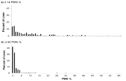
Figure 6: Polymorphonuclear cell (PMN) percentage distribution in uterine cytology samples from dairy cows at a) day 14, and b) day 42 postpartum. ... 36 Figure 7: Association between polymorphonuclear cell (PMN) group and the proportion of cows
ovulated by a specified day postpartum. The PMN groups are based on uterine cytology results from samples collected on day 14 postpartum: Low PMN (≤ 1% PMN); Medium PMN (2 to 13% PMN); High PMN (≥ 14% PMN). Ovulation was defined as the first sample day postpartum that progesterone concentration was > 1 ng/mL. Raw means and the maximum standard error of the difference (Max SED) are presented. † - There was a trend (P = 0.09) for the Low PMN group to have a higher proportion of cows ovulated by 28 days postpartum than the Medium or High PMN groups, but not at other times. ... 37 Figure 8: Effect of a non-steroidal anti-inflammatory drug (NSAID) treatment, and the interaction
with (A) Low, (B) Medium (MED), and (C) High polymorphonuclear cell (PMN) groups, on the proportion of cows pregnant by a specified week after the planned start of mating. Uterine cytology results from samples collected on day 14 postpartum were used to retrospectively create three PMN groups: Low PMN (≤ 1% PMN); Medium PMN (2 to 13% PMN); High PMN (≥ 14% PMN). The weekly pregnancy proportions have been calculated using the estimated conception date, which was calculated using the final pregnancy test results and mating data. There was an interaction (P = 0.04) between NSAID treatment and PMN group 4 weeks after the planned start of mating and there was a trend for an interaction at week 5 (P = 0.06), 8 (P = 0.07), 9 and 10 (P = 0.09); the interaction reflects an increase in pregnancy rate in the High PMN group treated with NSAID, but not the Low or Medium PMN groups. ... 38 Figure 9: Plasma concentrations of (a) total protein (b) albumin (c) globulin (d) the
albumin:globulin ratio (e) aspartate aminotransferase (ASAT) and (f) glutamate dehydrogenase (GDH) for the polymorphonuclear cell (PMN) groups based on uterine cytology results from samples collected on day 14 postpartum: Low PMN (≤ 1% PMN); Medium PMN (2 to 13% PMN); High PMN (≥ 14% PMN). Raw means and the maximum standard error of the difference (Max SED) are presented. ... 39 Figure 10: Plasma concentrations of (A) NEFA (B) Mg and (C) Ca for the polymorphonuclear cell
postpartum: Low PMN (≤ 1% PMN); Medium PMN (2 to 13% PMN); High PMN (≥ 14% PMN). Raw means and the maximum standard error of the difference (Max SED) are presented. ... 40 Figure 11: Regression of actual polymorphonuclear cell (PMN) % values against fitted PMN % for
the week +1 model. The actual PMN % values were obtained from Dataset 1, and the fitted PMN % values were calculated from Dataset 1 using the equation for the week +1 model. The week +1 model was generated from serum metabolite and cow body condition score data collected one week after calving. ... 48 Figure 12: Regression of actual polymorphonuclear cell (PMN) % values against predicted PMN %
for models (week relative to calving): a) week -1, b) week +1, and c) week +2. The actual PMN % values were obtained from Dataset 2, and the predicted PMN % values were calculated from Dataset 2 using the Dataset 1 equations. The models were generated from a single week’s serum metabolite and cow body condition score data that was collected weekly relative to calving. The P-value displayed is for the
correlation between the actual and predicted PMN % values. ... 48 Figure 13: Sensitivity and specificity for predicting cows at risk of subclinical endometritis using
five polymorphonuclear cell (PMN) % fitted value thresholds obtained from a receiver operating characteristic curve. The fitted values for PMN % were calculated from a model based on serum metabolite and cow body condition score data
obtained one week after calving (week +1 model). ... 49 Figure 14: The potential pathways by which a non-steroidal anti-inflammatory drug (NSAID)
treatment increased pregnancy rate in cows with subclinical endometritis. The blue arrows represent the pathway discussed for scenario 1, the red arrows represent the pathways discussed for scenario 2, and the dashed black and purple arrows represent the pathways discussed for scenario 3. The box labelled ‘reproductive axis’
represents components of the reproductive axis such as the hypothalamus and the ovaries. PMN = polymorphonuclear cells. ... 52 Figure 15: Hypothetical example of potential differences in polymorphonuclear cell (PMN) % rate
of decline from 14 to 42 days postpartum between cows that were treated with a non-steroidal anti-inflammatory drug (NSAID) and those that were not (Control). The shaded area is the period during which the NSAID treatment was administered. ... 53
List of abbreviations
AI Artificial insemination
AGR Albumin:globulin ratio
-APP Negative acute phase protein
+APP Positive acute phase protein
AS Acetylsalicylate
ASAT Aspartate aminotransferase
BCS Body condition score
CL Corpus luteum
COX Cyclooxygenase enzyme
CV Coefficient of variation
D 0 Day of calving
D 4 Four days after calving
DIM Days in milk
DMI Dry matter intake
FSH Follicle stimulating hormone
GDH Glutamate dehydrogenase
GnRH Gonadotrophin releasing hormone
LH Luteinising hormone
LPS Bacterial lipopolysaccharide
NEFA Non-esterified fatty-acids
NSAID Non-steroidal anti-inflammatory drug
PGD2 Prostaglandin D2
PGE2 Prostaglandin E2
PGF2α Prostaglandin F2α
PMN Polymorphonuclear cell(s)
PPAI Postpartum anovulatory interval
ROC Receiver operator characteristic curve
SCE Subclinical endometritis
SED Standard error of the difference
Chapter 1
Introduction
New Zealand’s dairy industry is a major contributor to the New Zealand economy as it is the
country’s biggest export earner, accounting for more than 25% of national export earnings (Burke
and Verkerk 2010). As greater than 90% of the milk produced is exported at world market prices
without subsidies, to remain profitable, tight economic constraints are placed on New Zealand dairy
farmers (Holmes et al. 2002). To remain profitable within these economic constraints, the cost of
milk production must be kept down. In New Zealand the cost of production is kept low by the use of
grazed pasture as the main constituent of dairy cow diets, because pasture grazed in situ is the
lowest cost feed available (Holmes et al. 2002; Blackwell et al. 2010). Therefore, to maximise pasture
utilisation, and in turn profitability, the changing herd feed demands need to be synchronised with
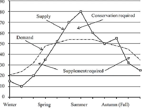
the growth pattern of pasture as pasture growth also varies throughout the year (Figure 1) (Parsons
1988; Burke and Verkerk 2010). This synchrony between pasture availability and herd feed demand is
achieved by seasonal calving, where cows calve in spring when herd feed demand is greatest and
pasture growth is maximal (Holmes et al. 2002; Blackwell et al. 2010).
Figure 1: Representation of annual herd feed demand and pasture supply for a pasture-based
seasonal calving system. Burke and Verkerk (2010), reproduced with permission from
Reproduction in Domestic Ruminants 7, 2010, published by the Society of Reproduction
In New Zealand, more than 95% of milk produced comes from strictly seasonal calving, grazed
pasture systems (Blackwell et al. 2010). To maximise the number of days in milk and thus milk
production, cows must calve every year in a tight calving pattern and have a 365 day inter-calving
interval (Blackwell et al. 2010). This means that farm productivity is directly related to cow
reproductive performance. To achieve this 365 day inter-calving interval, cows must conceive by 83
days after calving (Rhodes et al. 2003). This requires cows to resume cycling, display oestrous
behaviour, be mated during the early part of the breeding period, and in a seasonal calving system,
conceive by 83 days after the herd’s planned start of calving irrespective of time postpartum (Rhodes
et al. 2003; Blackwell et al. 2010). Cows that cannot maintain this 365 day inter-calving interval are at
risk of being culled (Blackwell et al. 2010).
The New Zealand dairy industry has set targets to achieve optimal reproductive performance. These
targets are a78% 6-week in-calf rate (78% of the cows pregnant after 6 weeks of mating), 90%
3-week submission rate (90% of cows mated within the first 3 3-weeks of the mating period), 60%
conception rate, and 6% not-in-calf rate after 12 weeks of mating (6% of cows not pregnant by the
end of the mating period; empty rate) (Burke et al. 2007). However, at an industry level, these
targets are not being met; in fact herd reproductive performance has been declining (Harris et al.
2006). The reduction in reproductive performance is partially attributed to the increase in the
number of cows that are anoestrous at the start of the mating period (Verkerk et al. 2000). This
increase in the number of anoestrous cows has a 2-fold impact on reproductive performance, in that
both the 3-week submission rate and the conception rate would be negatively affected. The 3-week
submission rate would be reduced because there would be fewer cows cycling and thus submitted
for mating within the first 3 weeks of the mating period. The conception rate would be reduced
because fertility increases as the number of oestrous cycles postpartum increases (up to ~4 cycles)
(Thatcher and Wilcox 1973), therefore, the cows that were anoestrous at the start of the mating
period will not have cycled four times prior to mating, and will therefore have reduced conception
rates. The reduction in submission and conception rates may result in these cows being culled for not
getting in calf, or for being a late calver in the current season or the subsequent season.
The increased risk of culling as a result of being anoestrous at the start of the mating period
highlights the fact that cows need to calve in the early part of the calving season and resume cycling
as early postpartum as possible. One factor that can delay the resumption of oestrous activity, and
negatively affect subsequent conception and pregnancy rates, is uterine disease. The uterine disease
that is investigated in this thesis, as a part of a larger programme of work, is subclinical endometritis
(SCE). Subclinical endometritis is a uterine pathology characterised by an increased proportion of
polymorphonuclear cells (PMN) present in the uterus after calving. International literature has
knowledge gap in the New Zealand context around this disease, the impact of SCE on dairy cow
reproductive performance in New Zealand was investigated. Previous studies by Burke et al. (2010)
and McDougall et al. (2011) reported that this disease was present in New Zealand and that it was
reducing reproductive performance. However, the mechanism by which SCE affected reproductive
performance in New Zealand dairy cows appeared to be different from that reported in the
international literature. This has provided the basis for the research reported in this thesis, which
sought to investigate the mechanism by which SCE reduces reproductive performance in New
Zealand dairy cows.
In addition to the need for further research into how SCE is reducing reproductive performance, the
need for a practical method to detect and diagnose SCE has also been identified. In New Zealand, it is
common practice for farmers to check their herd for clinical endometritis (Burke et al. 2007). They
are able to do this using the MetricheckTM device. However, the MetricheckTM device cannot detect
SCE (McDougall et al. 2011), therefore, currently there is no practical and cost-effective method for
on-farm detection and diagnosis of SCE. This has led to the investigation of using biological markers
in serum and cow body condition score to predict cows at risk of SCE.
The objectives of the research reported in this thesis are two-fold: (1) to attempt to determine if the
inflammation associated with SCE (both uterine and systemic) is the mechanism by which
reproductive performance is reduced in cows with SCE, and (2) to validate the usefulness of
biological markers in serum and cow body condition score for the identification of cows at risk of
SCE. The specific hypotheses for the research reported in this thesis are: (1) that treatment with
non-steroidal anti-inflammatory drug (NSAID) between 21 and 31 days postpartum will reduce the
severity (average polymorphonuclear cell (PMN) %) of uterine pathology at 42 days postpartum,
without lengthening the postpartum anoestrous interval (PPAI), and will mitigate the negative
association of SCE on reproduction and milk production by reducing inflammation and improving
liver function, and (2) that metabolic and physical characteristics could be used to predict cows at
risk of SCE and enable the identification of a subset of cows for cytological examination or
Chapter 2
Literature review
2.1
Introduction
As explained in the previous section, the research described in this thesis was designed to test two
different hypotheses. Because of this, the literature review covers two essentially separate, but
related, subjects: i.e. postpartum uterine disease and validation of biological markers. The topics that
are reviewed in the postpartum uterine diseases section are: endometritis (specifically SCE), its
prevalence, the effect of this disease on reproductive performance and milk production, the
proposed mechanisms of how factors associated with SCE reduce reproductive performance, and the
various treatment options that have been investigated for SCE. In the validation of biological markers
section, investigations of markers that have been trialled to detect/diagnose SCE are discussed.
2.2
Postpartum uterine diseases
Uterine disease can occur in the early postpartum period and manifests as conditions such as
metritis, pyrometra, or endometritis. An infection of the uterus within the first two weeks after
calving is called metritis (Sheldon et al. 2006). Metritis involves inflammation in all the layers of the
uterus: endometrial mucosa and submucosa, muscularis, and serosa (Bondurant 1999). Cows with
metritis show signs of systemic illness, e.g. fever, depression, weakness, inappetence, and decreased
milk yields (Bondurant 1999; Sheldon et al. 2006). Pyrometra is a chronic infection of the uterus, with
accumulation of purulent or mucopurulent material in the uterine lumen in the presence of a corpus
luteum (CL) (Sheldon et al. 2006; Parkinson et al. 2007).
Endometritis is defined as superficial inflammation of the endometrium, extending no deeper than
the stratum spongiosum (Bondurant 1999), that occurs more than two to three weeks postpartum
(Sheldon et al. 2006; LeBlanc 2008). The reason for stipulating a time frame for this disease is that
during the two to three weeks immediately after calving, a cow’s uterus undergoes an involution
process involving tissue remodelling and inflammation. The reported time to gross uterine and
cervical involution varies greatly from 25 to 47 days in milk (LeBlanc 2008). Thus it is inflammation of
the uterus that extends beyond two to three weeks postpartum that is termed endometritis (Sheldon
et al. 2006). Endometritis can be classified as either clinical or subclinical.
2.2.1
Clinical endometritis
Clinical endometritis in cows is defined as inflammation and infection of the uterus more than two to
purulent material on the tail and/or vulva, purulent (21 days postpartum) or mucopurulent (26 days
postpartum) material accumulated in the vagina, a cervical diameter > 7.5 cm after 20 days
postpartum, and malodorous mucus (LeBlanc et al. 2002a; Barlund et al. 2008; McDougall et al.
2011). It must be noted that the criteria for diagnosis, e.g. cervix size, changes with increasing time
postpartum (LeBlanc et al. 2002a).
Endometritis, both clinical and subclinical, causes significant economic losses because of decreased
reproductive performance and reduced milk yield (discussed in later sections), and increased culling
(Pleticha and Heuwieser 2009), therefore it is a condition that needs to be addressed. Clinical
endometritis is easy to detect and it is common practice for whole herds to be routinely checked
with the MetricheckTM device (Burke et al. 2007); therefore cows with clinical endometritis will most
likely be detected and treated. However, SCE is not easy to detect, which, therefore, means it can go
untreated, resulting in a greater probability of poor reproductive performance for the affected cows.
It is for this reason that the research reported in this thesis, where possible, is focused on SCE.
2.2.2
Subclinical endometritis
Subclinical endometritis is defined as cows with no systemic signs of illness or clinical infection
(Barlund et al. 2008), but that have inflammation of the endometrium, which is characterised by an
elevated population of PMN present in the uterus postpartum (Sheldon et al. 2009). Subclinical
endometritis has also been described as ‘cytological endometritis’ (Gilbert et al. 2005; Dubuc et al.
2010). Dubuc et al. (2010) defined cytological endometritis as “an increased proportion of PMN in
endometrial cytology samples obtained by endometrial cytobrush or low-volume uterine lavage”.
The authors indicated that cytological/subclinical endometritis and clinical endometritis may actually
represent different manifestations of reproductive tract disease and inflammation, rather than a
difference in severity of the same disease. Cows in the Dubuc et al. study were classified as having
cytological endometritis only, clinical endometritis only, or both cytological and clinical endometritis.
The results demonstrated that cytological endometritis only and clinical endometritis only had similar
detrimental effects on reproductive performance. However, for cows that had concurrent cytological
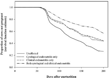
and clinical endometritis, the detrimental effects of both conditions were additive (Figure 2). This
finding is important as it may have implications for the way either form of endometritis is detected
Figure 2: The effect of cytological endometritis, clinical endometritis, or both, on the proportion of
cows pregnant. Dubuc et al. (2010), reproduced with permission from Elsevier.
As mentioned above, SCE is superficial inflammation of the endometrium (Sheldon et al. 2006;
LeBlanc 2008). The inflammation is present because inflammation is, as part of the innate immune
system, the body’s response to injury or infection (Serhan et al. 2010). Some of the processes that
occur during an inflammatory event include recognition of an injury or a foreign invading particle
(antigen), activation of endothelial cells to release pro-inflammatory cytokines and chemokines,
influx of polymorphonuclear cells (in particular neutrophils) to the site of the injury/antigen via
chemotaxis in response to the cytokines and chemokines, stimulation of release of acute-phase
proteins from the liver in response to proinflammatory cytokines, and phagocytosis of foreign
invading particles by PMN (Hussain 1989; Sheldon et al. 2009; Serhan et al. 2010; Galvão et al. 2011).
Inflammation is induced by prostaglandins that stimulate the release of cytokines (Bos et al. 2004).
Prostaglandins are bioactive lipids that are derived from the conversion of arachidonic acid by the
cyclooxygenase enzyme (COX) into PGH2 (Figure 3), which is then further metabolised into various
prostaglandins such as series F, E, I, and D prostaglandins (Mitchell et al. 1993; Sales and Jabbour
2003; Drillich et al. 2007). The COX-1 enzyme is constitutively expressed in cells; COX-2 is not, but its
expression is induced by some cytokines, endotoxins and inflammation (Mitchell et al. 1993; Drillich
Figure 3: The production of prostaglandins (PGE2, PGF2α, PGI2, PGD2) via the conversion of
arachidonic acid by the cyclooxygenase enzyme (COX) -1 and -2 (Sordillo et al. 2009).
In the case of bacterial infection, an inflammatory response is elicited by bacteria infecting the
uterine endometrium (Galvão et al. 2011). Once a pathogen has come into contact with the
endometrium, the endometrium is stimulated to produce cytokines and chemokines that attract
immune cells, in particular PMN, into the uterus and activate these cells once they are there (Galvão
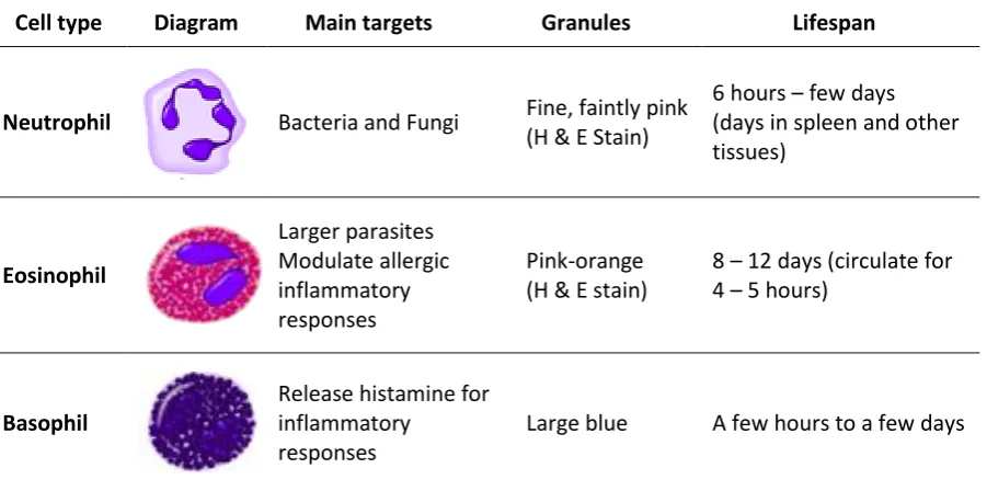
et al. 2011). There are three kinds of PMN: neutrophils, eosinophils and basophils (Table 1).
Neutrophils are the main PMN involved in uterine inflammation (Barlund et al. 2008). Neutrophils
are adept at seeking out and ingesting (phagocytising) bacteria and dead cells (Solomon et al. 2004).
Most of the cytoplasmic granules in neutrophils contain enzymes that digest the ingested material
(Solomon et al. 2004; Serhan et al. 2010).
Table 1: The three types of polymorphonuclear cells1
Cell type Diagram Main targets Granules Lifespan
Neutrophil Bacteria and Fungi Fine, faintly pink (H & E Stain)
6 hours – few days (days in spleen and other tissues)
Eosinophil
Larger parasites Modulate allergic inflammatory responses
Pink-orange (H & E stain)
8 – 12 days (circulate for 4 – 5 hours)
Basophil
Release histamine for inflammatory
responses
Large blue A few hours to a few days
1
(Alberts et al. 2007; Serhan et al. 2010)
The main function of PMN is phagocytosis and killing of invading bacteria. This involves four stages:
chemotaxis, adherence and attachment, ingestion, and digestion (Hussain 1989). Chemotaxis is the
Arachidonic acid
COX 1 and 2
PGE2 PGF2α
PGI2 PGD2
directed movement of a cell towards or away from a chemical source (Hussain 1989). When PMN are
exposed to cytokines and chemokines, the PMN become polarised and move towards the invading
bacteria (Hussain 1989; Galvão et al. 2011). It appears that the main cytokine involved in recruiting
PMN into the uterus is interleukin 8 (Ghasemi et al. 2012). Ghasemi et al. (2012) reported that cows
with SCE had a 50-fold higher gene expression of interleukin 8than cows without SCE. Once at the
site of infection, PMN attach to and ingest each bacterium by enclosing it in a phagosome (Hussain
1989). The bacteria are digested within phagosomes by lysosomal enzymes that are contained in the
cytoplasmic granules (Hussain 1989). It is because of these cytoplasmic granules that PMN are also
known as granulocytes. Polymorphonuclear cells are of particular interest with respect to SCE
because PMN are used to diagnose SCE.
2.3
Diagnosis of subclinical endometritis
Subclinical endometritis is diagnosed by determining the proportion of cells that are PMN within an
uterine cytology sample, i.e. PMN % (Sheldon et al. 2009). Uterine cytology is the collection and
counting of cells obtained from the uterus, and has become the standard to which other techniques
are compared (Barlund et al. 2008; Dubuc et al. 2010). There are two cytological techniques
commonly used to diagnose SCE (Kasimanickam et al. 2005a), the cytobrush method and the uterine
flushing/lavage method. Briefly, the cytobrush method involves a cytobrush being passed through
the cervix and gently rotated against the endometrium; the cellular material is then rolled on a slide
for microscopic evaluation to determine the proportion of cells that are PMN within the sample
(Kasimanickam et al. 2004; Barlund et al. 2008; McDougall et al. 2009). Uterine flushing involves the
uterine lumen being flushed with a small volume of saline solution, which is recovered and examined
by microscopy for the proportion of cells that are PMN within the sample (Gilbert et al. 2005).
Kasimanickam et al. (2005a) concluded that the cytobrush technique is a more consistent and
reliable technique than uterine lavage, the latter having a 17% failure rate in retrieving samples, a
reduced number of PMN recovered, and greater distortion of the recovered cells. For this reason, the
cytobrush technique will be used for the research conducted in this thesis.
To determine whether a cow has SCE or not, a diagnostic threshold for PMN % must be set. The PMN
% threshold used to define cows as having SCE varies depending on the time postpartum that the
cytology sample was collected and the outcomes being investigated. In general, the PMN % threshold
used decreases as time postpartum increases (Kasimanickam et al. 2005a; Gabler et al. 2010). This is
because uterine inflammation decreases as time postpartum increases as a result of the progression
of the involution process towards completion (Gilbert et al. 2005; Senosy et al. 2009). Studies have
used different approaches to determine what PMN threshold to use, such as using a PMN % that was
associated with a negative impact on reproductive performance or grouping cows into quartiles
analysis to determine the lowest PMN % that was associated with a negative effect on reproductive
performance; it was determined that > 18% PMN at 20 to 33 days postpartum and > 10% PMN at 34
to 47 days postpartum defined a cow as having SCE. Burke et al. (2010) used the second approach,
where the PMN % threshold was determined by categorising the cows into quartiles; the highest
PMN % quartile (> 6% PMN at day 42 postpartum) was used to identify cows with SCE. Both
approaches have positives and negatives. The first approach is good in that it determines its PMN %
threshold based on a tangible outcome, e.g. pregnant or not by a specified time after the start of the
mating period, rather than using an arbitrary cut-off. However, as many factors other than PMN %,
such as time from calving to mating and breeding management (Barlund et al. 2008; LeBlanc 2008),
will influence whether a cow gets pregnant by the specified time, a threshold based on reproductive
parameters may only be relevant for that specific study population. The benefit of the second
approach is that it avoids the circular argument of determining a threshold based on the
reproductive performance of the study population and then using that threshold to assess the
reproductive performance of that population (McDougall et al. 2011), but there is the potential that
the upper quartile PMN % may not be associated with reproductive performance at all. Therefore,
both approaches have merit, but which approach is best will be determined by the research question
that is being investigated.
Polymorphonuclear cells are used to diagnose SCE because they are a good indicator of the stage and
the degree of uterine inflammation as they are the predominant inflammatory cells found in
intrauterine fluid accumulations (Barlund et al. 2008). Anderson et al. (1985) and Kluciński et al.
(1990) indicated that there is almost a doubling (~90%) in the number of PMN present in the uterus
during clinical and subclinical uterine infection. These cells are a part of the body’s innate immune
system and provide the first line of defence against invading pathogenic organisms (Paape et al.
2002). Thus, when uterine infection or damage occurs, there is an influx of PMN into the uterine
lumen (Kasimanickam et al. 2004).
2.4
Risk factors for endometritis
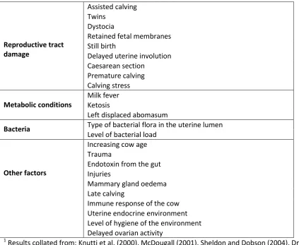
The main risk factors for the establishment of uterine infection and inflammation can be grouped
into four broad categories: reproductive tract damage, metabolic conditions, bacteria, and other
factors (Table 2). The most common risk factors reported are those associated with calving. Cows
that experience a difficult calving are at risk of developing endometritis postpartum. This is because
physical barriers such as the cervix, vagina, and vulva, which are the first line of defence against
bacteria, are breached and may be damaged during parturition (Sheldon and Dobson 2004). This
provides the opportunity for bacteria to invade and establish infection (Sheldon and Dobson 2004).
dystocia and may result in tissue that is more susceptible to bacterial infection (Parkinson et al.
2007).
Table 2: Risk factors for the establishment of endometritis1
Reproductive tract damage
Assisted calving Twins
Dystocia
Retained fetal membranes Still birth
Delayed uterine involution Caesarean section Premature calving Calving stress Metabolic conditions Milk fever Ketosis
Left displaced abomasum
Bacteria Type of bacterial flora in the uterine lumen
Level of bacterial load
Other factors
Increasing cow age Trauma
Endotoxin from the gut Injuries
Mammary gland oedema Late calving
Immune response of the cow Uterine endocrine environment Level of hygiene of the environment Delayed ovarian activity
1
Results collated from: Knutti et al. (2000), McDougall (2001), Sheldon and Dobson (2004), Drackley
et al. (2005), Drillich et al. (2005), Sheldon et al. (2006), LeBlanc (2008), Trevisi and Bertoni (2008),
McDougall et al. (2009), Sheldon et al. (2009), Burke et al. (2010), Cheong et al. (2011a), McDougall
et al. (2011).
The distinction must be made, however, between contamination and infection, as not all cows that
have bacteria present in their uterus have an infection (Sheldon et al. 2006). An infection requires
adherence of pathogenic organisms to the mucosa, colonisation or penetration of the epithelium by
the bacteria, and/or the release of bacterial toxins that lead to establishment of uterine disease
(Janeway et al. 2001). The establishment of infection depends on the bacterial load and species, as
well as the immune response of the cow (Sheldon et al. 2006; Parkinson et al. 2007).
The bacterial load within the uterus depends on two main factors; level of contamination and species
of bacteria (Parkinson et al. 2007). Contamination of the uterus with bacteria occurs during calving
for most cows. However, cows that suffer from peripartum disorders, such as dystocia and retained
risk of SCE. It must be noted though that although many species of bacteria may have entered the
uterus after calving, only certain species of bacteria are associated with SCE. Five main bacterial
species have been identified as the most common pathogenic bacteria isolated from the uterus in
total mixed ration (TMR) -based farm systems: Arcanobacterium pyogenes, Prevotella
melaninogenica, Escherichia coli, Fusobacterium necrophorum and Proteus spp. (Sheldon and Dobson
2004; Williams et al. 2007). The most common bacterial species for pasture-based farm systems have
not been reported. Of these five species, LeBlanc (2008) reported that commonly cows with
endometritis will have an infection predominated by Escherichia coli in the first week postpartum
and Arcanobacterium pyogenes in the second week. However, whether the invasion of the uterus by
these bacterial species results in infection depends on the immune response of the cow.
The immune response of a cow depends on many factors including her overall health status and
endocrine environment (Sheldon et al. 2006; Parkinson et al. 2007). A cow with compromised health
may be at greater risk of infection as she may not be able to mount a sufficient immune response
(Kasimanickam et al. 2004). Thus, the metabolic conditions and other factors mentioned in Table 2
could negatively affect a cow’s immune status and immune function, allowing bacteria to create an
infection. However, it is not only cows showing systemic signs of illness that may be at risk of SCE. It
has been reported by Green et al. (2009) and Burke et al. (2010) that cows that are retrospectively
classified as having SCE (based on PMN %) have indications of systemic inflammation and impaired
liver function. The indicators reported were a lower albumin concentration, a lower albumin:globulin
ratio, and elevated plasma concentrations of aspartate aminotransferase (ASAT) and glutamate
dehydrogenase (GDH). This systemic inflammation and impaired liver function may be negatively
affecting immune function, but this assertion has yet to be tested or reported. The endocrine
environment exerts its influence on immune function through progesterone and oestrogens.
Progesterone is immunosuppressive, which impairs the cows’ ability to mount an appropriate
immune response to bacteria, whereas oestrogens support immune function (Hussain 1989; Lewis
2003; LeBlanc 2008). This is in part because high oestrogen levels intensify PMN function, whereas
high progesterone levels inhibit PMN function (Hussain 1989; LeBlanc 2008). The interaction
between bacterial factors and cow immune function will affect the prevalence of SCE in the herd.
2.5
Prevalence of subclinical endometritis
Studies over the past 10 years in both pasture-based and TMR-based dairy systems have reported
Table 3: The prevalence of subclinical endometritis diagnosed by cytobrush or lavage cytology
reported in the literature
1
DIM = days in milk
2
Threshold = polymorphonuclear cell (PMN) % cut-off value used for diagnosing subclinical
endometritis
3
SCE = subclinical endometritis
From Table 3 it can be seen that the prevalence of SCE, in general, decreases with increasing time
postpartum. This is because many affected cows recover spontaneously (Gautam et al. 2009; Burke
et al. 2010). Green et al. (2009) reported a self-cure rate of 73% for cows with SCE. This can occur up
to four (LeBlanc et al. 2002b) to eight (Bretzlaff 1987) weeks postpartum. The spontaneous
resolution of endometritis is usually due to oestrus. When a cow ovulates, there is a spike of
oestrogens which stimulates uterine motility and PMN function, helping to clear uterine infection
(Hussain 1989; LeBlanc 2008). The high rate of self-cure in cows with endometritis is a factor that
needs to be taken into account when interpreting any results reported for the treatment of
endometritis.
It is important to note that although the inflammation/infection may have been resolved, its negative
impact on cow performance may still remain.
DIM1 Threshold2 SCE3 prevalence Study # of cows
Cytobrush cytology
18-38 > 5% PMN 38% Plöntzke et al. (2010) 194 (3 herds)
18-24 > 18% PMN 35% Lopdell et al. (2011) 46
20-33 > 18% PMN 35.1% Kasimanickam et al. (2004) 228
21-31 > 18% PMN 13.5% Heidarpour et al. (2012) 90
21-33 > 8% PMN 21.5% Madoz et al. (2013) 418 (4 herds)
28-41 > 8% PMN 11.8% Barlund et al. (2008) 221 (8 herds)
31-38 > 6% PMN 19.3% Dubuc et al. (2010) 1044 (6 herds)
32-52 > 5% PMN 19% Plöntzke et al. (2010) 194 (3 herds)
34-47 > 10% PMN 34% Kasimanickam et al. (2004) 228
34-47 > 6% PMN 16% Madoz et al. (2013) 418 (4 herds)
35 > 18% PMN 12.4% Kaufmann et al. (2010a) 209
39-45 > 18% PMN 7% Lopdell et al. (2011) 46
48-62 > 4% PMN 16% Madoz et al. (2013) 418 (4 herds)
53-59 > 4% PMN 11.1% Dubuc et al. (2010) 1018 (6 herds)
Lavage cytology
25-31 > 25% PMN 51.8% Hammon et al. (2006) 83
28-41 > 8% PMN 15.8% Barlund et al. (2008) 221 (8 herds)
31-38 > 8.5% PMN 38.2% Galvão et al. (2009a) 445 (5 herds)
40-60 > 5% PMN 53% Gilbert et al. (2005) 141 (5 herds)
46 > 10% PMN 25.9% Cheong et al. (2011a) 812 (39 herds)
2.6
The effect of subclinical endometritis on milk production
It was first hypothesised that endometritis would have no effect on milk production. This hypothesis
was initially supported by the results of Fourichon et al. (1999) who reported no loss of milk
production in cows with endometritis, and Dubuc et al. (2011) reported no reduction in milk
production for cows with SCE. However in studies conducted by Burke et al. (2010) and McDougall et
al. (2011), daily milk yield in cows with SCE was reduced by 0.6 – 1.03 kg/cow/day, and differences in
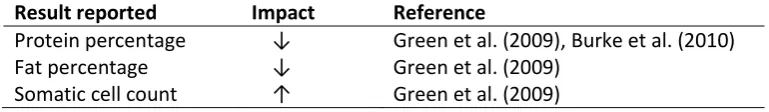
milk composition and somatic cell counts were also reported (Table 4).
Table 4: The impact of endometritis on milk composition and somatic cell count
Result reported Impact Reference
Protein percentage ↓ Green et al. (2009), Burke et al. (2010)
Fat percentage ↓ Green et al. (2009)
Somatic cell count ↑ Green et al. (2009)
The exact mechanism and nature of the link between endometritis and reduced milk production is
yet to be established. Preliminary theories revolve around reduced dry matter intake (DMI) for cows
with endometritis, which results in reduced milk production (Bertoni et al. 2008). The proposed
mechanism is that proinflammatory cytokines, which induce the inflammation associated with
endometritis, induce anorexia which reduces DMI and, therefore, milk production (Johnson and Finck
2001). This theory was supported by the reduced DMI and milk production observed in cows with
inflammation or uterine infection (Bell and Roberts 2007; Huzzey et al. 2007; Bertoni et al. 2008). It
therefore seems plausible that reducing inflammation would increase milk production. Support for
this hypothesis is provided by a study reported by Trevisi and Bertoni (2008). Half of the cows in the
study were untreated controls, the other half were treated with acetylsalicylate, an
anti-inflammatory drug, for the first five days postpartum. The treated cows had higher milk production,
higher peak milk yield (46.3 vs 40.9 kg/day), and reached peak daily milk production earlier (50.5 vs
57 days in milk) than control cows. These results indicate that the reduction of inflammation in the
early postpartum period is beneficial to milk production.
Compared with the inconsistent effect of SCE on milk production, there is substantial evidence that
SCE has a negative effect on reproductive performance.
2.7
The effect of subclinical endometritis on reproductive performance
The aim of reproductive management is to achieve good reproductive performance, specifically that
cows become pregnant at a biologically optimal time, in an efficient manner, and at an economically
beneficial interval after calving (LeBlanc et al. 2002a; Sheldon et al. 2006). In the New Zealand
seasonal calving system, this means cows must conceive by 83 days postpartum to maintain the
few weeks after calving (Opsomer et al. 2000; Rhodes et al. 2003). The ability to resume cycling, and
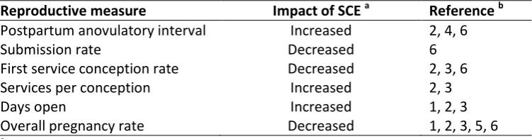
other reproductive parameters, has been reported to be negatively affected by SCE (Table 5).
Table 5: The impact of subclinical endometritis on reproductive performance
Reproductive measure Impact of SCE a Reference b
Postpartum anovulatory interval Increased 2, 4, 6
Submission rate Decreased 6
First service conception rate Decreased 2, 3, 6
Services per conception Increased 2, 3
Days open Increased 1, 2, 3
Overall pregnancy rate Decreased 1, 2, 3, 5, 6
a
SCE = subclinical endometritis
b
Results collated from: 1) Kasimanickam et al. (2004), 2) Gilbert et al. (2005), 3) Barlund et al. (2008),
4) Burke et al. (2010), 5) Dubuc et al. (2010), 6) McDougall et al. (2011).
Despite being a subclinical disease, there is comprehensive evidence that SCE has a negative effect
on reproductive performance (Table 5). These negative effects have been reported in both
pasture-based and pasture-based dairy systems. In a study reported by Gilbert et al. (2005), conducted in a
TMR-based dairy system, cows with SCE had a lower overall pregnancy rate at 300 days postpartum (63%
vs 89%), reduced probability of first-service conception (11% vs 36%), delayed days to first service
(median 101 vs 80 days), more services per conception required (median 3 vs 2 services; Figure 4)and
more median days open (206 vs 118 days), compared with cows without SCE.
Figure 4: The number of services required per conception for cows with and without subclinical
endometritis. Gilbert et al. (2005), reproduced with permission from Elsevier. No endometritis
Subclinical endometritis
Number of services
Pr
o
p
o
rti
o
n
n
o
t p
re
gn
an
In a study reported by McDougall et al. (2011), conducted in a pasture-based dairy system, cows with
SCE had a lower proportion (0.57 vs 0.97) pregnant overall, decreased pregnancy to first service, and
took longer to conceive (56.1 vs 32.6 days from the planned start of mating), compared with cows
without the condition.
From the studies reported in this section it is clear that SCE has a negative effect on reproductive
performance; the question is, how?
2.8
Factors associated with endometritis that impair reproductive
performance
The mechanisms by which SCE/endometritis reduces reproductive performance are still being
elucidated. This is because the interactions among infectious uterine bacteria, the immune system
and inflammatory response, and ovarian and uterine function are complex and not entirely
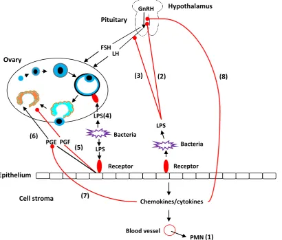
understood. Parts of these complex interactions are summarised in Figure 5, and explored in greater
detail in subsequent sections. The proposed mechanisms can be grouped into two broad categories:
those associated with bacteria, and those associated with the inflammation that accompanies SCE.
Figure 5: Mechanisms by which bacteria and inflammation affect reproductive performance in
cows (modified, with permission, from Sheldon et al. (2009)). (1) Chemokines and
cytokines induce polymorphonuclear cells (PMN) to leave the blood and enter the uterine (8)
(7)
(1) (6)
(5)
(4)
(2) (3)
GnRH
LH FSH
Pituitary
Hypothalamus
Ovary
Receptor LPS
Bacteria
Receptor LPS
Bacteria LPS
PGF PGE
Epithelium
Cell stroma Chemokines/cytokines
Blood vessel
endometrium. (2&3) Bacterial lipopolysaccharide (LPS) inhibits the secretion of
gonadotrophin releasing hormone (GnRH) and luteinizing hormone (LH), respectively, but
has no effect on follicle stimulating hormone (FSH). (4) Bacterial LPS binds to a receptor
on the ovary and suppresses the production of oestradiol. (5&6) Binding of LPS to
endometrial cells causes these cells to secrete E series prostaglandins (PGE2) instead of
the F series prostaglandins (PGF2α). (7) Cytokines reduce the mRNA expression for PGE2.
(8) Cytokines inhibit the secretion of GnRH.
2.8.1
Bacteria
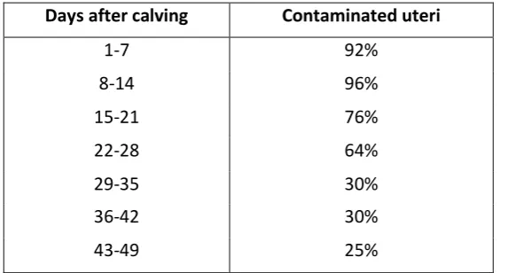
Parkinson et al. (2007) reported that > 90% of cows have bacterial contamination of the uterus for
the first two weeks after calving, with the prevalence decreasing with increasing time postpartum
(Table 6).
Table 6: The proportion of cows’ uteri that are contaminated with bacteria during the postpartum
period1
Days after calving Contaminated uteri
1-7 92%
8-14 96%
15-21 76%
22-28 64%
29-35 30%
36-42 30%
43-49 25%
1
Parkinson et al. (2007), reproduced, with permission, from 2007 Proceedings of the Society of Dairy
Cattle Veterinarians of the New Zealand Veterinary Association, published by VetLearn, Wellington,
New Zealand.
It has been widely reported that the invasion of the uterus by infection-causing bacteria has a
negative impact on reproductive performance. Bacteria exert their impact on reproductive
performance in a number of ways: by directly damaging the uterine endometrium (Herath et al.
2007; Williams et al. 2007) which delays uterine involution (McDougall 2001), through bacterial
lipopolysaccharide (LPS) disruption of follicle development and steroidogenesis which affects
subsequent ovulation (Herath et al. 2007), through disruption of the hypothalamus or pituitary gland
secretions (Battaglia et al. 1999), and through stimulation of the endometrium to secrete
prostaglandin E2 (PGE2) which leads to persistence of the CL (Williams et al. 2007; Sheldon et al.
Infection of the uterus with bacteria does not disrupt secretion of follicle-stimulating hormone (FSH),
thus, the first wave of ovarian follicles emerge approximately 10 days after parturition (Macmillan
1998; Sheldon et al. 2002). However, cows with endometritis have slower growth of dominant
follicles and lower peripheral plasma oestradiol concentrations, which reduces the likelihood of
ovulation (Sheldon et al. 2002). Oestrogens are essential for ovarian follicular growth, development
and function, and have a central role in nurturing the oocyte and ovulation (Schams and Berisha
2002). The granulosa cells that surround the oocyte produce oestradiol by aromatisation of
androstenedione derived from the theca cells, under the regulation of FSH (Fortune 1994). Bacteria
suppress production of oestradiol from the granulosa cells via the LPS molecules they express on
their surface (Herath et al. 2007). The LPS migrates to the ovary where it binds to the Toll-like
receptor-4 /CD14/MD-2 receptor complex, that is expressed by granulosa cells, and down-regulates
the expression of aromatase genes which results in suppression of oestradiol production and failure
of ovulation (Herath et al. 2007).
Another way that bacteria potentially disrupt follicle ovulation is by inhibiting the release of
gonadotrophin-releasing hormone (GnRH) and luteinising hormone (LH) from the hypothalamus and
the pituitary gland, respectively (Battaglia et al. 1999; Williams et al. 2001; Herath et al. 2009). Prior
to ovulation, plasma oestradiol concentrations rise; this signals the hypothalamus and the pituitary
gland to secrete the pre-ovulatory GnRH and LH surges which precede ovulation of the dominant
follicle (Moenter et al. 1990). However, the signal from oestradiol required for the surges of GnRH
and LH can be blocked by LPS (Battaglia et al. 1999), thus preventing ovulation. The mechanism by
which LPS block the oestrogen signal has not been reported. However, as LPS was administered
intravenously in this study, perhaps the LPS gained accessed the hypothalamus and pituitary to
supress the surges through the hypophysial portal blood system. It must be noted though, that LPS
only suppresses the secretion of gonadotrophins if the signal from the oestradiol is blocked before
the onset of GnRH/LH surge release. Once the surges have begun, LPS has no effect (Battaglia et al.
1999).
Once cows have successfully completed their first ovulation after calving, it is common for those with
uterine infections to have prolonged luteal phases, thus preventing the next ovulation from occurring
and delaying conception (Opsomer et al. 2000; Williams et al. 2007). If the infection is not cleared
during the follicular phase and remains during the following dioestrus, the CL becomes persistent
and has prolonged progesterone secretion (Parkinson et al. 2007). Progesterone suppresses immune
function and propagates continued infection (Parkinson et al. 2007), further reducing reproductive
function. Persistence of the CL is due to insufficient secretion of prostaglandin F2α (PGF2α), which is
required to regress the CL and terminate the luteal phase (Opsomer et al. 2000; Parkinson et al.
endometrial epithelial cells and is luteolytic, whereas PGE2, which is also secreted by endometrial
epithelial cells, is luteotrophic (Arosh et al. 2004). Insufficient secretion of PGF2α can be due to LPS
binding to endometrial cells and stimulating them to secrete PGE2 instead of PGF2α (Herath et al.
2009). This results in low concentrations of PGF2α and persistence of the CL.
In contrast to potentiating CL function, Williams et al. (2007) reported reduced luteal function in
cows with uterine infections. In this study, cows with uterine infections, that had ovulated, had
smaller corpora lutea and lower peripheral plasma concentrations of progesterone than normal
fertile animals. Lower levels of progesterone would inhibit conception and the survival of embryos,
as sufficient concentrations of progesterone are required to maintain pregnancy (McLeod and
Phillips 1998). The CL of cows with uterine infections were smaller because cows with uterine
infections ovulate smaller first dominant follicles, which produce smaller CL (Williams et al. 2007;
LeBlanc 2008).
While several studies reporting how bacteria negatively affect reproduction have been discussed in
this section, not all studies investigating the mechanisms by which endometritis affects reproduction
have found a link with bacteria. In contrast to the results reported in the TMR studies, the results
reported by McDougall et al. (2011) indicated that the five most common pathogenic bacteria
isolated from the uterus in TMR-based farm systems (A. pyogenes, P. melaninogenica, E. coli, F.
necrophorum and Proteus spp.) were not the major contributing factor to the poor reproductive
performance observed in cows with SCE in New Zealand dairy systems. This contrasting result may be
due to differences in management practices and environmental conditions. The authors concluded
that in cows with SCE, uterine inflammation (as measured by PMN %) is more closely related to the
negative reproductive outcomes associated with SCE than with isolation of bacteria from the uterus.
This conclusion is also supported by Barański et al. (2012). Therefore, the results reported by
McDougall et al. (2011) and Barański et al. (2012) suggest that there are inflammatory mechanisms
which reduce reproductive performance that are not necessarily related directly to bacteria.
2.8.2
Inflammation
Inflammation can be considered as a healing process if it is restrained within certain limits (e.g.
length of time the inflammation remains and the severity of the inflammatory response) but might
be harmful if those limits are exceeded (Grimble 2001). These limits appear to be exceeded in some
cows, as uterine inflammation has been reported to disrupt ovarian function, hypothalamic and
pituitary gland function, and reduce embryo survival.
The potential disruption of ovarian function by uterine inflammation was reported by Sheldon et al.
second dominant follicles in the ovary ipsilateral to the previously gravid horn, reduced dominant
follicle growth rates and decreased oestradiol secretion. The reduced dominant follicle growth rate
and oestradiol secretion would most likely result in failure of the dominant follicle to ovulate. A
possible reason for the reduced oestradiol secretion recorded in the Sheldon et al. (2002) study may
be that the release of proinflammatory cytokines by endometrial cells impairs granulosa cell
steroidogenesis (Spicer and Alpizar 1994; Sheldon et al. 2009). It has also been postulated that the
cytokines secreted by the inflamed endometrium are the reason for the reduced peripheral plasma
progesterone levels of cows with uterine inflammation (Sheldon et al. 2009). This is because bovine
luteal cells are highly responsive to a range of cytokines (Petroff et al. 2001; Okuda and Sakumoto
2003; Sheldon et al. 2009).
Rivest et al. (1993) reported that cytokines disrupted the hypothalamic-pituitary axis. In a study using
rats, the cytokine interleukin-1ß was reported to exert an effect on the hypothalamic-pituitary axis
by altering GnRH neuronal activity and inhibiting the release of hypothalamic GnRH. This would
prevent occurrence of the pre-ovulatory surge of GnRH that is essential for ovulation.
Uterine inflammation has been shown to have the potential to reduce embryo survival. Cows with
uterine inflammation have a suboptimal uterine environment for embryo survival, which contributes
to poor reproductive performance through embryonic loss. In a study conducted by Hill and Gilbert
(2008) embryos were cultured in media conditioned with the products recovered from either an
inflamed or a normal cow endometrium. The embryos cultured in inflamed conditioned media had
reduced blastocyst quality as determined by lower total trophectoderm cell numbers (83.1 vs 99.8
cells for inflamed vs non-inflamed conditioned media). This reduction in trophectoderm cell numbers
would be expected to result in reduced embryo viability and conception rate. The authors proposed
that the reduction in trophectoderm cell numbers could leave the inner-cell mass of the blastocyst
susceptible to damage from components such as inflammatory cytokines, and affect placental size
and function. This hypothesis is supported by Soto et al. (2003) who found that tumour necrosis
factor-α increased blastomere apoptosis and can thus compromised the development of the
resultant embryo.
A potential mechanism for the reduction in embryo quality in cows with uterine inflammation/SCE
was reported by Back et al. (2011). The cows with SCE in the study had increased concentrations of
histidine, alanine, aspartate, and serine in follicular fluid. Histidine is important in initiation of the
inflammatory response and can reduce the production of anti-inflammatory molecules; the other
three amino acids affect the function of cells of the innate immune system (Li et al. 2007). Back et al.
concluded that these changes in the amino acid profile could potentially result in altered oocyte