This may be the author’s version of a work that was submitted/accepted
for publication in the following source:
Maitland, Norman,
Chambers, Karen
, Georgopoulos, Lindsay,
Simpson-Holley, Martha, Leadley, Regina, Evans, Helen, Essand, Magnus,
Daniels-son, Angelika, van Weerden, Wytske, de Ridder, Corrina, Kraaij, Robert,
& Bangma, Chris
(2010)
Gene transfer vectors targeted to human prostate cancer: Do we need
better preclinical testing systems?
Human Gene Therapy
,
21
(7), pp. 815-827.
This file was downloaded from:
https://eprints.qut.edu.au/55861/
c
Consult author(s) regarding copyright matters
This work is covered by copyright. Unless the document is being made available under a Creative Commons Licence, you must assume that re-use is limited to personal use and
that permission from the copyright owner must be obtained for all other uses. If the docu-ment is available under a Creative Commons License (or other specified license) then refer to the Licence for details of permitted re-use. It is a condition of access that users
recog-nise and abide by the legal requirements associated with these rights. If you believe that this work infringes copyright please provide details by email to qut.copyright@qut.edu.au
Notice:
Please note that this document may not be the Version of Record
(i.e. published version) of the work. Author manuscript versions (as
Sub-mitted for peer review or as Accepted for publication after peer review) can
be identified by an absence of publisher branding and/or typeset
appear-ance. If there is any doubt, please refer to the published source.
Gene Transfer Vectors Targeted to Human Prostate Cancer:
Do We Need Better Preclinical Testing Systems?
Norman Maitland,1Karen Chambers,1Lindsay Georgopoulos,1Martha Simpson-Holley,1Regina Leadley,1 Helen Evans,1 Magnus Essand,2Angelika Danielsson,2Wytske van Weerden,3Corrina de Ridder,3
Robert Kraaij,3Chris H. Bangma,3and members of the GIANT FP6 Consortium
Abstract
Destruction of cancer cells by genetically modified viral and nonviral vectors has been the aim of many research programs. The ability to target cytotoxic gene therapies to the cells of interest is an essential prerequisite, and the treatment has always had the potential to provide better and more long-lasting therapy than existing che-motherapies. However, the potency of these infectious agents requires effective testing systems, in which hy-potheses can be explored bothin vitroandin vivobefore the establishment of clinical trials in humans. The real prospect of off-target effects should be eliminated in the preclinical stage, if current prejudices against such therapies are to be overcome. In this review we have set out, using adenoviral vectors as a commonly used example, to discuss some of the key parameters required to develop more effective testing, and to critically assess the current cellular models for the development and testing of prostate cancer biotherapy. Only by developing models that more closely mirror human tissues will we be able to translate literature publications into clinical trials and hence into acceptable alternative treatments for the most commonly diagnosed cancer in humans.
Introduction
T
he limited long-term in vivo efficacy of biologicaltherapies for human prostate cancer (Maitland et al., 2004) can be ascribed to a number of different causes: First, the entire concept could be wrong, that is, the gene or pathway to be exploited, perhaps discovered in cell line experiments, is simply not differentially expressed to the same extentin vivo; second, the agent itself could be wrong or inactive, perhaps unable to survive in thein vivoenvironment, or unable to penetrate cells while retaining its activity against the primary target; last, and often neglected, is that although the concept might be strong, and the agent active, the testing system used to define efficacy (and safety) could be defective.
Validity of Cell Lines That Represent Prostatic Disease
Prostate cancer has been poorly served by the establish-ment of cell lines. In general, new agents for prostate cancer are tested mainly in three cell types; PC3, LNCaP, and DU145. A review of the gene therapy literature suggests there are 2650 PubMed references that include the terms
prostate cancer andgene therapy. Of those, 562 employ only LNCaP cells; PC3 cells are used in 153 and DU145 cells in 179. The remainder employ a mixture of all three. In reality, only relatively few published studies make it through to the clinic. The Journal of Gene Medicine database (March 2009; http:==www.wiley.co.uk=genmed=clinical=) lists the actual number of prostate cancer clinical trials as considerably lower (105): a conversion rate of 4%. In fact, many of the reported trials are derivative, so the actual rate is closer to 1%. Later stage clinical trials have been dominated by vac-cine approaches to augment antitumor responses without any need to target the tumor itself (Smallet al., 2004).
The LNCaP, PC3, and DU145 cell lines were derived al-most 30 years ago from therapy-resistant metastatic cancers (Table 1). The TSU-Pr1 cell line was also considered to be from prostate, but was subsequently shown to be derived from the human bladder cancer cell line T24 (van Bokhoven et al., 2001). What is frequently lacking from these models is any representation of untreated low Gleason grade tumors within the prostate (Gleason, 1966), which have a high de-gree of normal prostate differentiation, or less structured,
1
Yorkshire Cancer Research Unit, Department of Biology, University of York, York YO10 5YW, United Kingdom. 2Clinical Immunology, Rudbeck Laboratory, Uppsala University, SE-75185 Uppsala, Sweden.
3Department of Urology, Josephine Nefkens Institute, Erasmus MC, 3000CA Rotterdam, The Netherlands.
DOI: 10.1089=hum.2009.210
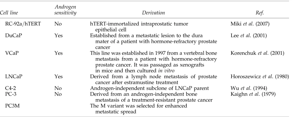
Table1. Cell Lines from Human Prostate for Vector Testing
Cell line
Androgen
sensitivity Derivation Ref.
Normal/ nonmalignant
PNT2 No Derived from normal prostate epithelium by transfection of anorimutant of SV40
Cussenotet al. (1991)
PNT1 No Derived from normal prostate epithelium by transfection of anoriSV40 plasmid
Cussenotet al. (1991)
BPH1 Noa Benign prostatic tissue after SV40 large-T antigen immortalization of primary cells
Haywardet al. (1995)
PZ-HPV-7 No Derived from epithelial cells cultured from normal tissue from the peripheral zone
of the prostate by transfection with HPV18 DNA
Weijermanet al. (1994)
RWPE-1 Noa Peripheral zone of a histologically normal adult human prostate transfected with a single copy of HPV18
Belloet al. (1997)
PWR-1E Noa Human prostatic epithelial cells, derived
from a normal prostate with mild hyperplasia, were immortalized with an adenovirus 12--SV40 hybrid virus (Ad12-SV40)
Webberet al. (1996)
WPMY-1 ND A myofibroblast stromal cell line derived from stromal cells from the peripheral zone of the histologically normal adult prostate, immortalized with SV40 large-T antigen gene
Webberet al. (1999)
RC156N=hTERT No hTERT-immortalized nonmalignant human prostate epithelial cell
Mikiet al. (2007)
Premalignant
P4E6 No Derived from Gleason 2þ2 (low-grade cancer) by retrovirus-mediated introduction of an HPV16 E6 gene
Maitlandet al. (2001)
RWPE-2 Noa Derived from RWPE-1 cells by transformation with Ki-ras, using the Kirsten murine sarcoma virus (Ki-MuSV): RWPE2-W99 was further selected by cloning in soft agar to select cells that show high expression of Ki-ras
Belloet al. (1997)
WPE1-NA22 WPE1-NB14 WPE1-NB11 WPE1-NB26
Noa WPE1-Nxx cells were derived from RWPE-1 cells after
exposure toN-methyl-N-nitrosourea
Webberet al. (2001)
WPE1-NB26-64 WPE1-NB26-65
ND WPE1-NB26-64 and WPE1-NB26-65 (CRL-2890; ATCC) were derived from a subcutaneous tumor in a nude mouse injected with WPE1-NB26
Rivetteet al. (2005)
WPE-stem WPE-int
ND WPE-stem and WPE-int cells were derived from the RWPE-1 cell line after two consecutive cycles of single-cell cloning
Tokaret al. (2005)
Carcinoma
MDA PCa 2b Yes MDA PCa 2b was established from a bone metastasis of androgen-independent adenocarcinoma
of the prostate
Navoneet al. (1997)
NCI-H660 ND Has the appearance and many (but not all) of the
properties of small-cell carcinoma of the prostate Gazdar and Minna (1996) LAPC-4 Yes Grown from androgen-sensitive lymph node
metastasis of prostate cancer
Kleinet al. (1997)
CA-HPV-10 Yes CA-HPV-10 was derived from cells from a prostatic adenocarcinoma of Gleason grade 4=4. The
cells were transformed by transfection with HPV18 DNA
Weijermanet al. (1994)
DU145 No Cultured from a brain metastasis of prostate cancer Stoneet al. (1978) 22Rv1 Yes 22Rv1 is a human prostate carcinoma epithelial cell
line derived from a xenograft of a primary prostate cancer that was serially propagated in mice after castration-induced regression and relapse of the parental, androgen-dependent CWR22 xenograft
Sramkoskiet al. (1999)
untreated but ‘‘undifferentiated’’ higher Gleason grade tu-mors. As screening and diagnostic techniques improve for prostate cancer (Schro¨der et al., 2009), ‘‘clinical’’ prostate cancers will be downgraded to lower Gleason grades, which will comprise the primary target for new therapies. At present, however, most biotherapies are used as a ‘‘last re-sort’’ after failure of hormone therapy, radiotherapy, and chemotherapy.
It is common in studies of new agents for prostate cancer to demonstrate their efficacy on one or more cancer cell lines, but only rarely do the experiments offer any estimation of damage to the normal prostate, as the prostate is considered a nonessential organ. Nonmalignant cell lines (Maitlandet al., 2001; Peehl, 2005) are listed in Table 1 along with many of the existing cancer cell lines. Primary cell cultures (see later), grown in a serum-free or low-serum medium, are frequently used as a comparator with the established cancer cell lines. These primary cultures are of a basal cell type, unless in-duced to differentiate by growth in a medium supplemented with serum, calcium, and androgens (Collins et al., 2005). Similarly, most nonmalignant epithelial cell lines from pros-tate are also basal in phenotype, and express cytokeratin-5=14, but not the androgen receptor.
Thus, when studying vector-mediated gene transfer effi-ciencies in prostate cell cultures, one must be careful which parameter is under study, for example, between androgen-sensitive and androgen-inandrogen-sensitive cancers. In this case the comparison is frequently between LNCaP and PC3=DU145. This is more precisely a comparison of luminal cells and basal cells, and a better comparison would be LNCaP with one of the many derived androgen receptor-negative sub-clones such as C4-2 (Wuet al., 1994). The comparison of the more recently established wild-type androgen expressing PC346C with one of its androgen receptor-negative subclones provides an even more clinically relevant alternative (Marqueset al., 2005, 2006).
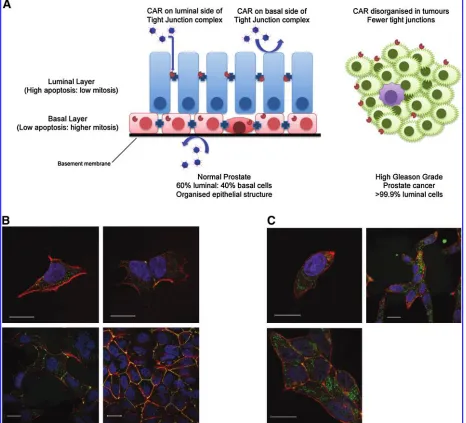
It was the aim of the GIANT (Gene Therapy: An In-tegrated Approach to Neoplastic Treatment) program
(www.giant.eu.com) to assess the validity of the preclinical testing procedures for near-to-patient testing. As mentioned previously, cell culture models of prostate epithelium often lack the luminal differentiation present in tumors, unless derived from a later stage metastatic tumor. As illustrated in Fig. 1A, a normal human prostate acinus consists of an upper luminal layer (60% of epithelial cells), in close contact with a basal cell layer (40% of epithelial cells). These proportions are grossly disrupted in higher Gleason grade prostate cancers. Most untreated hormone-naive prostate cancers have a lu-minal phenotype (express androgen receptor and lulu-minal cytokeratin-18) and lack or contain <1% basal cells (no p63=cytokeratin-5=14 expression). The LNCaP cell line (Horoszewicz et al., 1980) is frequently used to study hor-mone-sensitive (luminal) cancer, but LNCaP was isolated from a patient who had failed a primitive form of anti-hormone therapy (estramustine chemotherapy) and the cells express a mutant androgen receptor, which responds equally well to other steroid hormones (Veldscholteet al., 1990).
Toward Better Preclinical Systems: Vector Adhesion in Two-Dimensional Cell Cultures
Initial testing of infectious agents in cell cultures is often optimized for viral replication or infectivity, for example, attachment of viruses to the cell surface. Many viruses use adhesion molecules as receptors and adenovirus is no exception (reviewed in Greber and Gastaldelli, 2007). For example, the main adenovirus receptor, the coxsackievirus– adenovirus receptor (CAR), forms homodimers between contacting cells and serves as a cell adhesion molecule (van Raaijet al., 2000), but adenoviruses also require a secondary receptor in the form of integrins for high-efficiency trans-duction. Of these,avb3is probably the key integrin involved
in virus uptake (Wickhamet al., 1993; Nemerow, 2000). CAR protein is also expressed in mobile membrane lipid rafts (Ashbourne Excoffonet al., 2003) and coimmunoprecipitates with ZO-1, a tight junction protein (Cohenet al., 2001).
Table1. Continued
Cell line
Androgen
sensitivity Derivation Ref.
RC-92a=hTERT No hTERT-immortalized intraprostatic tumor epithelial cell
Mikiet al. (2007)
DuCaP Yes Established from a metastatic lesion to the dura mater of a patient with hormone-refractory prostate cancer
Leeet al. (2001)
VCaP Yes This line was established in 1997 from a vertebral bone metastasis from a patient with hormone-refractory prostate cancer. It was passaged as xenografts in mice and then culturedin vitro
Korenchuket al. (2001)
LNCaP Yes Derived from a lymph node metastasis of prostate cancer after estramustine treatment
Horoszewiczet al. (1980)
C4-2 No Androgen-independent subclone of LNCaP parent Wuet al. (1994) PC-3 No Derived from an androgen-independent bone
metastasis of a treatment-resistant prostate cancer
Kaighnet al. (1979)
PC3M The M variant was selected for enhanced metastatic spread
Abbreviations:ATCC, American Type Culture Collection; HPV, human papilloma virus; hTERT, human telomerase reverse transcriptase; ND, not determined; SV40, simian virus 40.
In normal prostate tissues, CAR is located at the lateral junctions between luminal cells, and at the apical border, whereas in basal cells mostly cytoplasmic expression is ob-served (Rauenet al., 2002). Prostate cancer is mainly luminal in phenotype, which suggests it should express more CAR, as application of hormones to rat seminal vesicle and prostate epithelium results in a reorganization and differentiation-regulated increase in junction formation (Ortiz and Ca-vicchia, 1990; Mitra et al., 2006). However, in sections of prostate cancer, the expression of CAR is lost at the cell
junctions and also decreases at the protein level (Liet al., 1999; Okegawa, 2000). Therefore the viral vectors may find alterative means of entry, for example, adenovirus may enter through direct contact of the penton base with integrins or other receptors. The loss of CAR was confirmed in our lab-oratory, where we could not detect expression at the apical border of primary prostate cancer samples (K. Chambers, unpublished data). Although the common productive cell line for adenoviral culture, HEK-293, has extensive intracel-lular CAR (Fig. 1B), in prostate cancer cell lines, such as
LNCaP, CAR is present at lateral junctions but is expressed mainly in the cytoplasm of single cells without adhesions (Fig. 1C; and K. Chambers, unpublished data). CAR is, however, still detectable in subconfluent prostate cancer cell cultures even after disruption of the junctions for flow cy-tometric analysis (Pandhaet al., 2003).
Because of the expression of CAR at the tight junctions, the availability of the receptor from the luminal surface is blocked by their proximity to or incorporation into tight intracellular junctions (Balda and Matter, 1998; Cohenet al., 2001). Access and spread can be governed by restricted access to receptor complexes, as the mean distance between cells in tight and gap junctions is 1.5 and 4 nm, respectively, whereas the full diameter of many nonviral particles is at least 50 nm and that of the rigid adenoviral particle is 100 nm.
With prostate cancers, there is also the question of the cell surface available to be infected in a structured and polarized epithelium (present in most lower Gleason grade tumors). In vivo, it is likely that infection will occur through the lu-minal surface (see Fig. 1A) rather than through basal contact via the basement membrane, which adenoviruses can fail to penetrate, although genetic modification of the virus by re-placement of the E3 gene to encode the relaxin-degradative enzyme can improve penetration (Kim et al., 2006). Other strategies to improve infectivity via CAR include internali-zation of tight junction proteins such as occludin to expose the tight junction receptor CAR, without affecting the dis-tribution of other tight junctional proteins (Coyne et al., 2007), and increased exposure of CAR through actin re-modeling (Coyne and Bergelson, 2005).
In tissues, the damage induced by direct injection results in effective infection restricted to a few millimeters around needle tracks (Patel et al., 2009). Therefore it will probably never be possiblein vivoto saturate a tumor with prodrug-activating cells after gene transduction by intratumoral in-jection of even high titers of the most efficient vectors, without facilitating intratumoral spread. It was for this rea-son that an oncolytic approach was taken for the initial trials in GIANT (Cheng et al., 2006), where intracellular spread occurs after infected cell lysis.
However, in immunocompetent patients such spread is a matter of timing between the triggering of a potent immune response against adenoviruses and the ability of the virus to reinfect multiple times. Although the immune response may be restrictive in the spread of the virus (Paratoet al., 2005), there is evidence to suggest that patients who respond im-munologically to a therapeutic viral infection also mount a powerful immune response against tumor cell antigens. The strong adjuvant effect of high levels of viral antigens is thought to be responsible, resulting in regression of not only the primary tumor, but also of distant metastases (Freytag et al., 2002). This type of outcome emphasizes the need to use virotherapies in immunocompetent patients to achieve max-imal therapeutic efficacy. Predictably, in most phase 1 (in which safety assessment is the object) but more surprisingly in phase 2 (in which efficacy is sought) clinical trials of bio-therapies, this is seldom the case.
Primary Tissue Infections
An excellent model, which takes into account differential patient susceptibility to infection, is direct infection of needle
biopsies from fresh prostate cancer tissues, in which tissue architecture and viability can be preserved in culture for a period of up to 2 weeks in a basal medium. Such tests pro-vide, on a patient-to-patient basis, a measurement of sus-ceptibility not only to gene therapy agents but also to many common chemotherapies. However, ethical restrictions in obtaining fresh tissues, or the risk of compromising patho-logical diagnosis, can compromise the quantities of tissue available to do such studies in a rational and quantitative way.
A good alternative would be to establish and test the transduction of fragments of tissue as primary xenografts in immunocompromised mice. Short-term ‘‘culture’’ of human tissues is possible in xenografts, with maintenance of both tissue architecture and hormone responsiveness at subcuta-neous, subrenal capsule, and orthotopic mouse prostate sites. Although the production of such xenografts can be labor intensive, and they are limited in size and scope for the testing of agents, they do offer the best measurement of therapeutic effectiveness as a patient-specific medicine, the ‘‘gold standard’’ for sophisticated and targeted biotherapies. Within the GIANT program we have used xenograft models, established for many years (Table 2).
Primary Epithelial Cell Cultures
To at least partly restore luminal differentiation, recon-structions of the multilayered and multicellular prostatic epithelium can be achieved in two dimensions by allowing the epithelial cells to grow out of monolayer and form a bilayer. Induction of luminal differentiation is further en-hanced by addition of prostate androgen-responsive stroma and medium supplements such as dihydrotestosterone and calcium, resulting in a polarization of the epithelial layers. At this point the characteristic patterns of expression of cell surface receptors are found, as gene expression switches from basal to luminal cytokeratin expression in the upper (luminal) layer (Swiftet al., 2010).
Three Dimensions: Better Than Two?
Whereas two-dimensional cell culture presents a single surface for virus infection, tissues exist in vivo as multiple layers bordered by tight junctions and desmosomes, and
containing a multitude of different structurally differentiated cell types, which will inhibit vector penetration. One way to model this is to generate three-dimensional structures in cell culture. To assess the effects of differentiation, three-dimensional cultures can be induced to both polarize and differentiate by suspension in an appropriate matrix, the stimulus to polarize being provided by signals from prostate stromal cells (Langet al., 2001).
Adenoviruses have a limited ability to extravasate from blood vessels, unless the capillaries are severely damaged or are irregular in development, such as is the case with in-tratumoral capillaries (Wang and Yuan, 2006). When ade-novirus was administered via the intravenous route in mice xenografted with a human prostate tumor, infection was observed mainly in endothelial cells of healthy vessels in the tumor. Few infected tumor cells were detectable after intra-venous administration. However, when virus was adminis-tered intratumorally in the same model, many infected tumor cells were detected, although the distribution pattern remained concentrated around the needle track (R. Kraaij, unpublished). Complex three-dimensional structuresin vitro can also address the issue of penetration.
In another tissue, that is, infections of the eye, adeno-viruses were also unable to penetrate the outer layers of epithelium to infect the basal epithelium, whereas enveloped viruses of less rigid structure, such as baculovirus (mean diameter, 60 nm), could do so (Kinnunenet al., 2009). This suggests that particle diameter is not the only parameter to take into account in determining tissue penetration.
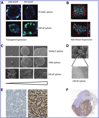
To model tissue penetration in the GIANT program, three-dimensional spheroids grown from a number of different prostate cell lines were employed. Different morphologies of spheroid were derived from the different cell types (Fig. 2). Some were rigid and heavily structured, whereas others were much more loosely associated, in which the require-ment to form structures and to communicate with other cells had been gradually lost either as a result of oncogenic changes, or through long-term culture. In such full three-dimensional models (Lang et al., 2001) it became apparent that most vectors, including adenoviruses, had difficulties in penetrating basement membranes such as Matrigel, which reflects restrictions on their spread in tissuesin vivoas well. However, even more striking was the inability of adeno-viruses to pass through and penetrate organized spheroids as shown in Fig. 2A and B. Furthermore, P4E6 cells (derived from a well-differentiated prostate cancer) form a highly organized spheroid (Fig. 2C), which also contains a stretched layer of thin epithelium to provide structural integrity. Both by immunofluorescence (Fig. 2A and B) and by electron mi-croscopy (Fig. 2D), virus replication could be seen within the outer layer of spheroid epithelium, but spread within the spheroid was almost totally absent. When yields of oncolytic virus in this system were compared with the yields from a replicating monolayer culture and subsequently with xeno-grafts (see later), the three-dimensional culture more closely matched the situation in xenograft models (L. Georgopoulos, H. Evans, and R. Nugent, unpublished data).
Cellular Heterogeneity in Prostate Cancer
For most cancer medicines, there has long been the as-sumption that all the cells in a tumor grow more quickly than
Table2. Human Prostate Cancer Xenograft Modelsa
Name
Derived from:
Androgen
responsive PSA AR
Year derived
PC-82 Prostate Yes Yes Yes 1977
PC-133 Bone No No No 1981
PC-135 Prostate No No No 1982
PC-EW Prostate Yes Yes Yes 1981
PC-295 LN Yes Yes Yes 1991
PC-310 Prostate Yes Yes Yes 1991
PC-324 TURP No No No 1991
PC-329 Prostate Yes Yes Yes 1991
PC-339 TURP No No No 1991
PC-346 TURP Yes Yes Yes 1991
PC-346I PC-346 No Yes Mutant 1992
PC-346B TURP Yes Yes Yes 1991
PC-346BI PC-346B No Yes Yes 1992
PC-374 Skin Yes Yes Yes 1992
TEN12 Prostate Yes Yes Yes 1985 LuCaP 23.1 Lymph
node
Yes Yes 1996
LuCaP 23.8 Lymph node
Yes Yes 1996
LuCaP 23.12 Liver Yes Yes 1996 LuCaP 35 Lymph
node
Yes Yes Yes 2003
LuCaP 35V LuCap 35 No 2003
LuCaP 49 Metastasis No No No 2002 LAPC-3 AI TURP No Yes=no Yes 1999
LAPC-4 AI LN Yes Yes Yes 1997
LAPC-9 AI Bone Met
Yes Yes Yes 2001
CWR22 AD Met Yes Yes Mutant 1993
CWR21 AD Met Yes Yes Yes 1993
CWR31 AD Met Yes Yes Yes 1993
CWR91 AD Met Yes Yes Yes 1993
MDA Pca-31 Liver N=A 1998
MDA Pca-40 Liver N=A 1998
MDA Pca-43 Adrenal N=A 1998
MDA Pca-44 Skin N=A 1998
Abbreviations:AD, androgen dependent; AI, androgen indepen-dent; AR, androgen receptor; LN, lymph node metastasis; Met, metastasis; N=A, not applicable; PC, prostate cancer; PSA, prostate-specific antigen; TURP, transurethral resection of the prostate.
their normal counterparts, and that they homogeneously express the tissue-specific mRNA patterns of a prostate tumor. The rapid emergence of therapy-resistant tumors, even for biotherapies, has been cited as proof of the existence of a preexisting therapy-resistant fraction, sometimes termed cancer stem cellsortumor-initiating cells(Diehnet al., 2009). In human prostate cancer, this fraction seems not to respond to androgens, which form the basis of much of the tissue-specific targeting strategies employed in virus targeting (Maitland and Collins, 2008). As suggested in a review (Short and Curiel, 2009), the development of new
genera-tions of vectors with the ability to eliminate cancer stem cells, as part of a combined gene therapy approach to shrink tu-mor bulk, should extend the effectiveness of such therapies.
Contamination of Cell Models
High-throughput testing of infectious agents, relying on both primary cells and cell lines, also presents a number of dangers with respect to the validity of the cells to be em-ployed. It is now mandatory for publication in some journals that all cell cultures be regularly genotyped. The standard
applied by the American Tissue Culture Collection (ATCC, Manassas, VA; http:==www.atcc.org=) is the PowerPlex sys-tem (a microsatellite genotyping kit; Promega, Madison, WI). All cells to be employed in therapy testing should be geno-typed initially as primary cells, in comparison with the original tissue DNA (or lymphocytes). If an established cell line is used the reference PowerPlex fingerprint is available from the ATCC. Even primary cultures, from normal or tu-mor tissues, have a limited life span as a true reflection of the original tumor, for example, glioblastoma cell cultures (Lee et al., 2006). This life span can be extended by purifying different cell populations, including stem cells, as described previously (Collinset al., 2005).
Second, cells derived from xenografts or primary cultures passaged on inactivated mouse stromal feeder layers require routine assessment of human cell content and the presence of murine cells and DNA, derived either from murine infiltrates in the xenografts or ‘‘breakthrough’’ of feeder cells resulting from inadequate inactivation, by polymerase chain reaction (PCR) using repetitive species-specific centromeric sequences (Beckeret al., 2002).
The last restriction when working with primary tissues, xenografts (in particular), and established cell lines, is the necessity to eliminate mycoplasma contamination, which frequently prevents vector attachment and penetration. A number of excellent commercial PCR-based assays are available for mycoplasma detection.
Ex VivoClinically Relevant Testing for Antivector Immunity
One aspect of gene therapy, which cannot be determined in a standard cell culture environment, concerns exposure of the infectious agent to the bloodstream and immune system of the host. The current standardin vivosystem, a xenograft of the original cell line used in cell culture in an immuno-compromised mouse, suffers 2-fold in this respect.
First, there is no strong humoral or T cell-mediated immune response against the infectious particles, as the commonly used nude mouse is athymic. The nonobese dia-betic-severe combined immunodeficient (NOD-SCID) mouse lacks more of the T cell immunity in addition to B cells, but is now being replaced in practical use by even more immu-nocompromised hosts such as the Rag2=gc double-negative hosts, which most notably also lack most natural killer (NK) cell functions (Le De´ve´decet al., 2009). These hosts still retain complement responses, but show different abilities to engraft with human tumor cells. However, all hosts share the in-ability to mount any immune response to tumor cell lysis products, acknowledged as one of the major influences on tumor destruction (see previously).
Second, infectious particles, in particular adenoviruses, interact in a different way with blood components in murine and human hosts. It has been shown that more than 90% of adenovirus administered to human blood ex vivobinds to human erythrocytes whereas only a negligible fraction (<0.1%) of adenovirus binds to blood cells in freshly isolated murine whole blood (Lyonset al., 2006). Human erythrocytes bind and inactivate adenovirus serotype 5 (Ad5) by expres-sing both the coxsackievirus–adenovirus receptor (CAR) and the complement receptor-1 (CR1) (Carlisle et al., 2009). Mouse erythrocytes do not express either CAR or CR1.
Be-cause complement receptors are important for induction of neutralizing antibodies (Seregin et al., 2009) mouse blood does not mimic the situation in human blood, that is, the effective dose of virus in humans is now calculated to be much lower than that originally predicted from the critical preclinical studies in mice.
As part of the GIANT project, we sought to overcome this by testing vectors in a human ex vivo blood loop system, which had previously been developed for the testing of other biomaterials (Hong et al., 2001). This system is a powerful preclinical model for studies of viral and nonviral vector interactions with all components of whole blood, omitting the need for high-level anticoagulants that can alter the re-sults by chelating the system of positive ions such as Ca2þ
and Zn2þ. Furthermore, in contrast to the use of sealed small-volume tubes, the loop system also benefits from reproduc-ing shear forces, which are important for platelet function and the continuous flow of the bloodstream.
We have previously shown (Georgopouloset al., 2009) that a major complement response in blood loops was mounted against a nonhuman virus, baculovirus. Significant binding of IgM and complement components was observed and strong immunoreactivity was characterized by blood clot formation. The importance of the complement responses to baculovirus, against which humans should have no immu-nological memory, was indicated by clot prevention by use of complement inhibitors such as compstatin (Nilssonet al., 1998). The life span of the vector particles could therefore be extended in serum by coinoculation with specific comple-ment inhibitors.
Within the GIANT network, we have used the blood loop system to assess the immune response in whole human blood to an Ad5 vector and immunologically ‘‘stealthed’’ PEGylated Ad5. Such PEGylation of Ad5-based vectors has proven efficient in reducing the immune response and in-creasing the circulation times in mouse models (O’Riordan et al., 1999; Gaoet al., 2007) and reducing virus uptake by mouse Kupffer cells (Moket al., 2005). PEGylation reduced Ad5 adhesion to blood cells, and both complement activation and cytokine release to a certain degree, especially when the neutralizing anti-Ad5 antibody titer was low (Danielsson et al., 2010). However, the reductions were lower than would have been predicted from mouse models (O’Riordanet al., 1999; Moket al., 2005; Gaoet al., 2007; Wortmannet al., 2008). The reason may well be the difference in cell adhesion of Ad5 to human and mouse blood cells and the fact that PEGylation has only a limited protective effect when the neutralizing anti-Ad5 antibody titers are high. This finding simply em-phasizes the importance of using a relevant model for eval-uating viral vectors.
Toward More Clinically RelevantIn VivoTesting
cells are not particularly susceptible to vectors targeting human cancers, and oncolytic adenoviruses do not grow in murine cells, testing systems are usually restricted to human xenografts.
Just as with cell culture lines, until the early 1990s, the limited number of xenograft models for prostate cancer re-presented predominantly late-stage disease, whereas models of untreated tumors were underrepresented (i.e., radical prostatectomy samples). The more recent establishment of ‘‘early-stage’’ xenograft models and cell lines has largely overcome this limitation (Tables 1 and 2). The latter xeno-graft models (van Weerdenet al., 1996, 2009; Marques, 2006) have become even more relevant as prostate cancer is being diagnosed at an earlier stage. Until more recently, for the testing of gene therapy vectors, the field still remained heavily dependent on the LNCaP, PC3, and DU145 cell lines.
PC3/DU145 Xenografts
PC3 cells form reproducible tumors at most murine sites of inoculation, as subcutaneous, subrenal capsule, and in-traprostatic orthotopic grafts (see review by Hoffman, 2007). The tumors are capable of killing the host with an inocula-tion of about half a million cells within 28 days. This ex-tremely rapid growth from an undifferentiated epithelial cell mass has been used extensively as the target for many prostate-specific therapies. DU145 cells are similarly tumor-igenic in immunocompromised animals, although they have been used less frequently (Nemethet al., 1999).
New variants expressing indicator genes such as luciferase and green fluorescent protein (GFP) have been developed, and more metastatic variants of PC3 have been selected by prolonged passage in nude mice. It could be argued that the lack of structure in the tumors and the rapid growth are not comparable with human prostate cancers, which are much slower growing and display elements of organization and structure even in their least differentiated forms, as described originally by Gleason (1966). One complication with PC3 is that bone tumors, which can be obtained by direct inocula-tion into bone (Nemethet al., 1999), are oncolytic rather than osteoblastic, as is commonly found in advanced human cancers (see review by Dotan, 2008).
LNCaP Xenografts
In contrast, the LNCaP cell line expresses androgen re-ceptor (albeit a mutant androgen rere-ceptor), which typifies luminal differentiation in well-differentiated common pros-tate cancers. However, xenografting with LNCaP can be technically difficult, in particular to subcutaneous sites. It is also capable of throwing off many substrains and the C4-2 series originally described by Thalmann and colleagues (1994) clearly has the capacity not only to engraft but also to induce metastasis.
The success of LNCaP xenografts can, however, be sub-stantially improved by coinoculation with prostate stromal cells (Tuxhornet al., 2002). This again mimicks much more closely the situation in humans.
Other Prostate Epithelial Cell Line Xenografts
The BPH1 cell line, which derived originally from benign prostatic hyperplasia epithelium by introduction of SV40T
antigen (Hayward et al., 1995), can be induced to form ag-gressive tumors by grafting subrenally in the presence of carcinoma-associated fibroblasts (CAFs) from prostate (Haywardet al., 2001). This again emphasizes the importance of the epithelial–stromal interaction in the development of prostate cancers bothin vitroandin vivo. The mechanism for this oncogenic transformation of a previously benign cell type simply by the presence of fibroblasts is thought to occur through a complex series of interactions, at least some of which involve transforming growth factor (TGF)-bsignaling (Ao et al., 2007). To authentically model lower grades of prostate cancer in the mouse environment will probably re-quire such multicellular grafts.
PC346C Xenografts
The more recently established PC346C cells reliably form undifferentiated epithelial tumors after subcutaneous or in-traprostatic orthotopic inoculation of 1 million cells within 20 days. Importantly, PC346C is androgen sensitive, expresses a wild-type androgen receptor, and secretes large amounts of prostate-specific antigen, which typifies well-differentiated common prostate cancers. Prolonged culture of PC346C cells in the absence of androgen and=or in the presence of the antiandrogen hydroxyflutamide, as well asin vivopassage in castrate recipient mice, has resulted in several castration-resistant variants (Marqueset al., 2005, 2006). Furthermore, orthotopic injection of PC346C has provided variant sublines derived from metastatic lesions (lymph node and lung) that are equally tumorigenic (W.M. van Weerden, unpublished data). At present, PC346C variants expressing indicator genes such as luciferase and GFP or RFP fluorescence are being developed, which will permit real-time monitoring of tumor cell fate.
Within the GIANT program we chose to exploit the latter xenografts (for a review see Van Weerdenet al., 2009), which have been selected to span a range of pathologies, from an-drogen dependency through to anan-drogen independency and a number of different morphologies and histological grades (see Table 2).
Tumors of early prostate cancer, as represented by the androgen-responsive PC82, PC295, and PC310 xenografts, are well-differentiated tumors with a cribriform growth pattern that continue to express the prostate antigens and more critically a wild-type androgen receptor (Fig. 2E). In contrast, xenografts that represent later stage disease, such as PC133, PC135, and PC339, are relatively undifferentiated.
The limitation of the xenograft panel largely lies in the fact that these tumors, except for PC346, can be propagated only subcutaneously, as we have been unable so far to establish permanent cell lines from these xenograft models. Because subcutaneously grown tumors generally do not metastasize, these tumors cannot be used to study metastasis from the primary site. However, PC346C cells that are inoculated into the prostate have been shown to spread to other organs (metastases in the lymph nodes and lung). For preclinical testing, the GIANT program has employed the PC346C tumor, which has the great advantage of being able to be grownin vitroand as a serially transplanted xenograft. The ability of PC346C cells to sustain virus growth has been previously demonstrated (Kraaij et al., 2005), and several prostate-specific oncolytic adenoviral vectors, such as Ad[I= PPT-E1A] (Cheng et al., 2006) and Ad-ZH=3 (Magnusson et al., 2007), have now been preclinically validated using the orthotopic PC346C xenograft model. (Fig. 2F; and R. Kraaij, unpublished).
Future Considerations/Outlook
The potential of gene therapy to provide an alternative treatment for prostate cancer has been discussed since the early 1990s (Sandaet al., 1994). The early promise has not yet been fulfilled, although we now know a great deal more about how to kill the correct cells, and perhaps more im-portantly how to target potent therapies to the correct cell types. Sadly, the testing strategies have failed to maintain the rapid pace of advance in agent development, which could account in part for the failure to convert our basic knowledge into clinical trials.
The translation from cell lines in culture, and of rapidly growing xenografts of the same cell lines, to a heterogeneous and relatively slowly growing tumor in an immunocompetent patient is rarely considered in vector development strategies. We have attempted to highlight the difficulties in achieving total tumor infection as a result of restrictions in vector spread by using adenoviral vectors as a common example. The re-striction of spread from a needle injection in tissues remains limited to at best a few millimeters, requiring precise location of the tumor to be infected (Patelet al., 2009). By treating the vector inoculum as a medicine, requiring formulation just like a small molecule, we can perhaps reduce thein vivo restric-tions. For example, blockade of the innate immune response, and incorporation of tissue disruptors such as relaxins (or equivalent small molecules) to open tight junctions and reveal receptors, would aid such spread.
However, the most important consideration must remain patient selection: the stage of prostate cancer to be targeted, and the critical realization that responses in each individual human will vary and also differ from those in mouse models. Increasingly, these responses can now be modeled in the laboratory, restricting unnecessary time and expenditure on premature clinical trials.
Acknowledgments
The authors thank all the collaborators on the EU-funded FP6 Project GIANT for discussions and contributions. The authors particularly thank Michelle Scaife for assistance in manuscript preparation. All original research reported was funded by the GIANT Integrated Project (contract number
LSHB-CT-2004-512087). N.J.M. also received invaluable core support from Yorkshire Cancer Research.
Author Disclosure Statement
No competing financial interests exist connected with the information conveyed in this review.
References
Ao, M., Franco, O.E., Park, D., Raman, D., Williams, K., and Hayward, S.W. (2007). Cross-talk between paracrine-acting cytokine and chemokine pathways promotes malignancy in benign human prostatic epithelium. Cancer Res. 67, 4244–4253. Ashbourne Excoffon, K.J., Moninger, T., and Zabner, J. (2003). The coxsackie B virus and adenovirus receptor resides in a distinct membrane microdomain. J. Virol. 77, 2559–2567. Balda, M.S., and Matter, K. (1998). Tight junctions. J. Cell Sci.
111, 541–547.
Becker, M., Nitsche, A., Neumann, C., Aumann, J., Junghahn, I., and Fichtner, I. (2002). Sensitive PCR method for the detection and real-time quantification of human cells in xeno-transplantation systems. Br. J. Cancer 87, 1328–1335.
Bello, D., Webber, M.M., Kleinman, H.K., Wartinger, D.D., and Rhim, J.S. (1997). Androgen responsive adult human prostatic epithelial cell lines immortalized by human papillomavirus 18. Carcinogenesis 18, 1215–1223.
Carlisle, R.C., Di, Y., Cerny, A.M., Sonnen, A.F.-P., Sim, R.B., Green, N.K., Subr, V., Ulbrich, K., Gilbert, R.J.C., Fisher, K.D., Finberg, R.W., and Seymour, L.W. (2009). Human erythro-cytes bind and inactivate type 5 adenovirus by presenting coxsackie virus–adenovirus receptor and complement recep-tor 1. Blood 113, 1909–1918.
Cheng, W.-S., Dzojic, H., Nilsson, B., To¨tterman, T.H., and Essand, M. (2006). An oncolytic conditionally replicating adenovirus for hormone-dependent and hormone-independent prostate cancer. Cancer Gene Ther. 13, 13–20.
Chung, L.W., Chang, S.M., Bell, C., Zhau, H., Ro, J.Y., and von Eschenbach, A.C. (1988). Prostatic carcinogenesis evoked by cellular interaction. Environ. Health Perspect. 77, 23–28. Cohen, C.J., Shieh, J.T., Pickles, R.J., Okegawa, T., Hsieh, J.T.,
and Bergelson, J.M. (2001). The coxsackievirus and adenovirus receptor is a transmembrane component of the tight junction. Proc. Natl. Acad. Sci. U.S.A. 98, 15191–15196.
Collins, A.T., Berry, P.A., Hyde, C., Stower, M.J., and Maitland, N.J. (2005). Prospective identification of tumorigenic prostate cancer stem cells. Cancer Res. 65, 10946–10951.
Coyne, C.B., and Bergelson, J.M. (2005). CAR: A virus receptor within the tight junction. Adv. Drug Deliv. Rev. 57, 869–882. Coyne, C.B., Shen, L., Turner, J.R., and Bergelson, J.M. (2007). Coxsackie virus entry across epithelial tight junctions requires occludin and the small GTPases Rab34 and Rab5. Cell Host Microbe 2, 181–192.
Cunha, G.R. (1984). Androgenic effects upon prostatic epithe-lium are mediated via trophic influences from stroma. Prog. Clin. Biol. Res. 145, 81–102.
Cussenot, O., Berthon, P., Berger, R., Mowszowicz, I., Faille, A., Hojman, F., Teillac, P., Le Duc, A., and Calvo, F. (1991). Im-mortalization of human adult normal prostatic epithelial cells by liposomes containing large T-SV40 gene. J. Urol. 146, 881–886. Danielsson, A., Elgue, G., Nilsson, B.M., Nilsson, B., Lambris,
De Marzo, A.M., Meeker, A.K., Epstein, J.I., and Coffey, D.S. (1998). Prostate stem cell compartments: Expression of the cell cycle inhibitor p27Kip1in normal, hyperplastic, and neoplastic cells. Am. J. Pathol. 153, 911–919.
Di Cristofano, A., Pesce, B., Cordon-Cardo, C., and Pandolfi, P.P. (1998). Pten is essential for embryonic development and tumour suppression. Nat. Genet. 19, 348–355.
Diehn, M., Cho, R.W., and Clarke, M.F. (2009). Therapeutic implications of the cancer stem cell hypothesis. Semin. Radiat. Oncol. 19, 78–86.
Dotan, Z.A. (2008). Bone imaging in prostate cancer. Nat. Clin. Pract. Urol. 5, 434–444.
Freytag, S.O., Khil, M., Stricker, H., Peabody, J., Menon, M., Deperalta-Venturina, M., Nafziger, D., Pegg, J., Paielli, D., Brown, S., Barton, K., Lu, M., Aguilar-Cordova, E., and Kim, J.H. (2002). Phase I study of replication-competent adenovirus-mediated double suicide gene therapy for the treatment of locally recurrent prostate cancer. Cancer Res. 62, 4968–4976.
Gao, J.-Q., Eto, Y., Yoshioka, Y., Sekiguchi, F., Kurachi, S., Morishige, T., Yao, X., Watanabe, H., Asavatanabodee, R., Sakurai, F., Mizuguchi, H., Okada, Y., Mukai, Y., Tsutsumi, Y., Mayumi, T., Okada, N., and Nakagawa, S. (2007). Effective tumor targeted gene transfer using PEGylated adenovirus vector via systemic administration. J. Control. Release 122, 102–110.
Gazdar, A.F., and Minna, J.D. (1996). NCI series of cell lines: An historical perspective. J. Cell. Biochem. Suppl. 24, 1–11. Georgopoulos, L.J., Elgue, G., Sanchez, J., Dussupt, V., Magotti,
P., Lambris, J.D., To¨tterman, T.H., Maitland, N.J., and Nilsson, B. (2009). Preclinical evaluation of innate immunity to bacu-lovirus gene therapy vectors in whole human blood. Mol. Immunol. 46, 2911–2917.
Gleason, D.F. (1966). Classification of prostatic carcinomas. Cancer Chemother. Rep. 50, 125–128.
Greber, U.F., and Gastaldelli, M. (2007). Junctional gating: The Achilles’ heel of epithelial cells in pathogen infection. Cell Host Microbe 2, 143–146.
Hall, J.A., Maitland, N.J., Stower, M., and Lang, S.H. (2002). Primary prostate stromal cells modulate the morphology and migration of primary prostate epithelial cells in type 1 colla-gen gels. Cancer Res. 62, 58–62.
Hayward, S.W., Dahiya, R., Cunha, G.R., Bartek, J., Deshpande, N., and Narayan, P. (1995). Establishment and characteriza-tion of an immortalized but non-transformed human prostate epithelial cell line: BPH-1. In Vitro Cell Dev. Biol. Anim. 31, 14–24.
Hayward, S.W., Wang, Y., Cao, M., Hom, Y.K., Zhang, B., Grossfeld, G.D., Sudilovsky, D., and Cunha, G.R. (2001). Malignant transformation in a nontumorigenic human pros-tatic epithelial cell line. Cancer Res. 61, 8135–8142.
Hoffman, R.M. (2007). Orthotopic metastatic mouse models of prostate cancer. In: Metastasis of Prostate Cancer, Vol. 10 of Cancer Metastasis—Biology and Treatment (Springer Nether-lands, Dordrecht, The Netherlands)pp.143–169.
Hong, J., Larsson, A., Ekdahl, K.N., Elgue, G., Larsson, R., and Nilsson, B. (2001). Contact between a polymer and whole blood: Sequence of events leading to thrombin generation. J. Lab. Clin. Med. 138, 139–145.
Horoszewicz, J.S., Leong, S.S., Chu, T.M., Wajsman, Z.L., Friedman, M., Papsidero, L., Kim, U., Chai, L.S., Kakati, S., Arya, S.K., and Sandberg, A.A. (1980). The LNCaP cell line: A new model for studies on human prostatic carcinoma. Prog. Clin. Biol. Res. 37, 115–132.
Isaacs, J.T., and Kyprianou, N. (1987). Development of androgen-independent tumor cells and their implication for the treatment of prostatic cancer. Urol. Res. 15, 133–138. Kaighn, M.E., Narayan, K.S., Ohnuki, Y., Lechner, J.F., and
Jones, L.W. (1979). Establishment and characterization of a human prostatic carcinoma cell line (PC-3). Invest. Urol. 17, 16–23.
Kim, J.-H., Lee, Y.-S., Kim, H., Huang, J.-H., Yoon, A.-R., and Yun, C.-O. (2006). Relaxin expression from tumor-targeting adenoviruses and its intratumoral spread, apoptosis induc-tion, and efficacy. J. Natl. Cancer Inst. 98, 1482–1493 Kinnunen, K., Kalesnykas, G., Ma¨ho¨nen, A.J., Laidinen, S.,
Holma, L., Heikura, T., Airenne, K., Uusitalo, H., and Yla¨-Herttuala, S. (2009). Baculovirus is an efficient vector for the transduction of the eye: Comparison of baculovirus- and adenovirus-mediated intravitreal vascular endothelial growth factor D gene transfer in the rabbit eye. J. Gene Med. 11, 382– 389.
Klein, K.A., Reiter, R.E., Redula, J., Moradi, H., Zhu, X.L., Brothman, A.R., Lamb, D.J., Marcelli, M., Belldegrun, A., Witte, O.N., and Sawyers, C.L. (1997). Progression of meta-static human prostate cancer to androgen independence in immunodeficient SCID mice. Nat. Med. 3, 402–408.
Korenchuk, S., Lehr, J.E., McLean, L., Lee, Y.G., Whitney, S., Vessella, R., Lin, D.L., and Pienta, K.J. (2001). VCaP, a cell-based model system of human prostate cancer. In Vivo 15, 163–168.
Kraaij, R., van Rijswijk, A.L., Oomen, M.H., Haisma, H.J., and Bangma, C.H. (2005). Prostate specific membrane antigen (PSMA) is a tissue-specific target for adenoviral transduction of prostate cancerin vitro. Prostate 15, 253–259.
Lang, S.H., Stark, M., Collins, A., Paul, A.B., Stower, M.J., and Maitland, N.J. (2001). Experimental prostate epithelial mor-phogenesis in response to stroma and three-dimensional Ma-trigel culture. Cell Growth Differ. 12, 631–640.
Le De´ve´dec, S., van Roosmalen, W., Maria, N., Grimbergen, M., Pont, C., Lalai, R., and van de Water, B. (2009). An improved model to study tumor cell autonomous metastasis programs using MTLn3 cells and the Rag2–=–gc–=– mouse. Clin. Exp. Metastasis 26, 673–684.
Lee, J., Kotliarova, S., Kotliarov, Y., Li, A., Su, Q., Donin, N.M., Pastorino, S., Purow, B.W., Christopher, N., Zhang, W., Park, J.K., and Fine, H.A. (2006). Tumor stem cells derived from glioblastomas cultured in bFGF and EGF more closely mirror the phenotype and genotype of primary tumors than do serum-cultured cell lines. Cancer Cell 9, 391–403.
Lee, Y.G., Korenchuk, S., Lehr, J., Whitney, S., Vessela, R., and Pienta, K.J. (2001). Establishment and characterization of a new human prostatic cancer cell line: DuCaP. In Vivo 15, 157– 162.
Li, Y., Pong, R.C., Bergelson, J.M., Hall, M.C., Sagalowsky, A.I., Tseng, C.P., Wang, Z., and Hsieh, J.T. (1999). Loss of adeno-viral receptor expression in human bladder cancer cells: A potential impact on the efficacy of gene therapy. Cancer Res. 59, 325–330.
Lyons, M., Onion, D., Green, N.K., Aslan, K., Rajaratnam, R., Bazan-Peregrino, M., Phipps, S., Hale, S., Mautner, V., Sey-mour, L.W., and Fisher, K.D. (2006). Adenovirus type 5 in-teractions with human blood cells may compromise systemic delivery. Mol. Ther. 14, 118–128.
Affibody molecule with specificity for tumor antigen HER2=neu. Cancer Gene Ther. 14, 468–479.
Maitland, N.J., and Collins, A.T. (2008). Prostate cancer stem cells: A new target for therapy. J. Clin. Oncol. 26, 2862–2870. Maitland, N.J., Macintosh, C.A., Hall, J., Sharrard, M., Quinn, G.,
and Lang, S. (2001). In vitromodels to study cellular differ-entiation and function in human prostate cancers. Radiat. Res. 155, 133–142.
Maitland, N.J., Stanbridge, L.J., and Dussupt, V. (2004). Target-ing gene therapy for prostate cancer. Curr. Pharm. Des. 10, 531–555.
Marques, R., Erkens-Schulze, S.E., de Ridder, C.M.A., Hermans, K.G., Waltering, K., Visakorpi, T., Trapman, J., Romijn, J.C., van Weerden, W.M., and Jenster, G. (2005). Androgen recep-tor modifications in prostate cancer cells upon long-term an-drogen ablation and antianan-drogen treatment. Int. J. Cancer 117, 221–229.
Marques, R., van Weerden, W.M., Erkens-Schulze, S.E., de Ridder, C.M.A., Bangma, C.H., and Jenster, G. (2006). The human PC346 xenograft and cell line panel: A model system for prostate cancer progression. Eur. Urol. 49, 245–257. Miki, J., Furusato, B., Li, H., Gu, Y., Takahashi, H., Egawa, S.,
Sesterhenn, I.A., McLeod, D.G., Srivastava, S., and Rhim, J.S. (2007). Identification of putative stem cell markers, CD133 and CXCR4, in hTERT-immortalized primary nonmalignant and malignant tumor-derived human prostate epithelial cell lines and in prostate cancer specimens. Cancer Res. 67, 3153–3161. Mitra, S., Annamalai, L., Chakraborty, S., Johnson, K., Song, X.-H., Batra, S.K., and Mehta, P.P. (2006). Androgen-regulated formation and degradation of gap junctions in androgen-responsive human prostate cancer cells. Mol. Biol. Cell 17, 5400–5416.
Mok, H., Palmer, D.J., Ng, P., and Barry, M.A. (2005). Evaluation of polyethylene glycol modification of first-generation and helper-dependent adenoviral vectors to reduce innate immune responses. Mol. Ther. 11, 66–79.
Navone, N.M., Olive, M., Ozen, M., Davis, R., Troncoso, P., Tu, S.M., Johnston, D., Pollack, A., Pathak, S., von Eschenbach, A.C., and Logothetis, C.J. (1997). Establishment of two human prostate cancer cell lines derived from a single bone metas-tasis. Clin. Cancer Res. 3, 2493–2500.
Nemerow, G.R. (2000). Cell receptors involved in adenovirus entry. Virology 274, 1–4.
Nemeth, J.A., Harb, J.F., Barroso, U., He, Z., Grignon, D.J., and Cher, M.L. (1999). Severe combined immunodeficient-hu model of human prostate cancer metastasis to human bone. Cancer Res. 59, 1987–1993.
Nilsson, B., Larsson, R., Hong, J., Elgue, G., Ekdahl, K.N., Sahu, A., and Lambris, J.D. (1998). Compstatin inhibits complement and cellular activation in whole blood in two models of ex-tracorporeal circulation. Blood 92, 1661–1667.
Okegawa, T., Li, Y., Pong, R.C., Bergelson, J.M., Zhou, J., and Hsieh, J.T. (2000). The dual impact of coxsackie and adeno-virus receptor expression on human prostate cancer gene therapy. Cancer Res. 60, 5031–5036.
O’Riordan, C.R., Lachapelle, A., Delgado, C., Parkes, V., Wadsworth, S.C., Smith, A.E., and Francis, G.E. (1999). PE-Gylation of adenovirus with retention of infectivity and pro-tection from neutralizing antibodyin vitroandin vivo. Hum. Gene Ther. 10, 1349–1358.
Ortiz, H.E., and Cavicchia, J.C. (1990). Androgen-induced changes in nuclear pore number and in tight junctions in rat seminal vesicle epithelium. Anat. Rec. 226, 129–134.
Pandha, H.S., Stockwin, L.H., Eaton, J., Clarke, I.A., Dalgleish, A.G., Todryk, S.M., and Blair, G.E. (2003). Coxsackie B and adenovirus receptor, integrin and major histocompatibility complex class I expression in human prostate cancer cell lines: Implications for gene therapy strategies. Prostate Cancer Prostatic Dis. 6, 6–11.
Parato, K.A., Senger, D., Forsyth, P.A.J., and Bell, J.C. (2005). Recent progress in the battle between oncolytic viruses and tumours. Nat. Rev. Cancer 5, 965–976.
Patel, P., Young, J.G., Mautner, V., Ashdown, D., Bonney, S., Pineda, R.G., Collins, S.I., Searle, P.F., Hull, D., Peers, E., Chester, J., Wallace, D.M., Doherty, A., Leung, H., Young, L.S., and James, N.D. (2009). A phase I=II clinical trial in localized prostate cancer of an adenovirus expressing nitroreductase with CB1954. Mol. Ther. 17, 1292–1299.
Peehl, D.M. (2005). Primary cell cultures as models of prostate cancer development. Endocr. Relat. Cancer 12, 19–47. Pienta, K.J., Abate-Shen, C., Agus, D.B., Attar, R.M., Chung,
L.W., Greenberg, N.M., Hahn, W.C., Isaacs, J.T., Navone, N.M., Peehl, D.M., Simons, J.W., Solit, D.B., Soule, H.R., Vandyke, T.A., Weber, M.W., Wu, L., and Vessella R.L. (2008). The current state of preclinical prostate cancer animal models. Prostate 68, 629–639.
Rauen, K.A., Sudilovsky, D., Le, J.L., Chew, K.L., Hann, B., Weinberg, V., Schmitt, L.D., and McCormick, F. (2002). Ex-pression of the coxsackie adenovirus receptor in normal pros-tate and in primary and metastatic prospros-tate carcinoma: Potential relevance to gene therapy. Cancer Res. 62, 3812–3818. Richardson, G.D., Robson, C.N., Lang, S.H., Neal, D.E., Maitland, N.J., and Collins, A.T. (2004). CD133, a novel mar-ker for human prostatic epithelial stem cells. J. Cell Sci. 117, 3539–3545.
Riethdorf, S., and Pantel, K. (2008). Disseminated tumor cells in bone marrow and circulating tumor cells in blood of breast cancer patients: Current state of detection and characteriza-tion. Pathobiology 75, 140–148.
Rivette, A.S., Tokar, E.J., Williams, D.E., Mackenzie, C.D., Ablin, R.J., and Webber, M.M. (2005). Selection of cell lines with enhanced invasive phenotype from xenografts of the human prostate cancer cell line WPE1-NB26. J. Exp. Ther. Oncol. 5, 111–123.
Sanda, M.G., Ayyagari, S.R., Jaffee, E.M., Epstein, J.I., Clift, S.L., Cohen, L.K., Dranoff, G., Pardoll, D.M., Mulligan, R.C., and Simons, J.W. (1994). Demonstration of a rational strategy for human prostate cancer gene therapy. J. Urol. 151, 622–628. Schro¨der, F.H., Hugosson, J., Roobol, M.J., Tammela, T.L.J.,
Ciatto, S., Nelen, V., Kwiatkowski, M., Lujan, M., Lilja, H., Zappa, M., Denis, L.J., Recker, F., Berenguer, A., Ma¨a¨tta¨nen, L., Bangma, C.H., Aus, G., Villers, A., Rebillard, X., van der Kwast, T., Blijenberg, B.G., Moss, S.M., De Koning, H.J., Auvinen, A., and ERSPC Investigators. (2009). Screening and prostate-cancer mortality in a randomized European study. N. Engl. J. Med. 360, 1320–1328.
Seregin, S.S., Aldhamen, Y.A., Appledorn, D.M., Schuldt, N.J., McBride, A.J., Bujold, M., Godbehere, S.S., and Amalfitano, A. (2009). CR1=2 is an important suppressor of adenovirus-induced innate immune responses and is required for induc-tion of neutralizing antibodies. Gene Ther. 16, 1245–1259. Short, J.J., and Curiel, D.T. (2009). Oncolytic adenoviruses
tar-geted to cancer stem cells. Mol. Cancer Ther. 8, 2096–2102. Small, E., Higano, C., Smith, D., Corman, J., Centero, A., and
with metastatic hormone-refractory prostate cancer (HRPC). J. Clin. Oncol. 22, 4565.
Sramkoski, R.M., Pretlow, T.G., Giaconia, J.M., Pretlow, T.P., Schwartz, S., Sy, M.S., Marengo, S.R., Rhim, J.S., Zhang, D., and Jacobberger, J.W. (1999). A new human prostate carci-noma cell line, 22Rv1. In Vitro Cell Dev. Biol. Anim. 35, 403– 409.
Stone, K.R., Mickey, D.D., Wunderli, H., Mickey, G.H., and Paulson, D.F. (1978). Isolation of a human prostate carcinoma cell line (DU 145). Int. J. Cancer 21, 274–281.
Swift, S.L., Burns, J.E., and Maitland, N.J. (2010). Altered ex-pression of neurotensin receptors is associated with the dif-ferentiation state of prostate cancer. Cancer Res. 70, 347–356. Thalmann, G.N., Anezinis, P.E., Chang, S.M., Zhau, H.E., Kim,
E.E., Hopwood, V.L., Pathak, S., von Eschenbach, A.C., and Chung, L.W. (1994). Androgen-independent cancer progres-sion and bone metastasis in the LNCaP model of human prostate cancer. Cancer Res. 54, 2577–2581.
Tokar, E.J., Ancrile, B.B., Cunha, G.R., and Webber, M.M. (2005). Stem=progenitor and intermediate cell types and the origin of human prostate cancer. Differentiation 73, 463–473.
Tuxhorn, J.A., McAlhany, S.A., Dang, T.D., Ayala, G.E., and Rowley, D.R. (2002). Stromal cells promote angiogenesis and growth of human prostate tumors in a differential reactive stroma (DRS) xenograft model. Cancer Res. 62, 3298–3307. van Bokhoven, A., Varella-Garcia, M., Korch, C., and Miller, G.J.
(2001). TSU-Pr1 and JCA-1 cells are derivatives of T24 bladder carcinoma cells and are not of prostatic origin. Cancer Res. 61, 6340–6344.
van Raaij, M.J., Chouin, E., van der Zandt, H., Bergelson, J.M., and Cusack, S. (2000). Dimeric structure of the coxsackievirus and adenovirus receptor D1 domain at 1.7 A˚ resolution. Structure 8, 1147–1155.
van Weerden, W.M., and Schro¨der, F.H. (2008). The use of PSA as biomarker in nutritional intervention studies of prostate cancer. Chemico-Biological Interactions 171, 204–211. van Weerden, W.M., De Ridder, C.M.A., Verdaasdonk, C.L.,
Romijn, J.C., van der Kwast, T.H., Schro¨der, F.H., and van Steenbrugge, G.J. (1996). Development of seven new human prostate tumor xenograft models and their histopathological characterization. Am. J. Pathol. 149, 1055–1062.
van Weerden, W.M., Bangma, C., and De Wit, R. (2009). Human xenograft models as useful tools to assess the potential of novel therapeutics in prostate cancer. Br. J. Cancer 100, 13–18. Veldscholte, J., Ris-Stalpers, C., Kuiper, G.G.J.M., Jenster, G., Berrevoets, C., Claassen, E., Vanrooij, H.C.J., Trapman, J., Brinkmann, A.O., and Mulder, E. (1990). A mutation in the ligand binding domain of the androgen receptor of human LNCaP cells affects steroid binding characteristics and re-sponse to anti-androgens. Biochem. Biophys. Res. Commun. 173, 534–540.
Wang, Y., and Yuan, F. (2006). Delivery of viral vectors to tumor cells: Extracellular transport, systemic distribution, and strategies for improvement. Ann. Biomed. Eng. 34, 114– 127.
Webber, M.M., Beuo, D., Kleinman, H.K.,Wartinger, D.D., Williams, D.E., and Rhim, J.S. (1996). Prostate specific antigen and androgen receptor induction and characterization of an immortalized adult human prostatic epithelial cell line. Car-cinogenesis 17, 1641–1646.
Webber, M.M., Trakul, N., Thraves, P.S., Bello-Deocampo, D., Chu, W.W., Storto, P.D., Huard, T.K., Rhim, J.S., and Wil-liams, D.E. (1999). A human prostatic stromal myofibroblast cell line WPMY-1: A model for stromal–epithelial interactions in prostatic neoplasia. Carcinogenesis 20, 1185–1192. Webber, M.M., Quader, S.T., Kleinman, H.K., Bello-Deocampo,
D., Storto, P.D., Bice, G., Demendonca-Calaca, W., and Wil-liams, D.E. (2001). Human cell lines as an in vitro=in vivo model for prostate carcinogenesis and progression. Prostate 47, 1–13.
Weijerman, P.C., Ko¨nig, J.J., Wong, S.T., Niesters, H.G., and Peehl, D.M. (1994). Lipofection-mediated immortalization of human prostatic epithelial cells of normal and malignant ori-gin using human papillomavirus type 18 DNA. Cancer Res. 54, 5579–5583.
Wickham, T.J., Mathias, P., Cheresh, D.A., and Nemerow, G.R. (1993). Integrinsavb3andavb5promote adenovirus internali-zation but not virus attachment. Cell 73, 309–319.
Wortmann, A., Vo¨hringer, S., Engler, T., Corjon, S., Schirmbeck, R., Reimann, J., Kochanek, S., and Kreppel, F. (2008). Fully detargeted polyethylene glycol-coated adenovirus vectors are potent genetic vaccines and escape from pre-existing anti-adenovirus antibodies. Mol. Ther. 16, 154–162.
Wu, H.C., Hsieh, J.T., Gleave, M.E., Brown, N.M., Pathak, S., and Chung, L.W. (1994). Derivation of androgen-independent human LNCaP prostatic cancer cell sublines: Role of bone stromal cells. Int. J. Cancer 57, 406–412.
Address correspondence to: Dr. Norman Maitland YCR Cancer Research Unit Department of Biology University of York York YO10 5YW,UK
E-mail:njm9@york.ac.uk
Received for publication November 16, 2009; accepted after revision December 23, 2009.
![[Chlorobis(p chlorophenyl)(p tolyl)tin] μ 1,2 bis(diphenylphosphoryl)ethane κ2O:O′ [bromobis(p chlorophenyl)(p tolyl)tin]](data:image/gif;base64,R0lGODlhAQABAIAAAP///wAAACH5BAEAAAAALAAAAAABAAEAAAICRAEAOw==)