Adhesins in Human Fungal Pathogens: Glue with Plenty of Stick
Piet W. J. de Groot,aOliver Bader,bAlbert D. de Boer,aMichael Weig,bNeeraj Chauhanc,d
Regional Center for Biomedical Research, Albacete Science and Technology Park, University of Castilla—La Mancha, Albacete, Spaina
; Institute for Medical Microbiology and German National Reference Center for Systemic Mycoses, University Medical Center Göttingen, Göttingen, Germanyb
; Public Health Research Institutec and Department of Microbiology and Molecular Genetics,d
New Jersey Medical School, University of Medicine and Dentistry of New Jersey, Newark, New Jersey, USA
Understanding the pathogenesis of an infectious disease is critical for developing new methods to prevent infection and diagnose or cure disease. Adherence of microorganisms to host tissue is a prerequisite for tissue invasion and infection. Fungal cell wall adhesins involved in adherence to host tissue or abiotic medical devices are critical for colonization leading to invasion and dam-age of host tissue. Here, with a main focus on pathogenicCandidaspecies, we summarize recent progress made in the field of adhesins in human fungal pathogens and underscore the importance of these proteins in establishment of fungal diseases.
P
athogenic fungi are an important cause of superficial mucosal and disseminated infections in humans. In the nosocomial setting, invasive fungal infections, which are difficult to diagnose, are increasingly common and cause high morbidity and mortality. Those most frequently occurring are infections caused byCandidaspp. (candidiasis), but many other species, includingCryptococcus neoformans,Aspergillus fumigatus, and dimorphic fungi causing endemic mycoses (e.g.,Histoplasma capsulatum,Coccidioides im-mitis,Blastomyces dermatitidis, andParacoccidioides brasiliensis), are medically important (1).Candida albicansis the most frequent cause of candidiasis, but in recent years non-albicans species have caused significant disease.Candida glabrata, for example, in some studies has been encountered in 20 to 24% of the human blood-stream infections that were caused byCandida(2,3).
One of the striking characteristics ofCandidaspp. and other pathogenic fungi is their ability to adhere tightly to different sur-faces, including the human skin, and to endothelial and epithelial mucosal host tissues. Adhesion is considered an important first step in the establishment of fungal infections.Candidaspp. also stick to inert abiotic surfaces such as intravascular and urinary catheters, prosthetic cardiac valves, and denture prostheses (4,5). In addition, interaction betweenCandida, a normal inhabitant of the human microflora, and other host microbes as well as between differentCandida cells (“flocculation”) occurs. Altogether, this may result in the formation of large surface-attached multi- or monospecies communities, designated biofilms. This form of
Candidagrowth is a significant medical problem due to reduced susceptibility to antifungal substances of cells inside biofilms (6). Both C. albicans and C. glabrata owe their success as a pathogen, in part, to a large repertoire of adhesins present on the cell surface (7–9). Fungal adhesins have been recognized as major virulence factors that contribute to pathogenesis of these organisms (10–14). The main focus of this review is on the biosynthesis, structure, and function of adhesins reported in pathogenic Candida spp. However, adhesion in additional pathogenic fungi is starting to be addressed, and, where data already exist, adhesins from other fungal pathogens are also discussed. Comparisons are made with studies in the model yeastSaccharomyces cerevisiae, which contains proteins that are involved in flocculation and agglutination in contrast to the adhesins in pathogenic fungi that are involved in binding to host tissues or abiotic medical devices.
ADHESINS ARE OUTER-SURFACE COMPONENTS OF THE FUNGAL CELL WALL
The cell wall of a fungal cell is responsible for its shape and pro-vides a number of essential functions, including protection against environmental stresses. An extensive literature exists on cell wall structure and biosynthesis of baker’s yeast,C. albicans, and some other species (15–20). Approximately 60 to 70% of the total cell wall mass inCandidaspp. is accounted for by the carbo-hydrates-1,3- and-1,6-glucan and chitin. In addition, the cell walls of many fungi, includingCandidaspp., contain a diversity of glycoproteins. InCandida, on average about 80 to 90% of the cell wall protein mass are mannose residues added byN-glycosylation,
O-glycosylation, and/or glycosylphosphatidylinositol (GPI) an-choring. The majority of the cell wall proteins are GPI proteins that are covalently bound to-1,6-glucan via a remnant of their GPI anchor. These proteins are mostly present in the outer part of the cell wall, and among them are several proteins that govern primary host-pathogen interactions, such as superoxide dismuta-ses, aspartyl proteadismuta-ses, phospholipadismuta-ses, and adhesins.
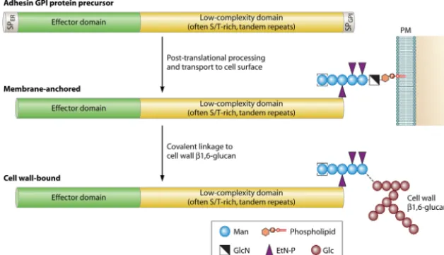
Most known fungal adhesins are GPI-modified wall proteins. The primary structure of GPI protein precursors includes con-served features, which therefore can be used to identify putative adhesins by bioinformatic means. At their N terminus, they have a signal peptide for entry into the endoplasmic reticulum (ER), and at their C-terminal end they have a peptide for anchoring to a preformed GPI lipid in the membrane of the ER (Fig. 1). Mature GPI proteins lack transmembrane domains. Most known mature adhesins are large proteins (usually⬎800 amino acids [aa]) with a modular structure; their N-terminal domain has a high complex-ity and mediates specific protein-protein, protein-sugar, or other protein-ligand interactions. These are believed to be largely re-sponsible for the specific interactions with their substrates, e.g., host cell surface proteins or carbohydrates (14,21,22). It is fol-lowed by a variable domain of low complexity that often is rich in serine/threonine (Ser/Thr) and usually contains tandem repeats (TRs). The repeat regions are subject to significant intraspecies
Published ahead of print8 February 2013
Address correspondence to Neeraj Chauhan, chauhan1@umdnj.edu. Copyright © 2013, American Society for Microbiology. All Rights Reserved. doi:10.1128/EC.00364-12
on September 8, 2020 by guest
http://ec.asm.org/
length polymorphisms due to slippage and/or recombination events during DNA replication (14, 23–25), which leads to re-moval or addition of repeat units. Longer repeat regions can con-fer greater adherence, while shorter repeat regions may result in decreased adhesion, possibly because the N-terminal effector do-main redo-mains buried in the cell wall (14).
Originally, binding specificities of adhesins were studied mostly by two complementary approaches. The first approach to assess gene function is to generate knockout mutant strains and study their phenotypes. However, fungal adhesin genes are often members of multigene families. Therefore, this approach is often hampered by functional redundancy as well as by compensatory mechanisms leading to upregulation of other adhesion genes whose products have similar or at least partially overlapping func-tions. Moreover, many adhesin genes show only low levels of ex-pression under the experimental conditions that are frequently used in the laboratory. To overcome these problems, a successful alternative approach is to heterologously express full or partial adhesins on the cell surface ofS. cerevisiaefor gain-of-function studies.S. cerevisiaestrains are available which lack a functional copy of the gene encoding flocculin-regulating transcription fac-tor Flo8, rendering them very poorly adherent to most substrates. This approach thus allows analysis of adherence to potential bind-ing ligands mediated by the gene of interest (22,26). For instance, heterologous expression studies inS. cerevisiaeshowed that adher-ence ofC. glabratato epithelial and endothelial cells is mediated, at least in part, by the proteins encoded by theEPAgene family (7,
22,27,28). Recently, fungal adhesin research has moved toward more structural studies using nanotechnology in which X-ray crystallography, nuclear magnetic resonance (NMR), and atomic force microscopy (AFM) are used to obtain detailed information with respect to the structure and ligand-binding specificities of adhesins inC. albicans,C. glabrata, andS. cerevisiae(termed floc-culins in the last-named organism) (29–33). Clearly, these high-resolution approaches largely improve our understanding of how fungal adhesins modulate adhesion, aggregation, biofilm forma-tion, and host-immune responses.
CANDIDA ALBICANSADHESINS
Until now, research mainly has addressed the three gene families
ALS,HWP, andIFF/HYRas adhesins ofC. albicans(listed inTable 1). They all conform to the domain organization described above and outlined inFig. 1. Additionally, a recent bioinformatics ap-proach (“FungalRV”) identified a plethora of proteins which have not previously been implicated in adhesion (81) but share at least some sequence and feature similarities with known adhesins. The value of this approach still needs to be validated experimentally. For example, one of the FungalRV top hits inC. albicans, Pga13, could not be confirmed to represent an adhesin in a recent study (82).
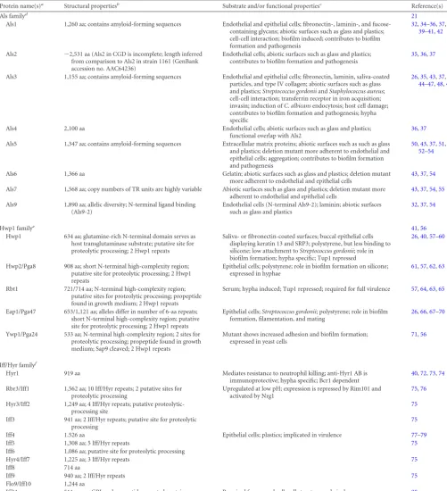
Als family.The Als family consists of eight large cell surface glycoproteins with a high degree of sequence similarity (Als1 to Als7 and Als9). The N-terminal parts of mature Als proteins com-prise tandem immunoglobulin (Ig)-like domains for mammalian protein interaction, followed by a threonine-rich conserved -sheet amyloid-forming “T region” and variable numbers of tan-dem repeats (TRs) (21). TRs seem to interact with hydrophobic surfaces and to facilitate aggregation (83, 84). A highly glyco-sylated serine- and threonine-rich spacer region in the C-terminal part extends the covalently attached proteins from the fungal sur-face into the environment and allows the ligand-binding domains to swivel and interact. Additional data onALSfamily composition and gene organization and the difficulties in the assembly of the
ALSgenes in theC. albicansgenome project are found in an ex-cellent review by Hoyer and colleagues (21).
Various disease models revealed host site-dependent expres-sion ofALSgenes indicating protein-specific functions. The con-tributions of Als1, Als2, and Als5 to pathogenesis were confirmed by mouse and reconstituted human oral epithelium (RHE) infec-tion models (34–36). Gene deletions, heterologous expression, and blocking experiments confirmed adhesive functions for, e.g., the family members Als1, Als2, Als3, and Als4 (36–38,43,50,85). Recently, the structure and ligand-binding properties of the N-terminal domain of Als1 and the protein encoded by the second allele ofALS9(NT-Als9-2) were resolved using NMR and X-ray
FIG 1Generic structure and posttranslational processing steps leading to cell wall incorporation of fungal adhesins. Abundant proteinN- and O-glycosylation, the latter especially taking place in the low-complexity domain, is not depicted for simplicity reasons. EtN-P, ethanolamine phosphate; Glc, glucose; GlcN, glucosamine; Man, mannose; PM, plasma membrane; PI, phosphatidylinositol.
on September 8, 2020 by guest
http://ec.asm.org/
TABLE 1Adhesins inC. albicans
Protein name(s)a
Structural propertiesb
Substrate and/or functional propertiesc
Reference(s)
Als familyd 21
Als1 1,260 aa; contains amyloid-forming sequences Endothelial and epithelial cells; fibronectin-, laminin-, and fucose-containing glycans; abiotic surfaces such as glass and plastics; cell-cell interaction; biofilm induced; contributes to biofilm formation and pathogenesis
32,34–36,37,38,
39–41,42
Als2 ⬃2,531 aa (Als2 in CGD is incomplete; length inferred from comparison to Als2 in strain 1161 (GenBank accession no. AAC64236)
Endothelial cells; abiotic surfaces such as glass and plastics; contributes to biofilm formation and pathogenesis
35,36,37
Als3 1,155 aa; contains amyloid-forming sequences Endothelial and epithelial cells; fibronectin, laminin, saliva-coated particles, and type IV collagen; abiotic surfaces such as glass and plastics;Streptococcus gordoniiandStaphylococcus aureus; cell-cell interaction; transferrin receptor in iron acquisition; invasin; induction ofC. albicansendocytosis; host cell damage; contributes to biofilm formation and pathogenesis; hypha specific
26,35,43,37,41,
44–47,48,49
Als4 2,100 aa Endothelial cells; abiotic surfaces such as glass and plastics; functional overlap with Als2
36,37
Als5 1,347 aa; contains amyloid-forming sequences Extracellular matrix proteins; abiotic surfaces such as such as glass and plastics; deletion mutant more adherent to endothelial and epithelial cells; aggregation; contributes to biofilm formation and pathogenesis
50,43,37,51,
52–54
Als6 1,366 aa Gelatin; abiotic surfaces such as glass and plastics; deletion mutant more adherent to endothelial and epithelial cells
43,37,54
Als7 1,568 aa; copy numbers of TR units are highly variable Abiotic surfaces such as glass and plastics; deletion mutant more adherent to endothelial and epithelial cells
43,37,54,55
Als9 1,890 aa; allelic diversity; N-terminal ligand binding (Als9-2)
Endothelial cells (N-terminal Als9-2); laminin; abiotic surfaces such as glass and plastics
32,37,54
Hwp1 familye 41,56
Hwp1 634 aa; glutamine-rich N-terminal domain serves as host transglutaminase substrate; putative site for proteolytic processing; 2 Hwp1 repeats
Saliva- or fibronectin-coated surfaces; buccal epithelial cells displaying keratin 13 and SRP3; polystyrene, but less binding to silicone; low attachment toStreptococcus gordonii; role in biofilm formation; hypha specific; Tup1 repressed
26,40,57–60
Hwp2/Pga8 908 aa; short N-terminal high-complexity region; putative site for proteolytic processing; 2 Hwp1 repeats
Epithelial cells; polystyrene; role in biofilm formation on silicone; expressed in hyphae
61,57,62,63
Rbt1 721/714 aa; N-terminal high-complexity region; putative sites for proteolytic processing; propeptide found in growth medium; 2 Hwp1 repeats
Serum; hypha induced; Tup1 repressed; required for full virulence 57,64,63,65
Eap1/Pga47 653/1,121 aa; alleles differ in number of 6-aa repeats; short N-terminal high-complexity region; putative site for proteolytic processing; 2 Hwp1 repeats
Epithelial cells;Streptococcus gordonii; polystyrene; role in biofilm formation, filamentation, and mating
26,66,67–70
Ywp1/Pga24 533 aa; N-terminal high-complexity region; 2 sites for proteolytic processing; propeptide found in growth medium; Sap9 cleaved; 2 Hwp1 repeats
Mutant shows increased adhesion and biofilm formation; expressed in yeast cells
71,56
Iff/Hyr familyf
Hyr1 919 aa Mediates resistance to neutrophil killing; anti-Hyr1 AB is immunoprotective; hypha specific; Bcr1 dependent
40,72,73,74
Rbr3/Iff1 1,562 aa; 10 Iff/Hyr repeats; 2 putative sites for proteolytic processing
Upregulated at low pH; expression is repressed by Rim101 and activated by Nrg1
75,76
Hyr3/Iff2 1,249 aa; 4 Iff/Hyr repeats; putative proteolytic-processing site
75
Iff3 941 aa; 2 Iff/Hyr repeats; putative site for proteolytic processing
75
Iff4 1.526 aa Epithelial cells; plastics; implicated in virulence 77–79
Iff5 1,308 aa; 5 Iff/Hyr repeats 75
Iff6 1,086 aa; putative site for proteolytic processing
Hyr4/Iff7 1,225 aa; 3 Iff/Hyr repeats 75
Iff8 714 aa
Iff9 940 aa; 2 Iff/Hyr repeats 75
Flo9/Iff10 1,244 aa
Iff11 511 aa; no GPI anchor peptide; secreted protein Required for normal cell wall structure and virulence 80
aProtein names are from theCandidaGenome Database (CGD) (http://www.candidagenome.org/). b
All listed proteins contain signal peptides for ER entry; all except Iff11 contain C-terminal signals for GPI anchoring. cOnly the most relevant phenotypes are listed.
d
Als family proteins share similar domain structures with N-terminal effector domains that are 55 to 90% identical across the whole family, central domains with tandem repeats (TR), and variable Ser/Thr-rich C-terminal domains.
e
Hwp1 family proteins have in common the presence of one or more Hwp1 repeats.
fIff/Hyr family proteins share a protein structure with a conserved putative N-terminal effector domain followed by a low-complexity C-terminal domain. The latter may contain a variable number of Iff/Hyr repeats.
on September 8, 2020 by guest
http://ec.asm.org/
crystallography (32). Salgado and coworkers showed that NT-Als9-2 is capable of binding flexible C termini of peptides in ex-tended conformations (32). This is consistent with results from earlier studies showing that Als proteins bind to a number of structurally unrelated proteins and peptides from randomly gen-erated sequences (51). In addition, the N-terminal part of Als1 protein specifically binds fucose-containing glycans (39). Adhe-sion through Als proteins can be activated and increased dramat-ically by amyloid nanodomain formation (see section below). Furthermore, Als1, Als2, and Als3 appear to be important for bio-film formation, and complementary roles in this process promot-ing monospecies biofilm formation were observed for Als1, Als3, and Hwp1 (36,40,41). Recently, single-molecule AFM uncovered the finding that, during the yeast-to-hypha transition, an increase in the distribution and adhesion of Als proteins is observed, ac-companied by dramatically increased surface hydrophobicity and the unfolding and extension of mannosylated Als proteins (29). Earlier work has shown a link between cell surface hydrophobicity and outer-chainN-mannosylation status (86). It was postulated that-1,2-oligomannosides present in the acid-soluble phospho-mannan part ofN-glycans might form a tight, inflexible helix that has a hydrophobic face. Consistent with this idea, consensusN -glycosylation sites are abundantly present in fungal adhesins, for instance, in repeat regions of Als proteins.
A high degree of allelic variability, especially in TR domains, is present in theALSfamily (21), and variations in tandem repeat copies ofALS3were shown to modulate protein function and adhesion (87). Beside a role in adhesion, effects on the fungal cell size were noticed for Als1 (21) and on host cell damage and cyto-kine induction for the hypha-specific Als3 (44). Als3 was also found to mediateC. albicansadherence toStaphylococcus aureus
and supported mixed-species biofilm formation with Streptococ-cus gordonii(26,45). Furthermore, the multifunctional Als3 pro-tein acts as an invasin by inducing endocytosis into host cells (46) and enables iron acquisition by binding transferrin (47).
Future research will provide mechanistic insight into the dif-ferent functions and binding specificities of the Als proteins and into whether this knowledge can be exploited to improve
anti-Candidatherapy, e.g., by immunization against the effector do-mains of Als proteins or by targeted inhibition of amyloid forma-tion (88).
Amyloid formation.Amyloids are fibrous protein structures present on the microbial cell surface. Recently, amyloids have been identified inC. albicansAls adhesins (66,89). Amyloid for-mation inC. albicanshas been analyzed primarily for the Als5 protein. Amyloid-forming sequences within the Als5 primary amino acid sequence were identified using the-aggregation pre-diction software TANGO (90). This software predicts the poten-tial of regions within a protein sequence to form-strand-rich aggregates based on inter- and intramolecular interaction energies (90). Sequences predicted to form amyloids are usually found within the T region of Als adhesins. The ability of peptides corre-sponding to putative amyloid sequences to form-aggregates and amyloid fibrils also has been demonstrated experimentallyin vitro
using transmission electron microscopy (TEM) and Congo red and thioflavin binding (66,89). Threonine, isoleucine, and valine residues present within the T regions ofC. albicansAls proteins are reported to have a significant role in amyloid formation (66,89). Similar clusters of threonine, isoleucine, and valine residues are widely present in fungal adhesins and are also present in C.
glabrata adhesins, though additional experimental evidence is needed to establish the role of these residues in aggregation and amyloid formation inC. glabrata. In addition to Als adhesins, functional amyloid formation has been reported for peptide frag-ments of theC. albicansadhesin Eap1 and the Flo1 and Flo11 flocculins ofS. cerevisiae(66).
Recent elegant work using AFM demonstrated that amyloid interactions provide cohesive strength toC. albicansAls proteins. Possibly, initial protein binding through the Ig-like domains and/or weak hydrophobic binding through the TR domains is followed by amyloid formation, thereby strengthening cell adhe-sion (84). Using AFM, it was demonstrated that long-lived protein interactions are enabled by Als amyloids that can function as a molecular zipper. Formation and propagation of Als5 adhesion nanodomains on the cell surface were observed in response to mechanical stimuli, which probably causes the T region to par-tially unfold and expose the amyloid-forming sequence (91). The formation of amyloid clusters could thus explain why Als proteins exhibit weak binding to many ligands but mediate strong adher-ence. These data provided evidence that Als-mediated adhesion largely depends on conformational modifications of existing ad-hesins rather than or in addition to signal transduction and ex-pression of new adhesin molecules (92).
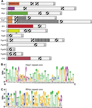
Hwp family.Although their N-terminal effector domains do not have sequence similarities, Hwp1, Hwp2 (“hyphal wall pro-tein”), and Rbt1 (“repressed by Tup1”) are considered to be part of a single family because they share a highly conserved 42-aa repeat unit (Fig. 2andTable 2) (61,101). Expression ofHWP1,
HWP2, andRBT1is hypha specific and also upregulated during mating of opaque cells (57). Their corresponding proteins are required for adhesion to host cell surface proteins, biofilm forma-tion, cell-cell aggregaforma-tion, and mating (41,57). Within this family, Hwp1 is unique in the way that its N terminus is highly enriched in glutamine residues. These are substrates for human host transglu-taminase enzymes, which covalently cross-link Hwp1 to extracel-lular matrix (ECM) proteins of epithelial host cells (58–60).
Since the adhesin Eap1 also features the conserved 42-aa do-main, it can be included in this family. In fact, seven other GPI proteins, namely, Pga6, Pga18, Pga38, Pga59, Pga62, Ywp1/Pga24, and Cht2, and Iff11, a secreted protein of the Iff family (see below), contain similar sequences (Fig. 2AandB) (102). The 42-aa-long repeat domain coincides with several amyloid-forming patches, which are thought to facilitate multimerization of adhesins (66). Moreover, the repeat unit features three conserved cysteine resi-dues. These might be involved in the formation of intermolecular disulfide bridges between proteins, which potentially promote protein aggregation or reinforcement of cell wall integrity. Al-though Pga59 and Pga62 are upregulated during biofilm forma-tion (103) and play a minor role in morphogenesis (102), their deletion mutants show no reduction in adhesion or biofilm-form-ing capacity (102). The repeat unit is also somewhat reminiscent of domains that have been shown to bind cell wall polymers (104), which might explain why the chitinase Cht2 has a similar domain. Thus, alternatively, these 42-aa domains might also be involved in interacting with cell wall polysaccharides and consequently con-tribute to cell-to-cell interactions.
EAP1 is expressed in both yeast and hyphal cells and is differ-entially regulated during yeast phenotypic switching. It is mainly specific to the white phase and contributes not only to biofilm formation and general epithelial cell and polystyrene adhesion
on September 8, 2020 by guest
http://ec.asm.org/
(67–69) but especially to cell-cell binding in response to␣ -pher-omone (69). The different domains of Eap1 mediate adhesion to different epitopes when expressed inS. cerevisiae(70): the N-ter-minal domain (Fig. 2, orange box) facilitates agar invasiveness, cell-cell contacts, and adhesion to mammalian cells, and the Ser/ Thr-rich repeat regions (Fig. 2A) contribute to polystyrene adhe-sion (70). Similar to Als3, Eap1 and Hwp1, but not Rbt1, can facilitate binding toStreptococcus gordonii, a colonizing bacterium of the oral cavity.S. gordoniifeatures proteins with similar amy-loid sequences on its surface, but binding to Als3 and Eap1 is independent of amyloid formation (26), indicating that this bind-ing is mediated through the N-terminal effector domain rather than the repeat units.
Strains deleted forYWP1are hyperadherent to several surfaces
and show increased biofilm formation. The Ywp1 protein is there-fore considered to be involved in the release of yeast cells from surfaces (71,105), countering the action of adhesins. The protein is processed by regulatory proteases of the Kex2 and Sap9/Sap10 group to generate 11-to-12-kDa propeptides which stay noncova-lently but tightly attached to the mature protein part (105). Inter-estingly, the Sap9 deletion mutant is also hyperadherent (106), and a peptide immediately downstream of the Ywp1 propeptide was released from purified yeastsap9⌬sap10⌬cell walls with re-combinant Sap9 (56), underlining the possible biological rele-vance of proteolytic activation for this protein. Similarly, Hwp1, Rbt1, and Hwp2 also contain predicted protease recognition sites within their effector domains (58,107) (Fig. 2A); thus, it can be speculated that regulatory proteolytic events also play a role in
FIG 2TheC. albicansHwp1 and Iff/Hyr families contain family-specific repeat sequences. (A) Diagram showing modular structures of the protein precursors of the Hwp1 family. Included are all 12C. albicansSC5314 proteins (genome assembly 21) containing at least one copy of the pattern T[ILV][ST]XCX(4)CX (16,20)TX[VYF][TV]T[YF]CP[ILV] (PROSITE format), which are indicated as diagonally striped boxes. N-terminal high-complexity domains of the mature proteins, believed to comprise effector domains, are presented in different colors because of their lack of sequence similarity. The twoEAP1alleles in strain SC5314 differ in length at a region that predominantly encodes repeats of a “PATEST” pattern (indicated by vertically striped boxes). A region with imperfect 41-to-50-aa serine-rich repeats in Pga18 is shown as the boxes with the thinner diagonal stripes. Signal peptides (SP) for ER entry and GPI anchoring are indicated. Putative and experimentally validated proteolytic Kex2 cleavage sites are depicted by open and black triangles, respectively. (B and C) Sequence logos (created athttp://weblogo.berkeley.edu/) of the Hwp1 group (B) and of the Iff/Hyr family repeats (75) (C). Amino acid color codes for both panels are as follows: purple, conserved tryptophan in the Iff group, cysteines, and prolines; green, amyloid forming (TLVIA); red, positively charged (KRH); blue, negatively charged (DE); orange, all other amino acids. Alignments used for building the logos have been communicated to Pfam (http://pfam.janelia.org/) for creation of Pfam hidden Markov models (HMM) entries. All Iff/Hyr repeats from reference75match the pattern WX(2)TX (7)TX(2)G[IV](2).
on September 8, 2020 by guest
http://ec.asm.org/
these proteins. A fragment of the potential propeptide of Rbt1, between the second and third putative recognition sites, was found in culture supernatants (64).
Iff/Hyr family.The 12 proteins encoded by theIFF/HYRgene family (“IFF” standing for “IPF family F” and “HYR” for “hy-phally upregulated protein”) have a high degree of sequence sim-ilarity in their N-terminal effector domains (Table 1). Six of them, Iff1/Rbr3, Iff2/Hyr3, Iff3, Iff5, Iff7/Hyr4, and Iff9, also share 41-to-51-aa-long Iff-specific tandem repeats (75) (Fig. 2C). The fam-ily includes the hypha-specific proteins Hyr1 and Iff11 (74,80). The latter represents a secretory protein that differs from other family members by lacking a GPI anchor. Deletion ofIFF11leads to significant alterations in the cell wall, suggesting that the pro-tein has a role in cell wall organization and/or enzymatic function (80). How these functions relate to otherIFF/HYRgenes on the molecular level is unknown, as studies have looked at these genes only at the phenotypical level (77,78). The molecular substrates of the Iff/Hyr family proteins have not yet been identified, but they are clearly of clinical relevance. Overexpression ofIFF4inC. albi-cansincreased adherence to plastic and epithelial cells (79). In animal models, both overexpression and underexpression ofIFF4
resulted in a reduction of virulence, indicating that a specific ex-pression level is required for maximal virulence (108). Hyr1 has been implicated in resistance to neutrophil killing (72), and an anti-Hyr1 antibody (AB) induced immunity to disseminated can-didiasis in mice (73).
ADHESINS IN NON-ALBICANSCTG-CLADECANDIDASPECIES Despite several phenotypic descriptions of adhesion, adhesins have not yet been investigated deeply at the molecular level in CTG-clade species other thanC. albicans. Genomic data indicate that the adhesin families described forC. albicans also exist in other species. The presence ofALS-like genes has been confirmed inCandida dubliniensis,Candida tropicalis,Candida parapsilosis,
Candida lusitaniae, andCandida guilliermondii(12,101,108), and theIFF/HYRgene family is also widely present in CTG-clade Can-didaspp. (101,109). Homologs of Rbt1 are present inC.
dublini-ensis, C. parapsilosis, C. tropicalis, and Candida orthopsilosis. NCBI-BLAST searches with the small N-terminal effector do-mains of Eap1 and Hwp2 revealed homologs only in the closely related speciesC. dubliniensis. A clear Hwp1 homolog is also pres-ent inC. dubliniensis, whereas a protein with only a short stretch of high sequence identity to Hwp1 is found inC. tropicalis. Of note, adhesins that fit the Als or Rbt1 definition are not present in non-CTG clade species such asC. glabrataorS. cerevisiae.
Comparative genomic analysis of theALSandIFF/HYRgene families of the different CTG-clade species showed high genetic variability, with apparent gene losses and multiplications that have occurred during evolution (75,101,110). For instance,C. dubliniensislacks an ortholog of the hyphally regulatedHYR1gene inC. albicansthat was lost during evolution (110). Also, theIFF/
HYR family shows duplications on three chromosomes in C. parapsilosis(101) but not inC. orthopsilosis(111). This is intrigu-ing, asC. parapsilosisstrains have the highest adhesion capacities of all clinically relevantCandida species and are mostly found growing in catheter-associated biofilms, pointing to the poten-tially high clinical importance of this gene family.
TheALSfamily is expanded to at least 13 members inC. tropi-calis(101). ForC. albicansand C. dubliniensis, there is a broad similarity between their ALS families. However, phylogenetic analysis showed that recombination between differentALSgenes has altered some sequences since speciation (110). Furthermore,
ALS5and the multifunctional and hypha-specificALS3are not present inC. dubliniensis, which instead has a duplicated copy of
ALS2. This high genetic variability and the recombination events occurring in fungal adhesin genes seem related to the frequent presence of intragenic tandem repeats. It should be noted, how-ever, that adhesin gene families are often located in subtelomeric regions, which may significantly contribute to the expansion of some of these adhesin gene families.
CANDIDA GLABRATAADHESINS
Analysis of the genome of theC. glabratastrain CBS138/ATCC 2001 revealed an exceptionally large number (66) of sequences
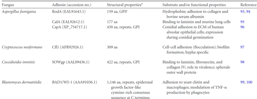
TABLE 2Adhesins in non-Candida human pathogenic fungi
Fungus Adhesin (accession no.) Structural propertiesa Substrate and/or functional properties Reference(s)
Aspergillus fumigatus RodA (EAL91643.1) 159 aa, GPI? Hydrophobin; adhesion to collagen and
bovine serum albumin
93,94
CalA (EAL92612.1) 177 aa Binding to laminin and murine lung cells 95
CspA (XP_754717.1) 430 aa, repeats, GPI Conidial adhesion to ECM of human alveolar epithelial cells; expression during conidial germination
96
Cryptococcus neoformans Cfl1 (AFR92926.1) 309 aa Cell-cell adhesion (flocculation); biofilm
formation; hypha specific
97
Coccidioides immitis SOWgp (AAL09436.1) 422 aa, repeats, GPI Binding to laminin, fibronectin, and
collagen IV; role in virulence; spherule outer wall protein
98
Blastomyces dermatitidis BAD1/WI-1 (AAA91036.1) 1,146 aa, repeats, epidermal
growth factor-like cysteine-rich consensus sequence at C terminus
Adhesion to yeast chitin and
macrophages; modulation of TNF-␣ production by phagocytes
99,100
a
All proteins have signal peptides for secretion. GPI, glycosylphosphatidylinositol-anchoring signal peptide. The GPI-anchor signal prediction in RodA is controversial as it includes one of the eight cysteines, which are believed to be crucial for correct folding of this amphipathic protein.
on September 8, 2020 by guest
http://ec.asm.org/
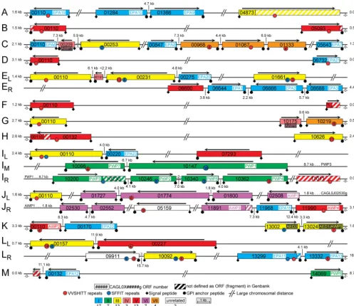
specifying GPI-modified adhesin-like wall proteins (8,9,112; see alsoFig. 3). All these proteins have the modular structure that is typical for adhesins: an effector domain followed by a low-com-plexity region that is often spiked with internal tandem repeats, termed “megasatellites,” inC. glabrata(113). Based on phylogenic analysis of their putative N-terminal ligand-binding regions, this large family can be divided into seven subfamilies (8). Only two proteins (CAGL0J05159g and CAGL0L09911g) fall outside these subfamilies, as their N-terminal domains are unrelated to those of any of the other putative adhesins (Fig. 3). The largest subfamily is
theEPAfamily, with 17 members in CBS138. Remarkably, earlier studies reported an even larger number ofEPAgenes (23) in strain BG2 (114,115). For instance, orthologs ofEPA4andEPA5, pres-ent in BG2 (116), are lacking in strain CBS138. Furthermore, none of the orthologousEPAgenes in either strain are 100% identical. Relevant in this respect is the fact that the tandem repeat regions, which usually are present in fungal adhesin-encoding genes, fre-quently show strain-dependent size reductions or expansions oc-curring during DNA replication (23–25,96). All together, these intraspecies gene variations underline the extremely large genetic
FIG 3Genomic organization of putative adhesin-encoding genes in the sequencedC. glabratastrain CBS138, modified from reference8. Chromosomes and open reading frames (ORFs) are numbered following Génolevures’s systematic ORF numbering, which is also used in theCandidaGenome Database. ORF sizes are to scale, but distances between ORFs are not. Many of the gene sequences, when translated, give rise to frameshifts, probably mostly due to sequencing and/or annotation errors or the presence of intronic sequences. Some unannotated ORF fragments (no ORF number), identified by BLASTX, are connected to incomplete ORFs. Colors indicate seven subfamilies, sharing homology in the N-terminal putative ligand-binding parts. Numbers of genes in each group are indicated. N-terminal domains of CAGL0L09911g and CAGL0J05170g (white) are unrelated to the other adhesins. For CAGL0E00187g (group IV, pink), only the GPI anchor peptide containing the C-terminal part was identified; its classification is therefore based on BLASTP analysis of this region. Numbers of nonadhesin ORFs separating adhesin-like ORFs and telomeres and distances of terminal adhesin-like genes to the end of the obtained telomeric DNA sequences are indicated. Numbers of the megasatellite signatures [VILF][VI][ST]H[IVS][TI][TGI] (“VVSHITT”) and SFFIT are specified only for ORFs whose protein sequences are complete in the databases. Arrows indicate directions of transcription.
on September 8, 2020 by guest
http://ec.asm.org/
plasticity with respect to fungal adhesin genes, including theEPA
family, inC. glabrata.
Epa family.The best-studied proteins in the Epa family are Epa1, Epa6, and Epa7. All three proteins mediate adherence to human epithelial and endothelial cells (7,22,27). Epa1 has also been reported to govern binding to innate immune cells (117). Remarkably, deletion of justEPA1reduces adherence to human epithelial cells in vitroto background levels, probably because otherEPAgenes are transcribed only at low levels when grown under laboratory conditions (116,118). This repression ofEPA
gene transcription is explained, in part, by the fact that most of the
EPAgenes are located within subtelomeric regions (Fig. 3) where they are subject to chromatin-based gene silencing (116, 118,
119). In fact, about two-thirds of the 66 putative adhesin genes in
C. glabrataare located within these subtelomeric regions (8) and in all subtelomeric areas of strain CBS138 at least one putative adhesin gene is present (Fig. 3).
The ligand-binding domains of Epa proteins are called PA14 (“anthrax protective antigen”) domains and have lectin proper-ties. Studies using carbochips (glycan arrays) have shown that Epa1, Epa6, and Epa7 bind to oligosaccharides that contain ter-minal galactose residues, such as those occurring in mucin-type
O-glycans (22,31). This is consistent with the idea that these pro-teins enable the fungus to bind to glycopropro-teins on the host cell surface.S. cerevisiaeflocculins (Flo proteins) contain N-terminal domains that exhibit weak similarity to the PA14 domains inC. glabrata(23). Structural analysis of theS. cerevisiae Flo family member Flo5 showed that this protein binds to high-mannose oligosaccharides (33), giving this glycoprotein self-binding prop-erties. Veelders et al. (33) described in detail that the crystal struc-ture of the Flo5 effector domain, complexed to cognate ligands, revealed a beta-sandwich core that utilizes a uniqueDcisD calci-um-binding motif for carbohydrate binding. Given the high abundance of high-mannose oligosaccharides in yeast cell walls, this confers to baker’s yeast the self-aggregating properties that lead to the formation of flocs and are exploited widely in the beer-and wine-making industries. Recently, the crystal structure of Epa1 complexed to cognate disaccharide ligands, including Gal1–3Glc and the T antigen (Gal1–3GalNAc), has been solved, showing a similar lectin fold andDcisDcalcium-binding motif (30,31). Further studies, using point mutants of Epa6 and Epa7, or using Epa1 variants with modified binding sites that cor-respond to Epa2, Epa3, and Epa6, showed that substrate specificity is governed by two inner loops, CBL1 and CBL2, involved in cal-cium binding as well as by three outer loops, L1, L2, and L3 (22,
31). The CBL2 loop was previously also shown to determine Flo/ NewFlo specificity (mannose binding versus mannose and glucose binding) inS. cerevisiae(120).
Pwp family.Of the other putative adhesins inC. glabrata, the N-terminal effector domains of one subfamily, with seven mem-bers, also show similarity to the PA14 domains of the Epa and Flo families. These sevenC. glabrataproteins were termed Pwp1 to Pwp7 (8). It is conceivable that Pwp proteins also are lectins with a role in aggregation, for instance, during biofilm formation, or host binding. In line with this, Pwp7 has been shownin vitroto play a role in adherence to human endothelial cells (112). How-ever, solid conclusions about the functionality of the Pwp family await further functional and structural studies to elucidate their ligand-binding properties.
Other putative adhesins.The N-terminal domains of the
re-maining 42 putativeC. glabrataadhesins have no clear homology with those of Epa or Pwp proteins, and, with the exception of Aed1, which, like Pwp7, mediates adherence to human endothe-lial cells (112), their functions and ligands are unknown.
In studies where the covalently bound cell wall proteome was studied using tandem mass spectrometry, Epa3 and Epa6 were identified as well as Awp1 to Awp6, six of the non-Epa adhesin-like wall proteins (8,121). The above-mentioned Aed1 (Awp5) is one of these proteins. Awp1 to Awp6 are members of four differ-ent adhesin subfamilies. Awp2 and Awp4 belong to a subfamily of putative adhesins with weak similarity to theC. albicansHyr/Iff family as well as to the haze-protective mannoproteins Hpf1 and Hpf1=inS. cerevisiae(122). Awp1 belongs to the same subfamily as Awp3. Except for Awp1, these (putative) adhesins were found by mass spectrometric analysis in isolated cell walls from strain CBS138, which was originally a feces isolate. So far, Awp1 has been detected only in a different clinical isolate (ATCC 90876, isolated from blood) grown to the stationary phase. Expression studies using a quantitative PCR (qPCR) approach showed that the cor-responding genes of most of the identified wall adhesins are sig-nificantly upregulated during biofilm development (121). Inter-estingly, mass spectrometric analysis identified Awp6 only in cell walls under biofilm-forming conditions. Thus, expression and wall incorporation of the adhesins seem to be dependent on the genetic strain background and growth conditions. Furthermore, the analysis supports the hypothesis thatC. glabratacontains a large repertoire of tightly regulated functionally diverse adhesins to enable rapid colonization of different host tissues under a vari-ety of host-defined conditions.
Many of theC. glabrataadhesin-like proteins also contain tan-dem repeats, such as repeated sequences containing VSHITT or SFFIT signatures, in the region downstream of the effector do-main (Fig. 3). The VSHITT repeat of about 46 amino acids is found in about half of the putative adhesins across the different subgroups, including Awp2 and Awp4 and the two unrelated ad-hesin-like proteins (8). Therefore, although the N-terminal effec-tor domains of proteins in different subfamilies may not show obvious similarities, these proteins do have structural (and prob-ably functional) relationships. Interestingly, the presence of (mul-tiple) VSHITT or SFFIT megasatellites seems specific for adhesins inC. glabrataas it does not occur in any other protein currently present in the NCBI protein database (123). Elucidation of the exact functions of the repeat regions remains largely elusive. It is generally believed that expansion of glycosylated repeat regions improves the surface exposure and ligand-binding propensities of the N-terminal domains of adhesins (25). On the other hand, a role in cellular aggregation through amyloid-forming properties has also been suggested (66).
ASPERGILLUS FUMIGATUSADHESINS
A. fumigatusis the main causative agent of invasive aspergillosis (IA), usually a fatal infection in patients with AIDS, solid-organ transplants, or chronic pulmonary diseases, including cystic fibro-sis and allogeneic bone marrow transplants, and in patients with various hematological malignancies.
One of the best-characterized proteins required for its role in adherence to host cells inA. fumigatusis RodA (Table 2). The RodA protein is expressed primarily on the surface of conidia and is required to form the surface rodlet layer (93). RodA extracted fromA. fumigatusconidia is immunologically inert, thus
on September 8, 2020 by guest
http://ec.asm.org/
ing immune recognition of fungal spores (94). Disruption of this gene decreased adherence to collagen and bovine serum albumin but not to pneumocytes, fibrinogen, or laminin (93). Although RodA is reported to be a GPI protein (94), it is not a classical wall adhesin. In fact, GPI modification of RodA would seem in conflict with the amphipathic fold of this hydrophobin, which relies on the formation of four disulfide bridges between eight conserved cys-teine residues (124), the last one of which is located in the pre-sumed GPI-anchoring signal peptide.
TheA. fumigatusadhesin CalA was identified by employing a bioinformatic approach (95) and is predicted to be a secreted pro-tein (Table 2). Recombinant CalA was shown to bind to laminin and mouse lung cells. Binding of anti-CalA antibodies toA. fu-migatusconidia suggested the presence of CalA on the conidial surface.
More recently, the GPI-modified wall protein CspA (“cell sur-face protein A”) was identified as a putative adhesin with internal repeats (Table 2), of which the number per gene is strain depen-dent (96). ThecspA-encoded cell wall protein is unmasked during conidial germination and is surface expressed during hyphal growth. Deletion of CspA, when combined with other cell wall proteins such as Ecm33 and Gel2, resulted in reduced conidial adherence to extracellular matrix (ECM) from human alveolar cells. Strains lacking CspA displayed cell wall weakening, whereas overexpression of CspA resulted in increased resistance to cell wall-degradative enzymes, suggesting that its main function is in cell wall integrity.
CRYPTOCOCCUS NEOFORMANSADHESINS
C. neoformansis an environmental fungus that is found usually in soil. Much of the insight gained about its pathogenesis has come from studies focusing on capsule and melanin production—two of the most prominent virulence factors of this fungus. Surpris-ingly, very little is known about adhesins fromC. neoformans. Recently, a secretory protein, named Cfl1 (cell flocculin 1), was reported to be an adhesin (Table 2) (97). TheC. neoformans Cfl1-encoding gene was found to be specifically expressed during the hyphal growth phase, and the protein was documented to regulate morphogenesis, cell-cell adhesion, biofilm formation, and viru-lence (97). Paradoxically, morphotype transition inC. neoformans
is typically observed during mating, but pheromone signaling components play no or minimal direct roles in virulence. Inter-estingly, Wang et al. have demonstrated that filamentation inC. neoformanscan occur independently of pheromone signaling and mating (97). Nevertheless, elucidation of the precise function of Cfl1 requires further investigation.
ADHESINS IN FUNGI CAUSING ENDEMIC MYCOSES AND RARE FUNGAL DISEASES
The causative agents of endemic mycoses are mostly dimorphic fungi that usually grow in a filamentous (mold) form at 25°C, while at 37°C they convert to yeast growth. From this group, only a few cell wall proteins mediating adherence inC. immitisandB. dermatitidishave been characterized (Table 2). RecombinantC. immitisadhesin SOWgp (“spherule outer wall glycoprotein”) was shown to bind to mammalian ECM proteins (laminin, fibronec-tin, and collagen IV) (98). Deletion ofSOWgpresulted in partial loss of binding ofC. immitisspherules (multinucleate round cells in the parasitic cycle of this fungus) to ECM and attenuation of virulence in a murine model of coccidioidomycosis.
In the case ofB. dermatitidis, BAD1/WI-1 (“Blastomyces adhe-sin”) has been identified as a yeast-phase-specific protein involved in adhesion. Studies performed with gene disruptants showed that BAD1 is critical for binding ofB. dermatitidisyeast cells to host cells and for pathogenesis in a murine model of pulmonary infec-tion (99). One of the major mechanisms by which the secretory protein BAD1 influencesB. dermatitidisis downregulation of ex-pression of proinflammatory cytokines such as tumor necrosis factor alpha (TNF-␣) (100).
InFusarium oxysporum, an opportunistic filamentous fungus that has the unique ability to infect both plant and mammalian hosts, four putative GPI-modified adhesins were identified in silico. However, none of these were identified during mass spec-trometric analysis of hyphal walls under adhesion-inducing con-ditions (125).
SUMMARY
The fungal cell wall and its constituents are of interest primarily due to its importance as a potential target for antifungal therapy and its role in pathogenesis. In recent years, tremendous progress has been made in identification and characterization of adhesins, largely by virtue of genome sequencing of many pathogenic fungi and development of novel genetic and molecular tools. Adhesins are shown to be important for disease establishment and progres-sion by helping in colonization. This correlation has been demon-strated by a number of studies, the most important of which are the construction of deletion mutants and the demonstration of their reduced adherence and virulence. The existence of large fam-ilies of adhesin proteins, e.g., the Als family inC. albicansand the Epa family inC. glabrata, with both overlapping and specific func-tions, conformational activation, differential gene expression pat-terns, and allelic variability, provides a versatile toolbox to these fungi. This may foster external reactions, for example, to enable coordinated adhesion to various tissues in specific host niches. However, there are still significant gaps in our knowledge about how fungal pathogens colonize and persist in the host. We antic-ipate that future studies focused on functional characterization of novel putative adhesins will not only provide new insights into their role in pathogenesis but will also help define their role related to the host niche where the organisms are found and thrive. More-over, mechanistic insights into the mode of action of adhesins obtained using nanotechnology-based approaches may provide valuable clues to their potential to serve as future diagnostic mark-ers or to improve antifungal therapies.
ACKNOWLEDGMENTS
P.W.J.D.G. is supported by an INCRECyT fellowship from Parque Cientí-fico y Tecnológico de Albacete. A.D.D.B. is the recipient of a postdoctoral fellowship from the junta de comunidades de Castilla—La Mancha (JCCM). M.W. was supported by the Deutsche Forschungsgemeinschaft (DFG) (WE 3537/1-2). N.C. is supported by PHRI startup funds.
REFERENCES
1.Perlroth J, Choi B, Spellberg B.2007. Nosocomial fungal infections: epidemiology, diagnosis, and treatment. Med. Mycol.45:321–346. 2.Pfaller MA, Diekema DJ.2007. Epidemiology of invasive candidiasis: a
persistent public health problem. Clin. Microbiol. Rev.20:133–163. 3.Borg-von Zepelin M, Kunz L, Ruchel R, Reichard U, Weig M, Gross
U.2007. Epidemiology and antifungal susceptibilities ofCandidaspp. to six antifungal agents: results from a surveillance study on fungaemia in Germany from July 2004 to August 2005. J. Antimicrob. Chemother.
60:424 – 428.
on September 8, 2020 by guest
http://ec.asm.org/
4.Busscher HJ, Rinastiti M, Siswomihardjo W, van der Mei HC.2010. Biofilm formation on dental restorative and implant materials. J. Dent. Res.89:657– 665.
5.Ramage G, Martinez JP, Lopez-Ribot JL.2006.Candidabiofilms on implanted biomaterials: a clinically significant problem. FEMS Yeast Res.
6:979 –986.
6.Ramage G, Culshaw S, Jones B, Williams C.2010. Are we any closer to beating the biofilm: novel methods of biofilm control. Curr. Opin. Infect. Dis.23:560 –566.
7.Cormack BP, Ghori N, Falkow S.1999. An adhesin of the yeast patho-genCandida glabratamediating adherence to human epithelial cells. Science285:578 –582.
8.de Groot PWJ, Kraneveld EA, Yin QY, Dekker HL, Groß U, Crielaard W, De Koster CG, Klis FM, Weig M.2008. The cell wall of the human pathogenCandida glabrata: differential incorporation of novel adhesin-like wall proteins. Eukaryot. Cell7:1951–1964.
9.Weig M, Jansch L, Gross U, De Koster CG, Klis FM, De Groot PWJ.
2004. Systematic identification in silico of covalently bound cell wall proteins and analysis of protein-polysaccharide linkages of the human pathogenCandida glabrata.Microbiology150:3129 –3144.
10. Bernhardt J, Herman D, Sheridan M, Calderone R.2001. Adherence and invasion studies ofCandida albicansstrains, using in vitro models of esophageal candidiasis. J. Infect. Dis.184:1170 –1175.
11. Calderone RA, Fonzi WA.2001. Virulence factors ofCandida albicans.
Trends Microbiol.9:327–335.
12. Hoyer LL.2001. TheALSgene family ofCandida albicans.Trends Mi-crobiol.9:176 –180.
13. Sundstrom P.2002. Adhesion inCandidaspp. Cell. Microbiol.4:461– 469.
14. Verstrepen KJ, Klis FM.2006. Flocculation, adhesion and biofilm for-mation in yeasts. Mol. Microbiol.60:5–15.
15. Chauhan N, Li D, Singh P, Calderone R, Kruppa M.2002. The cell wall ofCandidaspp., p 159 –175.InCalderone RA (ed), Candida and candi-diasis. ASM Press, Washington, DC.
16. Klis FM, De Groot P, Hellingwerf K.2001. Molecular organization of the cell wall ofCandida albicans.Med. Mycol.39(Suppl 1):1– 8. 17. Klis FM, Ram AFJ, De Groot PWJ.2007. A molecular and genomic view
of the fungal cell wall, p 95–117.InHoward RJ, Gow NAR (ed), The mycota, vol 8. Springer-Verlag, Berlin, Germany.
18. Lesage G, Bussey H.2006. Cell wall assembly inSaccharomyces cerevi-siae.Microbiol. Mol. Biol. Rev.70:317–343.
19. Orlean P.2012. Architecture and biosynthesis of the Saccharomyces cerevisiaecell wall. Genetics192:775– 818.
20. Latgé JP.2007. The cell wall: a carbohydrate armour for the fungal cell. Mol. Microbiol.66:279 –290.
21. Hoyer LL, Green CB, Oh SH, Zhao X.2008. Discovering the secrets of
theCandida albicansagglutinin-like sequence (ALS) gene family—a
sticky pursuit. Med. Mycol.46:1–15.
22. Zupancic ML, Frieman M, Smith D, Alvarez RA, Cummings RD, Cormack BP.2008. Glycan microarray analysis ofCandida glabrata ad-hesin ligand specificity. Mol. Microbiol.68:547–559.
23. Goossens K, Willaert R.2010. Flocculation protein structure and cell-cell adhesion mechanism inSaccharomyces cerevisiae.Biotechnol. Lett.
32:1571–1585.
24. MacCallum DM, Castillo L, Nather K, Munro CA, Brown AJ, Gow NAR, Odds FC.2009. Property differences among the four major Can-dida albicansstrain clades. Eukaryot. Cell8:373–387.
25. Verstrepen KJ, Fink GR. 2009. Genetic and epigenetic mechanisms underlying cell-surface variability in protozoa and fungi. Annu. Rev. Genet.43:1–24.
26. Nobbs AH, Vickerman MM, Jenkinson HF.2010. Heterologous ex-pression ofCandida albicanscell wall-associated adhesins in Saccharo-myces cerevisiaereveals differential specificities in adherence and biofilm formation and in binding oralStreptococcus gordonii.Eukaryot. Cell
9:1622–1634.
27. Domergue R, Castaño I, De Las Peñas A, Zupancic M, Lockatell V, Hebel JR, Johnson D, Cormack BP.2005. Nicotinic acid limitation regulates silencing ofCandidaadhesins during UTI. Science308:866 – 870.
28. Frieman MB, McCaffery JM, Cormack BP.2002. Modular domain structure in theCandida glabrataadhesin Epa1p, a1,6 glucan-cross-linked cell wall protein. Mol. Microbiol.46:479 – 492.
29. Beaussart A, Alsteens D, El-Kirat-Chatel S, Lipke PN, Kucharíková S,
Van Dijck P, Dufrêne YF.2012. Single-molecule imaging and func-tional analysis of Als adhesins and mannans duringCandida albicans
morphogenesis. ACS Nano6:10950 –10964.
30. Ielasi FS, Decanniere K, Willaert RG.2012. The epithelial adhesin 1 (Epa1p) from the human-pathogenic yeastCandida glabrata: structural and functional study of the carbohydrate-binding domain. Acta Crystal-logr. D Biol. CrystalCrystal-logr.68:210 –217.
31. Maestre-Reyna M, Diderrich R, Veelders MS, Eulenburg G, Kalugin V, Bruckner S, Keller P, Rupp S, Mösch HU, Essen LO.2012. Structural basis for promiscuity and specificity duringCandida glabratainvasion of host epithelia. Proc. Natl. Acad. Sci. U. S. A.109:16864 –16869. 32. Salgado PS, Yan R, Taylor JD, Burchell L, Jones R, Hoyer LL,
Mat-thews SJ, Simpson PJ, Cota E.2011. Structural basis for the broad specificity to host-cell ligands by the pathogenic fungusCandida albicans.
Proc. Natl. Acad. Sci. U. S. A.108:15775–15779.
33. Veelders M, Brückner S, Ott D, Unverzagt C, Mösch HU, Essen LO.
2010. Structural basis of flocculin-mediated social behavior in yeast. Proc. Natl. Acad. Sci. U. S. A.107:22511–22516.
34. Alberti-Segui C, Morales AJ, Xing H, Kessler MM, Willins DA, Wein-stock KG, Cottarel G, Fechtel K, Rogers B. 2004. Identification of potential cell-surface proteins inCandida albicansand investigation of the role of a putative cell-surface glycosidase in adhesion and virulence. Yeast21:285–302.
35. Zhao X, Oh SH, Cheng G, Green CB, Nuessen JA, Yeater K, Leng RP, Brown AJ, Hoyer LL.2004.ALS3andALS8represent a single locus that encodes aCandida albicansadhesin; functional comparisons between Als3p and Als1p. Microbiology150:2415–2428.
36. Zhao X, Oh SH, Yeater KM, Hoyer LL.2005. Analysis of theCandida albicansAls2p and Als4p adhesins suggests the potential for compensa-tory function within the Als family. Microbiology151:1619 –1630. 37. Aoki W, Kitahara N, Miura N, Morisaka H, Kuroda K, Ueda M.2012.
Profiling of adhesive properties of the agglutinin-like sequence (ALS) protein family, a virulent attribute ofCandida albicans.FEMS Immunol. Med. Microbiol.65:121–124.
38. Fu Y, Rieg G, Fonzi WA, Belanger PH, Edwards JE, Filler SG.1998. Expression of theCandida albicansgeneALS1inSaccharomyces cerevisiae
induces adherence to endothelial and epithelial cells. Infect. Immun.
66:1783–1786.
39. Donohue DS, Ielasi FS, Goossens KV, Willaert RG.2011. The N-ter-minal part of Als1 protein fromCandida albicansspecifically binds fu-cose-containing glycans. Mol. Microbiol.80:1667–1679.
40. Nobile CJ, Andes DR, Nett JE, Smith FJ, Yue F, Phan QT, Edwards JE, Filler SG, Mitchell AP.2006. Critical role of Bcr1-dependent adhesins in
C. albicansbiofilm formation in vitro and in vivo. PLoS Pathog.2:e63. doi:10.1371/journal.ppat.0020063.
41. Nobile CJ, Schneider HA, Nett JE, Sheppard DC, Filler SG, Andes DR, Mitchell AP.2008. Complementary adhesin function inC. albicans bio-film formation. Curr. Biol.18:1017–1024.
42. Finkel JS, Xu W, Huang D, Hill EM, Desai JV, Woolford CA, Nett JE, Taff H, Norice CT, Andes DR, Lanni F, Mitchell AP.2012. Portrait of
Candida albicansadherence regulators. PLoS Pathog.8:e1002525. doi:10 .1371/journal.ppat.1002525.
43. Sheppard DC, Yeaman MR, Welch WH, Phan QT, Fu Y, Ibrahim AS, Filler SG, Zhang M, Waring AJ, Edwards JE, Jr.2004. Functional and structural diversity in the Als protein family ofCandida albicans.J. Biol. Chem.279:30480 –30489.
44. Murciano C, Moyes DL, Runglall M, Tobouti P, Islam A, Hoyer LL, Naglik JR.2012. Evaluation of the role ofCandida albicans agglutinin-like sequence (Als) proteins in human oral epithelial cell interactions. PLoS One7:e33362. doi:10.1371/journal.pone.0033362.
45. Peters BM, Ovchinnikova ES, Krom BP, Schlecht LM, Zhou H, Hoyer LL, Busscher HJ, van der Mei HC, Jabra-Rizk MA, Shirtliff ME.2012.
Staphylococcus aureusadherence toCandida albicanshyphae is mediated by the hyphal adhesin Als3p. Microbiology158(Pt 12):2975–2986. 46. Liu Y, Filler SG.2011.Candida albicansAls3, a multifunctional adhesin
and invasin. Eukaryot. Cell10:168 –173.
47. Almeida RS, Brunke S, Albrecht A, Thewes S, Laue M, Edwards JE, Filler SG, Hube B.2008. The hyphal-associated adhesin and invasin Als3 ofCandida albicansmediates iron acquisition from host ferritin. PLoS Pathog.4:e1000217. doi:10.1371/journal.ppat.1000217.
48. Phan QT, Myers CL, Fu Y, Sheppard DC, Yeaman MR, Welch WH, Ibrahim AS, Edwards JE, Jr, Filler SG.2007. Als3 is aCandida albicans
on September 8, 2020 by guest
http://ec.asm.org/
invasin that binds to cadherins and induces endocytosis by host cells. PLoS Biol.5:e64. doi:10.1371/journal.pbio.0050064.
49. Silverman RJ, Nobbs AH, Vickerman MM, Barbour ME, Jenkinson HF.2010. Interaction ofCandida albicanscell wall Als3 protein with
Streptococcus gordoniiSspB adhesin promotes development of mixed-species communities. Infect. Immun.78:4644 – 4652.
50. Gaur NK, Klotz SA.1997. Expression, cloning, and characterization of a Candida albicans gene, ALA1, that confers adherence properties upon
Saccharomyces cerevisiaefor extracellular matrix proteins. Infect. Im-mun.65:5289 –5294.
51. Klotz SA, Gaur NK, Lake DF, Chan V, Rauceo J, Lipke PN.2004. Degenerate peptide recognition byCandida albicansadhesins Als5p and Als1p. Infect. Immun.72:2029 –2034.
52. Garcia MC, Lee JT, Ramsook CB, Alsteens D, Dufrêne YF, Lipke PN.
2011. A role for amyloid in cell aggregation and biofilm formation. PLoS One6:e17632. doi:10.1371/journal.pone.0017632.
53. Rauceo JM, De Armond R, Otoo H, Kahn PC, Klotz SA, Gaur NK, Lipke PN.2006. Threonine-rich repeats increase fibronectin binding in theCandida albicansadhesin Als5p. Eukaryot. Cell5:1664 –1673. 54. Zhao X, Oh SH, Hoyer LL. 2007. Deletion ofALS5,ALS6orALS7
increases adhesion ofCandida albicansto human vascular endothelial and buccal epithelial cells. Med. Mycol.45:429 – 434.
55. Zhang N, Harrex AL, Holland BR, Fenton LE, Cannon RD, Schmid J.
2003. Sixty alleles of theALS7open reading frame in Candida albicans:
ALS7is a hypermutable contingency locus. Genome Res.13:2005–2017. 56. Schild L, Heyken A, De Groot PWJ, Hiller E, Mock M, De Koster C, Horn U, Rupp S, Hube B.2011. Proteolytic cleavage of covalently linked cell wall proteins byCandida albicansSap9 and Sap10. Eukaryot. Cell
10:98 –109.
57. Ene IV, Bennett RJ.2009. Hwp1 and related adhesins contribute to both mating and biofilm formation in Candida albicans.Eukaryot. Cell
8:1909 –1913.
58. Staab JF, Bradway SD, Fidel PL, Sundstrom P.1999. Adhesive and mammalian transglutaminase substrate properties ofCandida albicans
Hwp1. Science283:1535–1538.
59. Staab JF, Bahn YS, Tai CH, Cook PF, Sundstrom P.2004. Expression of transglutaminase substrate activity onCandida albicansgerm tubes through a coiled, disulfide-bonded N-terminal domain of Hwp1 requires C-terminal glycosylphosphatidylinositol modification. J. Biol. Chem.
279:40737– 40747.
60. Ponniah G, Rollenhagen C, Bahn YS, Staab JF, Sundstrom P.2007. State of differentiation defines buccal epithelial cell affinity for cross-linking toCandida albicansHwp1. J. Oral Pathol. Med.36:456 – 467. 61. Hayek P, Dib L, Yazbeck P, Beyrouthy B, Khalaf RA.2010.
Charac-terization of Hwp2, aCandida albicansputative GPI-anchored cell wall protein necessary for invasive growth. Microbiol. Res.165:250 –258. 62. Younes S, Bahnan W, Dimassi HI, Khalaf RA. 2011. TheCandida
albicansHwp2 is necessary for proper adhesion, biofilm formation and oxidative stress tolerance. Microbiol. Res.166:430 – 436.
63. Sohn K, Urban C, Brunner H, Rupp S.2003.EFG1is a major regulator of cell wall dynamics inCandida albicansas revealed by DNA microar-rays. Mol. Microbiol.47:89 –102.
64. Sorgo AG, Heilmann CJ, Dekker HL, Bekker M, Brul S, De Koster CG, De Koning LJ, Klis FM.2011. Effects of fluconazole on the secretome, the wall proteome, and wall integrity of the clinical fungusCandida albicans.Eukaryot. Cell10:1071–1081.
65. Braun BR, Head WS, Wang MX, Johnson AD.2000. Identification and characterization ofTUP1-regulated genes inCandida albicans.Genetics
156:31– 44.
66. Ramsook CB, Tan C, Garcia MC, Fung R, Soybelman G, Henry R, Litewka A, O’Meally S, Otoo HN, Khalaf RA, Dranginis AM, Gaur NK, Klotz SA, Rauceo JM, Jue CK, Lipke PN.2010. Yeast cell adhesion molecules have functional amyloid-forming sequences. Eukaryot. Cell
9:393– 404.
67. Li F, Palecek SP.2003. EAP1, aCandida albicansgene involved in binding human epithelial cells. Eukaryot. Cell2:1266 –1273.
68. Li F, Svarovsky MJ, Karlsson AJ, Wagner JP, Marchillo K, Oshel P, Andes D, Palecek SP.2007. Eap1p, an adhesin that mediatesCandida albicansbiofilm formationin vitroandin vivo.Eukaryot. Cell6:931–939. 69. Sahni N, Yi S, Daniels KJ, Srikantha T, Pujol C, Soll DR.2009. Genes selectively up-regulated by pheromone in white cells are involved in bio-film formation inCandida albicans.PLoS Pathog.5:e1000601. doi:10 .1371/journal.ppat.1000601.
70. Li F, Palecek SP.2008. Distinct domains of theCandida albicansadhesin Eap1p mediate cell-cell and cell-substrate interactions. Microbiology
154:1193–1203.
71. Granger BL, Flenniken ML, Davis DA, Mitchell AP, Cutler JE.2005. Yeast wall protein 1 ofCandida albicans.Microbiology151:1631–1644. 72. Luo G, Ibrahim AS, Spellberg B, Nobile CJ, Mitchell AP, Fu Y.2010.
Candida albicansHyr1p confers resistance to neutrophil killing and is a potential vaccine target. J. Infect. Dis.201:1718 –1728.
73. Luo G, Ibrahim AS, French SW, Edwards JE, Jr, Fu Y.2011. Active and passive immunization with rHyr1p-N protects mice against hematog-enously disseminated candidiasis. PLoS One 6:e25909. doi:10.1371 /journal.pone.0025909.
74. Bailey DA, Feldmann PJF, Bovey M, Gow NAR, Brown AJP.1996. The
Candida albicans HYR1gene, which is activated in response to hyphal development, belongs to a gene family encoding yeast cell wall proteins. J. Bacteriol.178:5353–5360.
75. Boisramé A, Cornu A, Da Costa G, Richard ML.2011. Unexpected role for a serine/threonine-rich domain in theCandida albicansIff protein family. Eukaryot. Cell10:1317–1330.
76. Lotz H, Sohn K, Brunner H, Muhlschlegel FA, Rupp S.2004.RBR1, a novel pH-regulated cell wall gene ofCandida albicans, is repressed by
RIM101and activated byNRG1.Eukaryot. Cell3:776 –784.
77. Kempf M, Apaire-Marchais V, Saulnier P, Licznar P, Lefrançois C, Robert R, Cottin J.2007. Disruption ofCandida albicans IFF4gene involves modifications of the cell electrical surface properties. Colloids Surf. B Biointerfaces58:250 –255.
78. Kempf M, Cottin J, Licznar P, Lefrancois C, Robert R, Apaire-Marchais V.2009. Disruption of the GPI protein-encoding geneIFF4of
Candida albicansresults in decreased adherence and virulence. Myco-pathologia168:73–77.
79. Fu Y, Luo G, Spellberg BJ, Edwards JE, Jr, Ibrahim AS.2008. Gene overexpression/suppression analysis of candidate virulence factors of
Candida albicans.Eukaryot. Cell7:483– 492.
80. Bates S, De la Rosa JM, MacCallum DM, Brown AJP, Gow NAR, Odds FC.2007.Candida albicansIff11, a secreted protein required for cell wall structure and virulence. Infect. Immun.75:2922–2928.
81. Chaudhuri R, Ansari FA, Raghunandanan MV, Ramachandran S.
2011. FungalRV: adhesin prediction and immunoinformatics portal for human fungal pathogens. BMC Genomics 12:192. doi:10.1186/1471 -2164-12-192.
82. Gelis S, De Groot PWJ, Castillo L, Moragues MD, Sentandreu R, Gómez MM, Valentín E.2012. Pga13 inCandida albicansis localized in the cell wall and influences cell surface properties, morphogenesis and virulence. Fungal Genet. Biol.49:322–331.
83. Frank AT, Ramsook CB, Otoo HN, Tan C, Soybelman G, Rauceo JM, Gaur NK, Klotz SA, Lipke PN.2010. Structure and function of glyco-sylated tandem repeats fromCandida albicansAls adhesins. Eukaryot. Cell9:405– 414.
84. Lipke PN, Garcia MC, Alsteens D, Ramsook CB, Klotz SA, Dufrêne YF.2012. Strengthening relationships: amyloids create adhesion nano-domains in yeasts. Trends Microbiol.20:59 – 65.
85. Gaur NK, Klotz SA, Henderson RL.1999. Overexpression of the Can-dida albicansALA1gene inSaccharomyces cerevisiaeresults in aggrega-tion following attachment of yeast cells to extracellular matrix proteins, adherence properties similar to those ofCandida albicans.Infect. Im-mun.67:6040 – 6047.
86. Masuoka J, Hazen KC.1999. Differences in the acid-labile component
ofCandida albicansmannan from hydrophobic and hydrophilic yeast
cells. Glycobiology9:1281–1286.
87. Oh SH, Cheng G, Nuessen JA, Jajko R, Yeater KM, Zhao X, Pujol C, Soll DR, Hoyer LL.2005. Functional specificity of Candida albicans
Als3p proteins and clade specificity ofALS3alleles discriminated by the number of copies of the tandem repeat sequence in the central domain. Microbiology151:673– 681.
88. Ibrahim AS, Spellberg BJ, Avanesian V, Fu Y, Edwards JE, Jr.2006. The anti-Candidavaccine based on the recombinant N-terminal domain of Als1p is broadly active against disseminated candidiasis. Infect. Im-mun.74:3039 –3041.
89. Otoo HN, Lee KG, Qiu W, Lipke PN. 2008.Candida albicans Als adhesins have conserved amyloid-forming sequences. Eukaryot. Cell
7:776 –782.
90. Fernandez-Escamilla AM, Rousseau F, Schymkowitz J, Serrano L.