BOSNIAN JOURNAL OF BASIC MEDICAL SCIENCES 2007; 7 (3): 212-217&
Abstract
TIn our investigation,we used short-time model of myocardial infarction of rats induced by high dose of isoproterenol (ISP). We investigated cardiac troponin T blood level (cTnT) and histologi-cal characteristics of rat myocardium. ISP, single, intraperitoneal dose mg/kg was given to male, adult, Wistar rats (n=). Rats were distributed depending on their body weight in subgroups: ISP I (BW -g) and ISP II (BW -g).Control group (n=) was treated with intraperitoneal dose of , NaCl. Cardiac TnT was measured by electrochemiluminiscence (ECLA) sandwich immunoassay in rat serum hours after ISP application. Rats’ hearts were dissected and examined by qualitative histological method (HE). Statistical signifi cance was set at ,. Th ere was signifi cant diff erence in cTnT of ISP II (p=,) vs. control and ISP I (p<,) vs. control. Signifi cant diff erence was beetween ISP I and ISP II subgroups (p<.).Th e accent of histological changes of myocar-dium was on nuclei of cell. Cells showed acydophilic changes and nuclei disappearance as signs of coagulative necrosis development. Extensivity of histological changes were diff erent beetween ISP I and ISP II subgroup. Used dose of ISP induced development of myocardial necrosis in rats. Suben-docardial portion of myocardium was more vulnerability than subepicardial portion. Rats of ISP II had more extensive histological changes than these in ISP I. Administered doses of ISP enabled cTnT utilization as a marker of myocardial necrosis.
KEY WORDS: isoproterenol, cardiac troponin T, myocardial necrosis, histology
TROPONIN T AND
HISTOLOGICAL
CHARACTERISTICS
OF RAT MYOCARDIAL
INFARCTION INDUCED
BY ISOPROTERENOL
Sabaheta Hasić¹*, Radivoj Jadrić¹, Emina Kiseljaković¹, Zakira Mornjaković², Mira Winterhalter-Jadrić¹
¹ Institute for Physiology and Biochemistry, Faculty of Medicine, University of Sarajevo, Čekaluša ,
Sarajevo, Bosnia and Herzegovina
Institute for Histology and Embryology, Faculty of Medicine, University of Sarajevo, Čekaluša , Sarajevo, Bosnia and Herzegovina
BOSNIAN JOURNAL OF BASIC MEDICAL SCIENCES 2007; 7 (3): 212-217
Introduction
We’re witnesses of permanent changes in diagnosing and therapy of acute myocardial infarction patients. Permanent changes are result of experimental investi-gations and better understanding of molecular mecha-nisms occurred during myocardial necrosis develop-ment. Myocardial infarction can be induced chemically and non-invasivelly in small laboratory animals like rats. Commonly used non-invasive techniques for induction of rat myocardial necrosis are those with use of cat-echolamines. For this purpose, isoproterenol-synthetic catecholamine is the most often used. It’s β-adrenergic receptors agonist and causes severe stress in myocardi-um resulting in infarct-like lesion. Rona and coworkers published the fi rst results about ISP cardiotoxic eff ects (). Rat myocardial changes induced by ISP are a similar to human myocardium changes during myocardial in-farction (). ISP produces a relative ischemia or hypoxia because of myocardial hyperactivity, coronary hypoten-sion and cytosolic Ca+ overload. Th ere is evidence that ISP cardiotoxicity is result of catecholamine oxidation into aminochromes (). Degree of pathomorfological changes depends on used ISP dose (). Changes are present in subendocardial portion of myocardium, apex, left ventricle, papilar muscle and close to coronary artery. Troponins are highly sensitive and specifi c markers of cardiac cell damage (). Th ese contractile proteins are re-leased from myocardium in proportion to the degree of tissue injury. Cardiac troponin T (cTnT) belongs to the proteins of contractile apparatus that are unique for car-diac muscle. Except in myocardial infarction diagnosing, troponins should be included in the evaluation of car-diotoxicity and cardioprotective properties of new drugs (). Th ere is so little information about cTnT blood level and histological fi nding of myocardium in ISP induced rat model of myocardial infarction. There isn’t well-standardized short time animal model of rat myocardial infarction induced by ISP for studying of: a) histological and cardiac marker changes during myocardial infarc-tion, b) cardioprotective and c) cardiotoxic drugs inves-tigation. In this short time rat model, we examine his-tological characteristics of cardiac muscle damage and TnT blood level in rats hours after ISP administration.
Material and Methods
We used twelve, adult, male, Wistar rats as experimental group. Th ey were raised and housed in air-conditioned, humidity-controlled cages. Rats had free access to water and commercial food during experimental period.
Ethi-cal Committee of our Institution approved the experi-ment. Rats were distributed depending on their body weight in subgroups: ISP I (BW -g) and ISP II (BW -g). Rats of both subgroups were treated with single intraperitoneal (i.p.) dose of ISP ( mg/ kg BW). Control group (n=) was treated with intrap-eritoneal dose of , NaCl. Ketamin anesthesia (, ml/g BW) was performed in rats before of ISP ad-ministration. Isoproterenol hydrochloride was manufac-tured by Sigma Chemical Company, USA. Rats of ISP II died in diff erent intervals from ISP application within hours. Blood of these animals was taken by cardiac punc-tion. Rats of ISP I survived hours of experimental pe-riod and blood samples were drawn from tail wein. Th e blood was centrifuged for minutes at r.p.m. Th e sera were frozen and stored at –o until determination. Th e determination of cTnT was performed with Elecsys, electrochemiluminiscence (ECLIA) sandwich immu-noassay, manufactured by Roche Diagnostics. We used analyzer Elecsys , Roche. Values of cTnT are given in ng/ml. Th e chest cavities of the rats were opened to remove the heart shortly after blood samples were tak-en. Rats’ hearts were dissected for histological examina-tion. Left ventricular tissue was placed in buff ered formalin solution, embedded in paraffi n, sectioned at μm intervals and stained with hematoxylin-eosin (HE). Histological analysis was obtained by using microscope Nikon type E with installated digital camera. Results of histological analysis are presented by using qualitative histological analysis. Cardiac TnT blood level data were analyzed by two-tail, unpaired Students’t-test. Signifi-cance was set at , . Results are reported as mean ± SD.
Results
BOSNIAN JOURNAL OF BASIC MEDICAL SCIENCES 2007; 7 (3): 212-217Mean value was , ng/ml. Four hours of experimental period after ISP aplication, rats of ISP I subgroup were survived and ISP II died in diff erent intervals. Compared to control rats, cTnT blood level of ISP treated rats was signifi cantly higher (p<,). Mean value was , ng/ml (Figure ).
Signifi cance of diff erence of cTnT level in ISP I vs con-trol was p<,. ISP II had diff erence at higher level of signifi cans p=,. Mean values of cTnT in ISP I were , ± S.D., ng/ml and for ISP II subgroup TnT was , ± , ng/ml. There was significant differ-ence beetween ISP I and ISP II subgroups ( p<,) (Figure .).
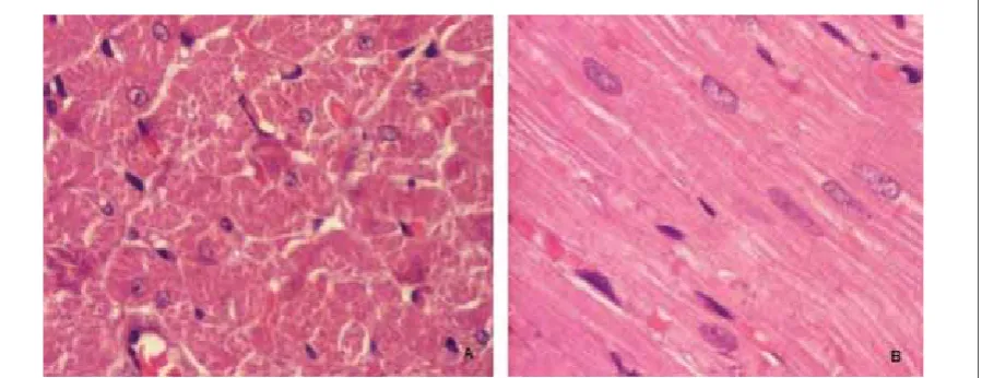
Figure . (A and B) presents normal myocardial tissue of control rats. Th e myocardium is composed of muscle fi bers. Each fi ber is enclosed by a sarcolemma. Th e nu-clei are generally located in the central portion of the fi ber. A net of reticular fi bers and fi ne collagenous fi bers surround each muscle fi ber. Th e myocardium is richly supplied with small vascular channels forming an intra-mural circulation.
Subendocardial myocardium of ISP I rats is presented with areas of necrotic changed myocytes. Myocites show variable degree of damage and accent of patho-logical changes are on nuclei. Cells are edematous, lost of the normal striation pattern and posses nuclei with inhomogeneous content (Figure .). Deposited material of hyperacidophilic features are presented close to sar-colemma (.B). In necrotic myocites myofi brillar lysis is complete. Deep subendocardial portion showes diff use arranged focal necrosis of myocites. Nuclei of these ar-eas are in phases of picnosis or lysis or completely dis-appearance. Blood vessels near necrotic area contain eo-sinophilic amorfi c material. Subepicardial myocardium of ISP I not deviate from normal myocardial tissue of control rats.
Rats of ISP II died in diff erent intervals from ISP applica-tion within hours. Large blood vessels involved in sub-endocardial tissue are strong dilated (Figure ). Histo-logical changes were more extensive compared to ISP I. Subendocardial portion is presented in form areas of massive necrosis, cellular arrangement disappeared, nu-clei disappeared or in form of massive vacuola. In con-trast to ISP I, rats of ISP II showes changes in subepicar-dial myocardium. Variable degree of nuclei cariolysis in subepicardial myocites is presented.
Discussion
stud-
BOSNIAN JOURNAL OF BASIC MEDICAL SCIENCES 2007; 7 (3): 212-217
ies in rats, it is known that high doses of ISP (- mg/ kg) induce acute myocardial damage that has conse-quence in increasing of blood cTnT (). We knew that cTnT determination with the cTnT assay, which was developed originally for human sera, was possible in the rat with the monoclonal antibodies used, because of cross-reaction between humans and rats cTnT (). Very small amount of cTnT circulates as result of natu-ral protein turnover. Minimal circulating cTnT values were obtained in our control rats. Detection of
BOSNIAN JOURNAL OF BASIC MEDICAL SCIENCES 2007; 7 (3): 212-217to obtained results of qualitative histological analysis. Degree of myocardium pathomorfological changes de-pends on used ISP dose (). Subcutaneous injection of ISP mg/kg causes rat myocardial necrosis due to prolonged tachycardia. Th e proposed mechanism for ISP myocardial necrosis are myocardial hypoperfusion, glycogen depletion, electrolyte imbalance, lipid accu-mulation and free radical damage (,). According to Preus and coworkers, hours after ISP application, cells showed acydophilic changes and nuclei disappear-ance as signs of coagulative necrosis development (). Th ere aren’t published data about using higher doses of ISP and expressed myocardial changes in fi rst hours af-ter application. By using ISP mg/kg dose, we noted extensive myocardial lesions hours from drug appli-cation. Results of histological analysis gave us clear evi-dence of necrogenic eff ect of ISP on heart. Coagulative necrosis induced by ISP, which we found in our study, is the same in case of heart damage caused by ischaemia and presented in human myocardial infarction. Massive myocardial necrosis characterizes human myocardial infarction while catecholamine model of myocardial necrosis is presented in form of focal coagulative ne-crosis. Joseph and Balasz described focal and multiple areas of myofi brillar necrosis after ISP application to rats (). Our results are in concordance with their fi ndings. Histological examination of the myocardial tissue in all ISP treated rats showed necrotic areas in the subendo-cardial layer of the left ventricle. Poorer vascularisation and oxygen supply of subendocardial portion is possible cause of more vulnerability. By qualitative histogical analysis, we noted the diff erence in extensivity of myo-cardial changes between ISP I and ISP II rats. Rats of ISP II had higher body weight (mean BW g) than ISP I rats (mean BW g). ISP I subgroup have had changed
subendocardial portion of myocardium, thus subepi-cardial portion didn’t show any changes. Lesions were presented in form of widespread focal necrotic areas. Cardiomyocites were most frequently without nuclei, lost of the normal myofi brilar striation pattern. Th ere were variable degrees of damage. Rats of ISP II had ex-pressed myocites changes in subepicardial and subendo-cardial portion of myocardium. Subendosubendo-cardial altera-tions were more extensive than in ISP I subgroup. Th ere were signs of massive necrosis with disappeared cellular arrangement and lost edematous and fragmented myo-cites. Large blood vessel in myocardium of ISP II group were dilated greatly and injected which present the sign of shock development and myocardial hypoperfusion. Cells’ nuclei have disappeared or presented in form of massive vacuola. Sarcoplasma of cells were fragmented. ISP II rats have had altered subepicardial portion of myocardial tissue in comparing with ISP I. Subepicardial myocites had variable degree of kariolysis. Sarcoplasma of isolated myocites showed separation and vacuoliza-tion of myofi brils. Lipolysis due to the adrenergic acvacuoliza-tion of ISP is a potential factor in ISP myocardial necrosis development. Th e heart suplies a signifi cant portion of its fatty acid substrates as free fatty acids derived by li-polysis from adipose tissue. Although lipid availability is important for the heart, excess level of fatty acids in cardiomyocites can be deleterious. According to Mo-han, lipids mobilized from the adipose depot reach their highest level in blood one hour after ISP administration and are cleared by hours (). Lipolysis as leading criti-cal biochemicriti-cal event in induced ISP myocardial necro-sis does not occur until hours after ISP injection, and this time frame sugests a critical role for lipolysis. Pos-sible mechanism of diff erence in extent of histological changes between ISP II and ISP I subgroup is lipolysis.
Conclusion
BOSNIAN JOURNAL OF BASIC MEDICAL SCIENCES 2007; 7 (3): 212-217
References
() Rona G. Catecholamine cardiotoxicity. J.Mol.Cell Cardiol. ; : -
() Geng B., Chang L., Pan C., Qi Y.,Zhao J. et al. Endogenous hydro-gen sulfi de regulation of myocardial injury induced by isoprotere-nol. Biochem. Biophys. Res.Commun. ; : -
() Remiao F., Carvalho M., Carmo H., Carvalho F., Bastos M.L. Cu+-induced isoproterenol oxidation into isoprenochrome in adult rat calcium-tolerant cardiomyocytes. Chem. Res. Toxicol. ; (): -
() Herman E., Zhang J., Knapton A., Lipshultz E.S., Rifai N. et al. Serum cardiac troponin T as a biomarker for acute myocardial injury induced by low doses of isoproterenol in rats. Cardiovasc. Toxicol. ; (-): -
() Babuin L., Jaff e A.S. Troponin: the biomarker of choice for the detection of cardiac injury. CMAJ ; (): -
() Wallace K.B., Hausner E., Herman E., Holt G.D., Macgregor J.T. et al. Serum troponins as biomarkers of drugs-induced cardiac toxicity. Toxicol. Pathol. ; :-
() O’ Brien P.J., Smith D.E.C., Knechtel T.J., Marchak M.A., Pruim-boom-Brees i et al. Cardiac troponin I is a sensitive, specifi c bio-marker of cardiac injury in laboratory animals. Lab. Anim. ; : -
() Herman E.H., Zhang J., Knapton A., Rifat N., Sistare F. et al. Th e utility of monitoring cardiac troponin T to detect cardiac injury induced by low doses of isoproterenol in rats. FDA SCIENCE Th e critical path from concept to consumer, th Annual FDA Science Forum .
() Bertinchant J.P., Robert E., Polge A., Marty - Double C., et al. Comparison of the diagnostic value of cardiac troponin I and T determination for detecting early myocardial damage and the relationship with histological fi ndings after isoprenaline-induced cardiac injury in rats. Clin. Chim. Acta. ; : -
() Acikel M., Buyukokuroglu M.E., Erdogan F., Aksoy H., Bozkurt E., Senocak H. Protective eff ects of dantrolene against myocardial injury induced by isoproterenol in rats: biochemical and histo-logical fi ndings. Int. J. Cardiol. ; : -
() Acikel M., Buyukokuroglu M.E., Aksoy H., Erdogan F., Erol M.K. Protective eff ects of melatonin against myocardial injury induced by isoproterenol in rats. J.Pineal. Res. ; :-
() Rajadurai M., Prince P.S.M. Comparative eff ects of Aegle marme-los extract and α-tocopherol on serum lipids, lipid peroxide and cardiac enzyme levels in rats with isoproterenol-induced myocar-dial infarction. Singapore Med. J. ; ():-
() Preus M., Bhargava A.S., Khater, Abd El R., Gunzel P. Diagnostic value of serum creatine kinase and lactate dehydrogenase isoen-zyme determinations for monitoring early cardiac damage in rats. Toxicol. Lett. ; :-
() Joseph X., Balazs T. Protection against isoproterenol-induced myocardial necrosis in rats by -mercaptopurine and -thiofua-nine or by irradiation. Res. Com. Chem. Path. Pharmacol. ; (): -