Abstract
Calcitonin is usually produced by the parafollicular cells of the thyroid. However in an immunohistochemistry experiment we observed that the cells of the serous acini of rat submaxillary gland tissue were stained positive with calcitonin antibodies. We further used immunocyto-chemistry and nucleic acid hybridization to localize the distribution of calcitonin protein and calcitonin mRNA respectively in cultivated cells of rat submaxillary glands. Th e results showed that the cytoplasm of the epithelial cells of the submaxillary glands had positive staining in immunocytochemistry using calcitonin monoclonal antibody and positive reaction in nucleic acid hybridization using calcitonin mRNA complementary DNA probe. For the fi rst time we found that the cells of the submaxillary glands of rats can produce the hormone calcitonin. © Association of Basic Medical Sciences of FB&H. All rights reserved
KEY WORDS: submaxillary gland, calcitonin, immunocytochemistry, nucleic acid hybridization
glands of rats
Yongli Zhang1*#, Baoli Li2#, Zhaoying Fu3#, Sheli Li1
1Department of Endocrinology, Affi liated Hospital of Yanan University, No. 43 North Street, 716000 Yanan, China. 2Department of
Pharmacology, Yanan University Medical College, No. 38 Guanghua Road, 716000 Yanan, China. 3Department of Immunology, Yanan
University Medical College, No. 38 Guanghua Road, 716000 Yanan, China.
INTRODUCTION
In recent years, researchers have discovered and extracted more than various bioactive substances in the submaxil-lary glands [-]. These bioactive substances were either secreted directly into the blood or secreted first into the digestive tract with saliva and then enter the blood stream through gastrointestinal absorption; they exert regula-tory roles on multiple tissues and cells to adjust their physi-ological activities [, ]. Some of these factors fully meet the standards of hormone, and it was put forward that they have dual functions of endocrine and exocrine [, ]. In doing an immunohistochemistry examination with calci-tonin antiserum, we accidently observed a positive staining in the rat tissue of submaxillary glands, which suggested that the submaxillary glands might secrete calcitonin. In order to clar-ify this phenomenon, we repeated the immunohistochem-istry experiment using calcitonin monoclonal antibody, and further cultivated rat submaxillary gland cells and performed
immunocytochemistry and nucleic acid hybridization to lo-calize the distribution of calcitonin protein and calcitonin mRNA respectively in the cultivated cells of the submaxil-lary glands. Our study for the fi rst time showed that the cells of the submaxillary glands of rats can produce the hormone calcitonin. This paper reports our study and the results.
MATERIAL AND METHODS
Materials and Animals
DMEM/F medium was the product of Fisher's Biological Chemical Products Co., LTD (Deputy in Beijing, China). Fe-tus bovine serum was the product of Lanzhou Rongye Bio-logical Science and Technology Limited Company (Lanzhou, China). Rabbit anti-CK (rat keratin-) monoclonal antibody was the product of Beijing BIOS Biological Technology Co., LTD (Beijing, China). Rabbit calcitonin monoclonal anti-body was the product of Th ermo Fisher (NeoMarkers, Shang-hai, China). Biotin labeled goat anti-rabbit IgG serum was the product of Zymed Laboratories (San Diego, California, Unit-ed States). SP immunocytochemical staining kit and DAB col-or-display reagent was the product of Beijing Zhongshanjinq-iao Biological Technology Co., LTD (Beijing, China). Nucleic acid hybridization detection kit was the product of Wuhan BOSTER Company (Wuhan, China). Digoxin labeled calci-tonin DNA oligonucleotide was made by Beijing AUGCT Biological Technology Limited Liability Company (Beijing,
#These authors contributed equally to the paper. * Corresponding author: Yongli Zhang, Department of Endocrinology, Affi liated Hospital of Yanan University, No. 43 North Street, 716000 Yanan, China
Phone: 86-13891196482; Fax: 0911-2881002 E-mail: [email protected]
Submitted: 17 April 2013 / Accepted: 3 December 2013
China). SD rats were from Experimental Animal Center of Xi’an Jiaotong University Medical School (Xi’an, China).
Preparation of rat submaxillary gland tissue
Male and female SD rats, weigh - g, were used in the ex-periment. Th e rats were anesthetized with ether, and the sub-maxillary glands quickly taken, fi xed in formalin solution. After conventional paraffi n embedding, the tissue blocks were
cut into continuous slices of μm thick. Th e experiment was performed in accordance to the international, national and institutional rules concerning animal experiments and rights.
Immunohistochemical staining of submaxillary gland tissue with anti- calcitonin antibody
After dehydration of paraffin wax, immunohistochemical staining SP (Streptavidin protein) method was performed []. : diluted rabbit anti-rat calcitonin monoclonal an-tibody (the fi rst anan-tibody) was applied and the slides were put into oC incubator and for the night. After
rewarm-ing at oC for min, biotin labeled goat anti-rabbit
se-rum (the second antibody) and horseradish peroxidase labeled Streptavidin were added in turn according to the instruction of the detection kits. The color was shown with DAB (, ,-diaminobenzidine). After conventional dehydration, transparent, and cementing, microscopic examination was done and photos were taken. For con-trols, PBS buffer was used instead of the first antibody.
Isolation and cultivation of epithelial cells of submaxillary glands
Th e rats were put to death by breaking the neck and the sub-maxillary glands were taken in sterile conditions. Th e newly removed submaxillary glands were put into ethanol for minutes. After careful removing of capsule, fat, blood vessels, and connective tissue, the obtained gland tissue was rinsed with PBS for three times. Th e submaxillary gland tissue was then cut into small masses of . mm~. mm, rinsed with PBS for times, and digested with ml of . EDTA (tryp-sin plus Ethylenediamine tetraacetic acid) at oC for min.
Th e digestion was terminated by adding ml culture medium (DMEM/F plus calf bovine serum) and the cell suspen-sion was collected and centrifuged at rpm for minutes. Th e pellet was taken and the process of digestion and centrif-ugation was repeated once more. Th en the cells were re-sus-pended with ml culture medium and the cell number was counted. After that, fi broblasts or fi brocytes were removed by differential attachment technique []. The cells were adjusted to x /ml concentration and cultivated at oC,
saturated humidity and CO. Two days later, the original culture medium was changed and eight days later the cells were transferred to a new passage of cultivation (subculture).
Identifi cation of cell type and demonstration of calcitonin dis-tribution in the cultured submaxillary cells with immunocyto-chemistry
Th e sub-cultivated cells were used to do immunocytochem-istry to show cell type as well as calcitonin localization with anti-cytokeratin (CK) antibody and anti-calcitonin an-tibody respectively. After day cultivation on cover glass, cells were fi xed with formaldehyde for min, saturated with . triton for min, and incubated with hydrogen peroxide for min to reduce nonspecifi c dyeing. Th en : rabbit anti-rat CK monoclonal antibody and : rabbit anti-calcitonin monoclonal antibody were added and all of these study materials were put into oC refrigerator for the
night. Next, biotin labeled goat anti-rabbit IgG serum was added and incubated at oC for min. Following that
the SP complex was added and after incubating at room temperature for min, color was shown with DAB. After re-dyeing, dehydration, transparency, and slide sealing, mi-croscopic examination was performed. For negative control, PBS was used to substitute rabbit anti-rat CK monoclonal antibody and rabbit anti-calcitonin monoclonal antibody.
Nucleic acid hybridization with calcitonin mRNA complemen-tary probe
Th e cover glass treated with poly-lysine was used in cell climb cultivation. When cells grew to cover - of the cover glass, it was removed and the cells were fi xed with poly-formaldehyde for min. After gradient alcohol hydration, pepsin μl was added followed with room temperature in-cubation for min. Gradient alcohol dehydration was done again and this was followed with addition of μl pre-hybrid solution and incubation at oC for min. Th en μl hybrid
solution containing the digoxin labeled calcitonin mRNA complementary DNA oligonucleotide probe ('-CCCATA-ATAGCCCAGAGAACAGCCAGAGAGG-') was ap-plied and followed with oC incubation for hours. After
times rinse with TTBS (Tris-Buff ered Saline and Tween . Contents: mM Tris, mM NaCl, . Tween ), - μl of blockers was added, which was followed with oC incubation for min. Again rinsed with TTBS for
times, enzyme labeled digoxin antibody μl was added and this was followed with oC incubation for min. Then
NBT/BCIP (Nitro-Blue-Tetrazolium/ -bromo--chloro--indolyl-phosphate) μl was added to show color with the reaction kept in dark place at oC for hours. After rinsing
with deionized water for min, nuclear solid red μl was added and allowed to react for min. Finally, after washing, dehydration, transparency, and slide sealing, a microscopic examination was performed. Th e hybrid solution used as neg-ative control contained the same hybrid buff er but no probes.
RESULTS
Immunohistochemistry with anti-calcitonin antibody demon-strated calcitonin positive cells in rat submaxillary gland
The immunohistochemistry results of rat submaxillary gland tissue with anti-calcitonin monoclonal antibody re-vealed calcitonin positive staining cells in the submaxil-lary glands of the rats, which was manifested as brown color. The anti-calcitonin immune reactive staining was mainly distributed in the cells of the serous acini while the cells of the mucous acini were only slightly stained. No positive staining or brown color was seen in the cells of the negative control. The anti-calcitonin immune reactiv-ity showed no sex differentiation in the cells of the sub-maxillary gland tissue of the rats. See Figure a, b and c.
Anti-CK antibody staining indicated that the cells obtained by primary isolation/cultivation were secretory epithelial cells
In preparing primary isolation/culture of rat submaxillary gland cells, care was taken to remove capsule, fat, blood ves-sels, and connective tissue of the originally obtained glands; this guaranteed that the obtained cells were mostly secretory epithelial cells. Diff erential attachment technique utilized be-fore the cultivation of the isolated cells removed fi broblasts or fi brocytes; this made the cell population more pure. Mi-croscopic observation showed that the shapes of the cultured cells suggested epithelial cells rather than fibroblasts or fi-brocytes (Figure a, b). Finally, immunocytochemistry with anti-CK monoclonal antibody showed that the cytoplasm of the cultured cells were positively stained (colored yellow or brown), indicating secretory epithelial cells (Figure a).
Immunocytochemistry showed the distribution of calcitonin staining in the cytoplasm of cultivated submaxillary gland cells
Immunocytochemical results in the cultivated cells of the submaxillary glands with anti-calcitonin
monoclo-FIGURE 1. The immunohistochemistry results of submaxillary gland tissue from male (A) and female (B) SD rats with anti-calcitonin monoclonal antibody. Calcitonin positive cells are stained brown. The positive staining is mainly distributed in the cells of the serous acini while the cells of the mucous acini are only slightly colored. The staining shows no sex diff erence. In the negative control (C), the cells are not stained by the antibody.
A B C
FIGURE 2a. Primary culture (8th days) of submaxillary gland cells observed with inverted microscope. ×400.
FIGURE 2b. Subculture (6th days) of submaxillary gland cells ob-served with inverted microscope. ×400.
nal antibody staining showed that the cells were calcito-nin positive as indicated by the easily recognizable yel-low or brown color in their cytoplasm (Figure b). The cytoplasm was grey or unstained in the negative control (Figure c). Th e nuclei were blue in color for both antibody stained and control cells, although with diff erent deepness.
Nucleic acid hybridization results showed that the cytoplasm of the cultivated submaxillary gland cells was calcitonin mRNA positive
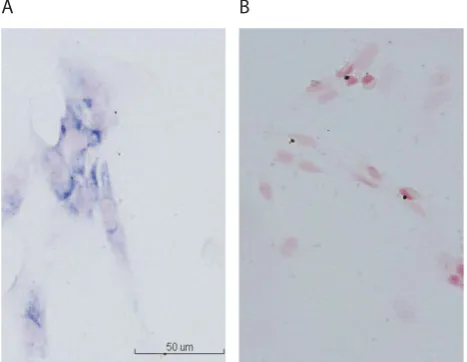
Th e cytoplasm of the positive cells in the nucleic acid hybrid-ization was blue or blue-violet in color and their nuclei were shallow red, which formed clear contrast with the control; the background was not colored (Figure a, b). Th e positive cells were very easy to identify and the mRNAs were wide-ly spread in the cytoplasm of the submaxillary gland cells.
DISCUSSION
In recent years, it was found that the granular convoluted tu-bule cells of rat submaxillary gland can synthesize, store, and release nearly kinds of bioactive peptides, such as epithelial growth factor, blood vessels relieve agent, insulin, endothe-lin, and liver cell growth factor [-]. Moreover, new bioac-tive substances were continually found in the submaxillary glands of rats. Fu et al. [] found that submaxillary glands have the expression of follicle-stimulating hormone and luteinizing hormone. Chen et al. [] found that submaxil-lary glands have the expression of luteinizing hormone and its receptors. And there were reports [] showing that go-nadotrophin releasing hormone and its receptors can coex-ist in submaxillary glands. However, no paper has ever re-ported that the submaxillary glands could produce calcitonin. Calcitonin is a -amino acid linear polypeptide hormone that primarily maintains the homeostasis of calcium and phosphate; it also inhibits the activity of osteoclast cells and promotes the activity of osteoblast cells. Calcitonin is usually produced by the parafollicular cells (C-cells) of the thyroid. When doing an immunohistochemical experiment on rat thyroid with anti-calcitonin serum, we observed unexpect-edly that the submaxillary glands of the animal contained positive staining cells. We carefully re-performed the immu-nohistochemistry examination of submaxillary gland tissue with anti-calcitonin monoclonal antibody using both male and female adult SD rats. Our results showed calcitonin posi-tive staining in the submaxillary glands of the rats which was the most obvious in the cells of the serous acini and which showed no sex diff erence. Th e results indicated that the sub-maxillary glands of rats do can synthesis and release calcitonin. We further investigated the cellular localization of calcito-nin protein and calcitocalcito-nin mRNAs in the cultivated cells (primary culture) of the submaxillary glands. With rab-bit anti-rat calcitonin monoclonal antibody as primary
FIGURE 3. Immunocytochem-istry results showing the cell type and calcitonin distribution in the cultivated submaxillary gland cells; (A) The cytoplasm of the isolated cells is positively stained by anti-CK8 antibody, indicating secretory epithelial cells; (B) The cytoplasm of the cultivated submaxillary gland cells stains brown or yellow by anti-calcitonin antibody; (C) In the negative control, the cyto-plasm is grey in color.
FIGURE 4. The nucleic acid hybridization result shows calcitonin mRNA positive cells in the submaxillary gland of rat; (A) The cyto-plasm of the positive cells is blue in the hybridization result (The shallow red regions are the nuclei); (B) In the negative control, only the nuclei are seen (shallow red).
A
A
B
B
C
antibody, the immunocytochemical results showed that the cytoplasm of the cultivated cells of the submaxillary glands contained calcitonin. By doing nucleic acid hybrid-ization with digoxin labeled calcitonin mRNA comple-mentary DNA as probe, we observed positive reaction in the cytoplasm of the cells, indicating the existence of calcitonin mRNAs in the cells of the submaxillary glands.
CONCLUSION
In summary, by using submaxillary gland tissue and pri-mary and secondary cell cultures and through detection of calcitonin protein and calcitonin mRNA, our investiga-tion demonstrated that the cells of the submaxillary gland of rats could synthesize and secrete calcitonin. We specu-late that the submaxillary glands of rats participate to a certain degree in the regulation of calcium and phosphate metabolism. Further studies are needed to address the problem of whether this is also the case in human beings.
ACKNOWLEDGMENT
Th e study was funded by Shaanxi Education Department re-search fund (JK).
DECLARATION OF INTEREST
Th e authors declare that there is no confl icts of interest with respect to the authorship or the publication of this article.
REFERENCES
[] Leone A, Spatola GF, Cucco D, Tessitore V, Bonaventura G, Uzzo ML. Immunohistochemical expression and distribution of orexin,
orphanin and leptin in the major salivary glands of some mammals. Folia Histochem Cytobiol. ; ():-.
[] Mathison RD, Davison JS, Befus AD, Gingerich DA. Salivary gland derived peptides as a new class of anti-infl ammatory agents: review of preclinical pharmacology of C-terminal peptides of SMR pro-tein. J Infl amm (Lond). ; :.
[] Zinzen KM, Hand AR, Yankova M, Ball WD, Mirels L. Molecular cloning and characterization of the neonatal rat and mouse sub-mandibular gland protein SMGC. Gene. ; :-. [] Myal Y, Iwasiow B, Yarmill A, Shiu RP. A new member of the
hor-monally regulated rodent submaxillary gland glycoprotein gene family: cDNA cloning and tissue specifi c expression. Mol Cell En-docrinol. Jul ; ():-.
[] Stoffers DA, Ouafik L, Eipper BA. Characterization of novel mRNAs encoding enzymes involved in peptide alpha-amidation. J Biol Chem. ; ():-.
[] Barka T. Biologically active polypeptides in submandibular glands. J Histochem Cytochem. ; ():-.
[] Morris KE, St Laurent CD, Hoeve RS, Forsythe P, Suresh MR, Mathison RD, Befus AD. Autonomic nervous system regulates se-cretion of anti-infl ammatory prohormone SMR from rat salivary glands. Am J Physiol Cell Physiol. ; ():C-.
[] Petrone C, Tremblay J, Gutkowska J. Natriuretic peptide system in the rat submaxillary gland. Jankowski M, Regul Pept. ; ():-.
[] He JC, Yao GG, Jia XM. Th e research progress of bioactive sub-stances in submaxilary gland. Sichuan Journal of Anatomy. ; (): -.
[] Gresik EW, Hosoi K, Kurihara K, Maruyama S, Ueha T. Th e rodent granular convoluted tubule cell --an update. Eur J Morphol. ; ():-.
[] Chen L, Bai HW, Sun XD, Yin SD, Wang AM, Zhang RQ. Th e dis-tribution, cloning and analysis of partial sequence of LH and its re-ceptor in submaxillary gland of rats. Acta Anatomica Sinica. ; ():-.
[] Zhang YL, Wang YL, Fan X. Improvement on primary culture method of rat submaxillary gland cells. Medical Information. ; ():-.
[] Fu JF, Huang WQ, Wang S, Chu CY. Studies on distribution of FSH, LH and colocalization with GnRHR in rat submaxilary grands. Acta Anatomica Sinica. ; ():-.
[] Yu H, Tang X, Lu BZ, Huang WQ, Zhao J. Studies of Localization and Distribution of Follicle-Stimulating Hormone Receptor in Rat Submandibular Gland and Stomach. Acta Anatomica Sinica. ; ():-.