IntroductIon
Wound healing is a complex, organized process that includes nitric oxide (NO) and many messenger molecules. NO is thought to trigger vasodilatation, angiogenesis, melano-genesis and activate various immune responses for the vari-able environmental factors directly and indirectly. There are several studies that prove the functional and critical role of NO on wound healing [1-6]. NO is also shown to have many regulatory roles in cellular proliferation, collagen synthesis and epithelization [4]. NO provides some of these effects by increasing soluble guanilate cyclase activity which forms cyclic guanosine monophosphate (cGMP) [1,7].
Sildenafil citrate is a powerful and selective inhibitor of cGMP specific phosphodiesterase type 5 (PDE5) which is the
dominant isoenzyme metabolizing cGMP [8]. Sildenafil also increases NO release by stimulating inducible nitric oxide syn-thase (iNOS) [2] and endothelial derived nitric oxide synsyn-thase (eNOS) at the mRNA and protein level [9,10]. Inhibition of iNOS by competitive inhibitors reduces collagen deposition and decrease wound tension strength in incisional wounds [5]. Use of NO donors increases incisional and excisional wound healing in rats [11]. NOS activity was shown to have a peak within 24 hours after injury, and then followed with a constant decrease line and a plateau line between 14 and 35 days [6].
Sildenafil citrate is being used for different indications as erectile dysfunction and pulmonary hypertension. There have been studies investigating the effect of sildenafil on flap survival, colon anastomosis, pressure ulcers, fracture, micro-vascular anastomoses, skeletal muscle injury and incisional wound healing [3,12-18]. Effect of sildenafil on wound healing by secondary intention has not been researched before. In this study, we searched the effect of sildenafil citrate gel, prepared by carbopol, on wound healing by secondary intention at dif-ferent dosages (1% & 5%).
*Corresponding author: Koray Gürsoy, M.D.
Ankara Research and Training Hospital, Plastic, Reconstructive and Aesthetic Surgery Clinic, Ankara, Turkey
Phone: +903125953651 E-mail: [email protected]
Submitted: 23 December 2013 / Accepted: 20 May 2014
Effect of topically applied sildenafil citrate on
wound healing: experimental study
Koray Gürsoy1*, Melike Oruç1, Yüksel Kankaya1, M. Gürhan Ulusoy1, Uğur Koçer1, Duygu Kankaya2,
R. Neslihan Gürsoy3, Özge Çevik3, Elmas Öğüş4, Vildan Fidanci4
1Ankara Research and Training Hospital, Plastic, Reconstructive and Aesthetic Surgery Clinic, Ankara, Turkey. 2Ankara University, Pathology
Department, Ankara, Turkey. 3Department of Pharmaceutical Technology, Faculty of Pharmacy, Hacettepe University, Ankara, Turkey. 4Ankara Research and Training Hospital, Biochemistry Clinic, Ankara, Turkey
Abstract
Wound healing is a complex process that necessitates organization of different cell types and several signalling molecules. The aim of this study is to evaluate the effect of different concentrations of sildenafil citrate, which decreases cGMP degradation, on wound healing by secondary intention. This study was performed using 25 Sprague Dawley rats weighing 200-250 grams. 4 dorsal defects were created. Four different treatment modalities which were 1% and 5% sildenafil citrate gel prepared with carbopol, pure carbopol gel without any drug in it and 0,9% NaCl solution; were applied to each lesion of the same rat. Randomly selected five rats (25 rats in total) were sacrificed on 3rd,
5th, 7th, 10th, and 14th days; and the effect of each modality was evaluated by means of defect area measurement, histopathological
examina-tion and measurement of tissue hydroxyproline levels. Sildenafil citrate gel applicaexamina-tion decreased the defect areas in a dose independent manner starting from 3rd day and dose dependent manner after 7th day. By means of vascularization, sildenafil citrate increased vascularity
starting from 3rd day. The strength of acute inflammation was superior in sildenafil groups starting from 5th day; and the amount and
mat-uration of granulation in the wound bed, as well as the strength of chronic inflammation were superior in defects treated with sildenafil citrate as early as 7th day.
KEY WORDS:wound healing, sildenafil citrate, cGMP
MAterIAls And Methods
Animals
In this study, 13 to 15 months old twenty-five female Sprague Dawley rats weighing 200 to 250 gr (mean 220 gr) were used. During the experiment, the rats were individually kept in separate cages in the same room with controlled light and room temperature (240C) and they received standard
food and water ad libitum. The study was in compliance with the principles stated by The National Institutes of Health in their Guide for Care and Use of Laboratory Animals. The study was approved by Local Ethics Committee on Animal Research.
Preparation of Pure Carbopol Gel
For preparation of 40 ml carbopol gel, 40 ml of ultra-pure water (pH 7.34) was taken in to the beaker with 40 mg meth-ylparaben and stirred over magnetic mixer at moderate speed. After methylparaben has been dissolved, carbopol was added to the beaker. Then two drops of triethanolamine was added to form clear viscous gel. Approximately 4 ml of 1 N NaOH was added to raise the pH to 7.
Preparation of Sildenafil Citrate Containing
Carbopol Gel
For preparation of 40 ml of gel, 150 ml of ultra-pure water was used and because sildenafil citrate dissolves at acidic mediums, the pH was decreased by 0.1 N HCl solution. 40 ml of this solution was taken into the beaker and 40 mg of meth-ylparaben was dissolved over magnetic mixer. Then either 0.4 g (for 1% gel) or 2 g (for 5% gel) sildenafil citrate and 0.8 g of carbopol was added and dissolved. Finally triethanolamine was added and stirred up to form gel. The pH of the gel was raised to 7 by adding 1 N NaOH. The gels were kept at 40C
during the experiment.
Surgical Procedure
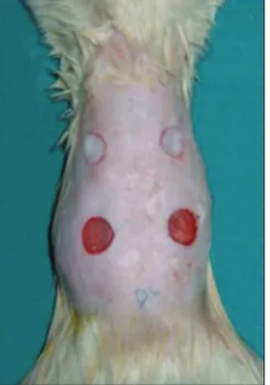
Anesthesia was obtained with Ketamine HCl (40 mg/kg) and Xylazin (5 mg/kg). On the dorsal regions of rats, four circu-lar skin-subcutaneous tissue and panniculus carnosus defects (1 cm apart from the midline and 2 cm from each other) were formed (Figure 1). We applied serum physiologic (Group 1) 0.2 cc/day for the left caudal defect, 5% sildenafil citrate gel (Group 2) 0.2 cc/day for the right cephalic defect, 1% sildena-fil citrate gel (Group 3) 0.2 cc/day for the left cephalic defect and pure carbopol gel (without sildenafil citrate) (Group 4) 0.2 cc/day for the right caudal defects. Randomly selected 5 rats were sacrificed on 3rd, 5th, 7th, 10th and 14th days. After
sac-rification of 25 rats, the defects were photographed and tissue
samples were taken from the base and edges of the defects for biochemical analysis and stored at -80 0C until the day of
biochemical analysis. For the histopathological analysis wedge excisions were taken from the borders of the defects and fix-ated in the 10% tamponed formalin solution at 40C.
Macroscopic Analysis
For assessment of the defect sizes, after sacrification, digital photographs were taken and each defect size was calculated by Digimizer Image Analysis Software (MedCalc Software, Belgium) on personal computer in cm2 as unit.
Histopathologic Analysis
The samples were embedded in paraffin blocks, sliced 3 micron thick, dyed with hematoxylin eosin and examined under light microscopy. Histopathology examination was made for the acute inflammation scores, chronic inflamma-tion scores, granulainflamma-tion tissue formainflamma-tion and maturainflamma-tion, re-epithelization and vascularization. Acute inflammation scores were evaluated according to the presence of the neu-trophils and chronic inflammation scores according to the presence of plasma cells and monocytes. Scoring system from 0 to 3 (0: none, 1: mild, 2: moderate, 3: severe) was used for the assessment of the acute inflammation score, chronic inflammation score, granulation tissue formation and mat-uration. For the assessment of the re-epithelization, scoring system from 0 to 4 was used. For assessment of the vascular-ization, numbers of vessels were counted under 1 high power field (1HPF) (x40). All the analysis was made by the same pathologist.
Biochemical Analysis
Hydroxyproline level which is the main marker of the col-lagen amount was used for assessment of colcol-lagen levels in the wound.
Statistical Analysis
The analysis of data was made with SPSS 16.0 program. Normally distributed data (area measurements and vascular-ization) was presented by mean and standard deviation while not normally distributed data was presented by median and interquartile range (25th-75th). Normally distributed data were
analysed by ANOVA. Tukey Multiple Comparison Test was used to detect the significant difference between groups for 3rd, 5th, 7th, 10th and 14th day results. Categorical variables such as
acute inflammation score, chronic inflammation score, granula-tion tissue formagranula-tion and maturagranula-tion and re-epithelizagranula-tion were evaluated by Pearson Ki-square Test for significant difference.
p value <0.05 was considered to indicate significant difference.
results
All of the rats survived until the day of sacrification. We have not encountered any complication or infection during and after the surgical procedure. Data derived from the exper-iment was analysed within each group. Results of 1% and 5% sildenafil citrate and pure carbopol groups were compared with each other and with the control group.
Macroscopic Analysis
After the surgical procedure, the mean defect size was approximately 1.05 cm2. No statistical difference was found
between the groups (p=0.999).
On 3rd and 5th days, no statistical difference was found
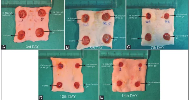
between 1% sildenafil and 5% sildenafil groups; and between pure carbopol and control groups (p=0.759). Defect sizes of sildenafil groups were smaller than the pure carbopol and control groups and this difference was found to be statistically significant (p=0.001) (Figure 2A and 2B).
After 5th day, the mean defect size of the 5% sildenafil group
was smaller than the other groups, and 1% sildenafil group was smaller than pure carbopol and control groups. The differ-ences were found to be statistically significant (p=0.001). No statistical difference was found between the pure carbopol and control groups (p=0.988) (Figure 2C-E, 3) (Table 1).
Histopathologic Analysis
3rd day results. All groups showed severe acute
inflam-mation score (Score 3), mild chronic inflaminflam-mation score and granulation tissue formation (Score 1). Vascularization of 5% sildenafil group was superior (16/1HPF) to the other groups; and 1% sildenafil group (14/1HPF) was superior to the pure carbopol (11/1HPF) and control groups (8/1HPF) (p=0.028). The statistical superiority of sildenafil groups was dose inde-pendent (p=0.194).
5th day results. Sildenafil groups showed severe acute and
chronic inflammation (Score 3) but pure carbopol and con-trol groups showed moderate acute and chronic inflammation (Score 2). Acute inflammation scores showed statistically sig-nificant difference between sildenafil groups and the others (p=0.001) whereas no difference could be found in terms of chronic inflammation scores (p=0.984). 5% sildenafil group showed severe (Score 3), 1% sildenafil and control groups showed moderate (Score 2) and pure carbopol group showed mild (Score 1) granulation tissue formation. Granulation tis-sue maturation was moderate on sildenafil groups (Score 2) and mild on control group (Score 1). The differences found for the granulation tissue formation and maturation were statis-tically significant (p=0.001). Vascularization of 5% sildenafil group was superior (24/1HPF) than the other groups; and 1% sildenafil group (16/1HPF) was superior to the pure carbopol (10/1HPF) and control groups (11/1HPF) (p=0.001). The statis-tical superiority of sildenafil groups was dose dependent.
Figure 2. Defect sizes on different time points after forming of panniculus carnosus defects and application of 1% and 5% sildenafil citrate gel or pure Carbopol gel. Defect sizes at day 3 (2A), 5 (2B), 7 (2C), 10 (2D) and 14 (2E) are shown
D
C B
A
7th day results. All groups showed moderate amount of acute inflammation scores (Score 2). Chronic inflammation scores were severe on sildenafil groups (Score 3), moderate on pure carbopol group (Score 2) and mild on control group (Score1); and these differences was found to be statistically sig-nificant. Granulation tissue formation and maturation scores were severe in sildenafil groups (Score 3) and moderate in pure carbopol and control groups (Score 2). The difference for granulation tissue formation and maturation was statistically significant (p=0.001). Re-epithelization was mild in sildenafil and control groups (Score 1) but not seen in pure carbopol group (Score 0). Vascularization of 5% sildenafil group was superior (26/1HPF) than the other groups; and 1% sildenafil group (21/1HPF) was superior to the pure carbopol (11/1HPF) and control groups (9/1HPF) (p=0.001). The statistical superi-ority of sildenafil groups was dose dependent.
10th day results. Acute and chronic inflammation scores
of sildenafil groups were moderate (Score 2) and others were mild (Score 1). Granulation tissue formation and maturation were severe in sildenafil groups (Score 3) and moderate in pure carbopol and control groups (Score 2). The difference was found to be statistically significant (p=0.001). 1% silde-nafil group showed more epithelization than the pure car-bopol group which was statistically significant. 5% sildenafil group and control group was not statistically different from the others. Vascularization of 5% sildenafil group was supe-rior (31/1HPF) than the other groups; and 1% sildenafil group (25/1HPF) was superior to the pure carbopol (15/1HPF) and control groups (10/1HPF) (p=0.001). The statistical superiority of sildenafil groups was dose dependent.
14th day results. Acute inflammation scores of all the
groups were mild (Score 1). Chronic inflammation scores of the sildenafil groups were moderate (Score 2) and the others
were mild (Score 1). Granulation tissue formations for all the groups were severe (Score 3) and granulation tissue matura-tion was severe in sildenafil groups (Score 3) and moderate for the others (Score 2). The difference for granulation tissue mat-uration was statistically significant (p=0.001). Control group showed better re-epithelization but not statistically different from the others (p=0.06). Vascularization of 5% sildenafil group was superior (31/1HPF) than the other groups; and 1% sildenafil group (21/1HPF) was superior to the pure carbopol (16/1HPF) and control groups (16/1HPF) (p=0.001). The sta-tistical superiority of sildenafil groups was dose dependent (Figure 4).
Biochemical Analysis
For the results of all days, tissue hydroxyproline levels of the sildenafil groups, dose dependently, were greater than of the pure carbopol and control groups, but difference was not statistically significant (p=0.955, 0.758, 0.901, 0.694, 0.387 for 3rd, 5th, 7th, 10th and 14th day results respectively) (Table 2)
(Figure 5).
dIscussIon
In order to understand wound healing, it is necessary to know the influence of a large number of intrinsic and extrinsic factors, much more than three stages of wound healing pro-cess which is composed of inflammation, proliferation and remodeling [19,20].
NO is synthesized inside the cell or around the cell mem-brane from its unique source, L-arginine, by a multifunctional enzyme complex called nitric oxide synthase (NOS). One of the main mechanisms of that enzyme complex to affect wound healing is to increase the amount of cGMP by activa-tion of soluble guanilate cyclase in cytosol [2,21,22]. The cellu-lar effects of cGMP depend not only on its synthesis but also on its rate of destruction. Destruction of cyclic nucleotides is carried out by phosphodiesterase enzymes [23].
The effect of L-Arginine/NO pathway in wound healing process was investigated by many researchers [4-6,11,24-26]. Dietary L-arginine supplementation, both in humans and ani-mals, is revealed to cause increased collagen deposition and wound tension strength [24,26]. In addition, several studies indicate the effect of L-arginine through the pathway of argi-nase and/or NOS [24-26]. The results obtained in L-arginine
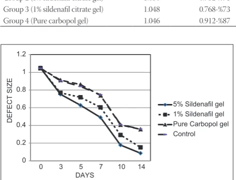
Table 1. Mean defect sizes (cm2) and their proportion with the beginning sizes.
Beginning 3rd day 5th day 7th day 10th day 14th day
Group 1 (control) 1.046 0.904-%86 0.840-%80 0.746-%71 0.410-%39 0.358-%34
Group 2 (5% sildenafil citrate gel) 1.050 0.752-%71 0.628-%60 0.490-%47 0.178-%17 0.086-%8
Group 3 (1% sildenafil citrate gel) 1.048 0.768-%73 0.718-%68 0.604-%57 0.292-%28 0.152-%14
Group 4 (Pure carbopol gel) 1.046 0.912-%87 0.862-%82 0.738-%70 0.406-%38 0.356-%34
'()(&76,=(
'$<6
6LOGHQDILOJHO 6LOGHQDILOJHO 3XUH&DUERSROJHO &RQWURO
studies [24,26] support the results obtained in our study with similar effects obtained in acute and chronic inflammation levels, vascularization, granulation tissue formation and mat-uration. Although statistically insignificant, we documented improved effects on tissue collagen levels and re-epitheliza-tion. These results may be due to the fact that improving effect of L-arginine on vascularity, inflammation scores, granula-tion tissue formagranula-tion and maturagranula-tion is mostly through the L-arginine-NO-cGMP pathway. Since increased collagen deposition seen in arginine supplementation studies [24] was not obtained in our study, we suggest this effect not to
be through cGMP pathway but through arginase pathway or through other effects of NO. Although collagen synthesis was shown to be along with NO synthesis [5], Glossman et al. [23], on the contrary, documented regional NO donor application to decrease while NOS suppression to increase the wound collagen levels.
Yamasaki et al. compared wound healing on dorsal exci-sional skin defects between iNOS-KO rats and normal rats. Delayed wound healing and a complete closure in iNOS-KO rats were seen within a time delay of approximately 5.3 days. Same effect was obtained with an iNOS inhibitor called N6-(iminoethyl)-L-lysine. Delay in wound healing of iNOS deficient rats was returned to normal by a single dose of adenoviral vector including human iNOS cDNA (AdiNOS) given at the time of injury [27]. The acceleration of wound clo-sure correlates with the ones in our study and suggest that NO production is important for wound closure. However, in the study of Most et al. who studied wound healing in iNOS-KO rats, no significant difference in wound tension strength and tissue hydroxyproline levels during the healing period was seen compared to control group [28]. Similar results for tissue hydroxyproline levels were also obtained in our study.
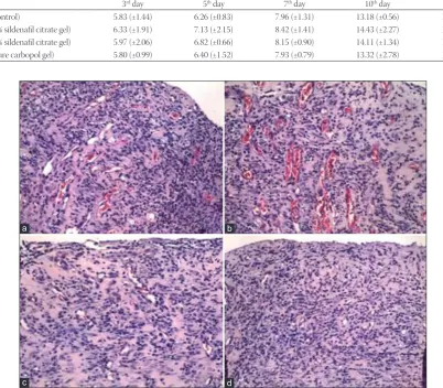
Table 2. Mean and SD of tissue hydroxyproline levels of the groups (μg/mg wet tissue).
3rd day 5th day 7th day 10th day 14th day
Group 1 (control) 5.83 (±1.44) 6.26 (±0.83) 7.96 (±1.31) 13.18 (±0.56) 15.79 (±0.90)
Group 2 (5% sildenafil citrate gel) 6.33 (±1.91) 7.13 (±2.15) 8.42 (±1.41) 14.43 (±2.27) 17.24 (±1.96)
Group 3 (1% sildenafil citrate gel) 5.97 (±2.06) 6.82 (±0.66) 8.15 (±0.90) 14.11 (±1.34) 16.60 (±1.81)
Group 4 (pure carbopol gel) 5.80 (±0.99) 6.40 (±1.52) 7.93 (±0.79) 13.32 (±2.78) 15.59 (±1.68)
+<'52;<352/,1(/(9(/
6
'$<6
6LOGHQDILOJHO 6LOGHQDILOJHO 3XUH&DUERSROJHO &RQWURO
Figure 5. Mean tissue hydroxyproline levels (μg/mg wet tissue) of the groups versus time (day)
Figure 4. Vascularization amounts of the 14th day. 1 % sildenafil (a) and 5 % sildenafil (b) groups were superior to control (c) and carbopol
(d) groups. (HE x20, x20, x20, x20)
d c
Derici et al. searched the effect of systemic sildenafil citrate on incisional wounds. Similar to our results, they reported insignificantly higher hydroxyproline level in silde-nafil group compared to controls. Their pathological evalua-tion on 4th, 14th, and 21st day also revealed better acute
inflam-mation scores in sildenafil compared to control groups, with the remarkable difference on 14th day. On the other hand,
they documented no significant difference in chronic inflam-mation score between two groups. In our study, compared to control and pure carbopol groups, acute inflammation scores were better in sildenafil group on 5th and 10th day and
chronic inflammation scores were better only after 7th day.
These effects were not associated with increase in sildenafil dose. As a result; wound healing in sildenafil group was faster from the beginning of the early phase and dose dependently increased. Derici et al. reported that although the difference was not statistically significant, granulation tissue forma-tion ratios on 4th, 7th, and 21st day were greater in sildenafil
group. We observed better granulation tissue formation on 5th, 7th and 10th days and it was only statistically significant
on 7th and 10th day results. Additionally, similar to the results
of our study, no significant difference between groups was discovered in terms of collagen deposition and re-epithe-lization. Neovascularization was the highest on 14th day and
similar with our study the difference between groups was significantly different [15].
In spite of the fact that we have not measured the cGMP levels, this study was designed to reveal increase in cGMP levels, by inhibiting its degradation by phosphodiesterase enzymes with sildenafil citrate. And finally effects of NO through cGMP were exposed. Based on the results obtained in our study, we suggest that, beside collagen deposition and re-epithelization, sildenafil citrate showed similar effects as previously seen in NO mediated studies.
The results of this study showed that topical sildenafil citrate application to excisional defects did not have any sig-nificant effect on keratinocytes. This might be because of min-ute effect of sildenafil on NO controlled TGF-β1 and EGF like growth factors.
conclusIon
In conclusion, sildenafil citrate gel provided statistically significant improvement in vascularity, inflammation, gran-ulation tissue formation and maturation in dose dependent manner. This effect of sildenafil may be emerged by the increase in cGMP levels, in parallel with previous literature.
The reason for sildenafil citrate not to act on collagen amount and epithelization in wound healing is possibly due to the fact that sildenafil only effects the last step of L-arginine-NO-cGMP pathway.
To reach higher concentrations in wound area and to pre-vent possible systemic adverse effects, sildenafil citrate was applied locally in this study. However, this was not previously studied. Detailed studies are needed to understand the mech-anisms of effects of locally applied sildenafil citrate.
Carbopol 940 which is used for preparation of sildenafil citrate gel is showed not to have any influence on wound heal-ing and for this reason it can be proposed for preparation of topically applied gels in similar studies.
declArAtIon of Interest
All the authors declare that they have no conflict of inter-est (employment, consultancies, stock ownership, honoraria, paid expert testimony, patent applications/registrations and grants or other funding).
references
[1] Salcido RS. Viagra and wound healing: the NO connection. Adv Skin Wound Care. 2008;21(3):106, 8-9.
[2] Wimalawansa SJ. Nitric oxide: new evidence for novel therapeutic indications. Expert Opin Pharmacother 2008;9(11):1935-1954. [3] Filippin LI, Cuevas MJ, Lima E, Marroni NP, Gonzalez-Gallego J,
Xavier RM. Nitric oxide regulates the repair of injured skeletal mus-cle. Nitric Oxide. 2011; 24(1):43-49.
[4] Efron DT, Most D, Barbul A. Role of nitric oxide in wound healing. Curr Opin Clin Nutr Metab Care. 2000; 3(3):197-204.
[5] Schaffer MR, Tantry U, Gross SS, Wasserburg HL, Barbul A. Nitric oxide regulates wound healing. J Surg Res. 1996; 63(1):237-240. [6] Lee RH, Efron D, Tantry U, Barbul A. Nitric oxide in the healing
wound: a time-course study. J Surg Res. 2001;101(1):104-108. [7] Farsaie S, Khalili H, Karimzadeh I, Dashti-Khavidaki S. An old drug
for a new application: potential benefits of sildenafil in wound heal-ing. J Pharm Pharm Sci. 2012;15(4):483-498.
[8] Corbin JD, Francis SH. Cyclic GMP phosphodiesterase-5: target of sildenafil. J Biol Chem. 1999; 274(20):13729-13732.
[9] Michell BJ, Chen ZP, Tiganis T, Stapleton D, Katsis F, Power DA, et al. Coordinated control of endothelial nitric-oxide synthase phos-phorylation by protein kinase C and the cAMP-dependent protein kinase. J Biol Chem. 2001;276(21):17625-17628.
[10] Sanchez FA, Rana R, Gonzalez FG, Iwahashi T, Duran RG, Fulton DJ, et al. Functional significance of cytosolic endothelial nitric-oxide synthase (eNOS): regulation of hyperpermeability. J Biol Chem. 2011; 286(35): 30409-30414.
[11] Shabani M, Pulfer SK, Bulgrin JP, Smith DJ. Enhancement of wound repair with a topically applied nitric oxide-releasing polymer. Wound Repair Regen. 1996;4(3):353-362.
[12] Barral SM, Araujo ID, Vidigal PV, Mayrink CA, Araujo AD, Costa PR. Effects of sildenafil on the viability of random skin flaps. Acta Cir Bras. 2011;26(4):314-319.
[13] Ayten R, Cetinkaya Z, Girgin M, Ozercan I, Ustundag B, Aygen E. The effects of intraperitoneal sildenafil administration on healing of left colonic anastomoses and intra-abdominal adhesion formation in the presence of intra-abdominal infection. Diseases of the colon and rectum. 2008;51(12):1837-1841.
[14] Histing T, Marciniak K, Scheuer C, Garcia P, Holstein JH, Klein M, et al. Sildenafil accelerates fracture healing in mice. J Orthop Res. 2011; 29(6):867-873.
[15] Derici H, Kamer E, Unalp HR, Diniz G, Bozdag AD, Tansug T, et al. Effect of sildenafil on wound healing: an experimental study. Langenbecks Arch Surg. 2010;395(6):713-718.
treatment of pressure ulcer: a randomised clinical trial. Int Wound J. 2013 Jun 4; doi: 10.1111/iwj.12104
[17] Pingarrón-Martín L, Arias-Gallo LJ. Sildenafil effect on preven-tion of thrombosis after microsurgical anastomosis: experimen-tal rat model of thrombotic suture. Oral Maxillofac Surg. 2013; 18(1):53-58
[18] Gravvanis A, Papalois A, Delikonstantinou I, Pentilas N, Zogogiannis I, Tsoutsos D, et al. Changes in arterial blood flow of free flaps after the administration of sildenafil in swine. Microsurgery. 2011;31(6):465-471.
[19] Mutschler W. [Physiology and pathophysiology of wound healing of wound defects]. Der Unfallchirurg. 2012;115(9):767-773. [20] Gethin G. Understanding the inflammatory process in wound
heal-ing. Br J Community Nurs. 2012; Suppl: S17-8, S20, S2.
[21] Daff S. NO synthase: structures and mechanisms. Nitric Oxide. 2010; 23(1): 1-11.
[22] Wu G, Bazer FW, Davis TA, Kim SW, Li P, Marc Rhoads J, et al. Arginine metabolism and nutrition in growth, health and disease. Amino acids. 2009; 37(1):153-168.
[23] Glossmann H, Petrischor G, Bartsch G. Molecular mechanisms of the effects of sildenafil (Viagra). Exp Gerontol. 1999; 34 (3): 305-318.
[24] Arbss MA, Ferrando JM, Vidal J, Quiles MT, Huguet P, Castells J, et al. Early effects of exogenous arginine after the implanta-tion of prosthetic material into the rat abdominal wall. Life Sci. 2000;67(20): 2493-2512.
[25] Kavalukas SL, Uzgare AR, Bivalacqua TJ, Barbul A. Arginase inhibi-tion promotes wound healing in mice. Surgery. 2012;151(2):287-295. [26] Debats IB, Wolfs TG, Gotoh T, Cleutjens JP, Peutz-Kootstra CJ, van
der Hulst RR. Role of arginine in superficial wound healing in man. Nitric Oxide. 2009;21(3-4):175-183.
[27] Yamasaki K, Edington HD, McClosky C, Tzeng E, Lizonova A, Kovesdi I, et al. Reversal of impaired wound repair in iNOS-de-ficient mice by topical adenoviral-mediated iNOS gene transfer. J Clin Invest. 1998;101(5):967-971.