Copyright © 2004, American Society for Microbiology. All Rights Reserved.
CbfA, the C-Module DNA-Binding Factor, Plays an Essential Role in
the Initiation of
Dictyostelium discoideum
Development
Thomas Winckler,
1* Negin Iranfar,
2Peter Beck,
1Ingo Jennes,
1Oliver Siol,
1Unha Baik,
1William F. Loomis,
2and Theodor Dingermann
1Institut fu¨r Pharmazeutische Biologie, Universita¨t Frankfurt (Biozentrum), Frankfurt am Main, Germany,1and Cell and
Developmental Biology, Division of Biology, University of California—San Diego, La Jolla, California2
Received 22 April 2004/Accepted 23 June 2004
We recently isolated fromDictyostelium discoideumcells a DNA-binding protein, CbfA, that interacts in vitro with a regulatory element in retrotransposon TRE5-A. We have generated a mutant strain that expresses CbfA at <5% of the wild-type level to characterize the consequences forD. discoideumcell physiology. We found that the multicellular development program leading to fruiting body formation is highly compromised in the mutant. The cells cannot aggregate and stay as a monolayer almost indefinitely. The cells respond properly to prestarvation conditions by expressing discoidin in a cell density-dependent manner. A genomewide microar-ray-assisted expression analysis combined with Northern blot analyses revealed a failure of CbfA-depleted cells to induce the gene encoding aggregation-specific adenylyl cyclase ACA and other genes required for cyclic AMP (cAMP) signal relay, which is necessary for aggregation and subsequent multicellular development. However, thecbfAmutant aggregated efficiently when mixed with as few as 5% wild-type cells. Moreover, pulsingcbfA
mutant cells developing in suspension with nanomolar levels of cAMP resulted in induction ofacaAand other early developmental genes. Although the response was less efficient and slower than in wild-type cells, it showed that cells depleted of CbfA are able to initiate development if given exogenous cAMP signals. Ectopic expres-sion of the gene encoding the catalytic subunit of protein kinase A restored multicellular development of the mutant. We conclude that sensing of cell density and starvation are independent of CbfA, whereas CbfA is essential for the pattern of gene expression which establishes the genetic network leading to aggregation and multicellular development ofD. discoideum.
Dictyostelium discoideumis a social amoeba that lives in soil
and feeds on bacteria. When the cells sense environmental conditions unfavorable for vegetative growth, they start to communicate with each other by means of extracellular cyclic AMP (cAMP). Individual cells move toward higher concentra-tions of cAMP and produce cAMP themselves, which is se-creted and serves to relay the signal in the aggregation field. Oscillatory secretion and perception of extracellular cAMP guide the cells into aggregation centers, in which they cooper-ate to form fruiting bodies consisting of stalk cells that support a mass of dormant spores (2, 23).
As the density of growing cells increases, aggregation com-petence is acquired in response to secreted quorum-sensing proteins referred to as prestarvation factor (PSF) and condi-tioned-medium factor (CMF) (7). The PSF protein induces the expression of a subset of early developmental genes, such as the extracellular matrix discoidin proteins and the cAMP re-ceptor CAR1 (8, 32). Extracellular accumulation of PSF also induces the expression of the protein kinase YakA, which interrupts the cell cycle and releases the translation block from the mRNA of the catalytic subunit of protein kinase A (PKA-C) (35, 36). CMF is not secreted by growing cells but is rapidly released when the cells starve (21). CMF is an 80-kDa protein that modulates signal transduction from CAR1 to its downstream effector, the aggregation-specific adenylyl cyclase
ACA. When CMF concentrations are low, ligand-induced ac-tivation of CAR1 cannot be transmitted to ACA even in the presence of high cAMP concentrations (3, 4). Thus, the cells sense starvation of neighboring cells as a function of the CMF level and can proceed to aggregate (17).
CAR1 is a typical serpentine receptor that couples via trim-eric G proteins to several intracellular effectors, including ad-enylyl cyclase, guanylyl cyclase, and phospholipase C (23, 29). The gene encoding the activatable adenylyl cyclase ACA, acaA, is rapidly induced when the cells start to respond to extracellular cAMP (30). Activation of ACA is complex and requires at least the G␥ complex of heterotrimeric G pro-teins, the cytosolic regulator of adenylyl cyclase (CRAC), and components of the Ras pathway (2, 23, 29). ACA activity is the major source of cAMP produced in early stages ofD. discoi-deum development (30). Most cAMP is secreted in order to relay the cAMP signal to nearby aggregation-competent cells. PKA is an essential regulator of all stages ofD. discoideum development (14, 25, 31, 33). PKA is a heterodimeric protein consisting of a catalytic and a regulatory subunit. Cytoplasmic cAMP binds to the regulatory subunit of PKA, which releases the catalytic subunit to phosphorylate downstream substrates that lead to alteration in gene expression (40). Extracellular cAMP is degraded by the product of pdsA, a cAMP-specific phosphodiesterase, whose expression increases dramatically following the initiation of development (13). This extracellular cAMP-specific phosphodiesterase is controlled by a specific inhibitor (PDI) whose expression is regulated in response to cAMP (15). Intracellular cAMP is broken down by the phos-phodiesterase RegA (34). The concerted action of many gene
* Corresponding author. Mailing address: Institut fu¨r Pharmazeu-tische Biologie, Universita¨t Frankfurt (Biozentrum), Marie-Curie-Strasse 9, D-60439 Frankfurt, Germany. Phone: 49-69-798-29648. Fax: 49-69-798-29662. E-mail: [email protected].
1349
on September 8, 2020 by guest
http://ec.asm.org/
products involved in the generation and perception of periodic nanomolar cAMP waves, including ACA, CAR1, PkaC, PdsA, RegA, and others, establishes a positive feedback loop that leads to the activation of genes required for aggregation and subsequent multicellular development. Inactivation of any gene involved in this regulatory network will result in mutants that cannot aggregate, and suppression of the aggregation block with genes involved in the autofeedback loop can be used to dissect the components of these regulatory pathways.
Genomewide DNA microarray analysis of expression pro-files is a powerful tool that extends our knowledge of the complex gene networks that regulateD. discoideum develop-ment (19, 20, 24, 39). Particularly helpful are genomewide comparative studies of wild-type cells versus mutant cells in which a single known developmental gene is disrupted (19, 24). Retrotransposons are autonomous genetic entities that can be amplified and inserted at different chromosomal locations in their hosts (11). Active retrotransposons are permanent sources of insertion mutagenesis. Thus, in compact genomes like that ofD. discoideum, retrotransposons must follow strat-egies to avoid disruption of host genes upon retrotransposition (45). One such strategy is the specific recognition of regions flanking tRNA genes as integration sites (45). Studies of the tRNA gene-targeting retrotransposon TRE5-A has led to the isolation of a host-encoded protein that binds specifically to a DNA sequence at the 3⬘end of TRE5-A, the C module (16, 18). This protein was named C-module-binding factor (CMBF). We have now changed the name of CMBF to CbfA in order to avoid confusion with CMF (conditioned-medium factor) and to conform to accepted Dictyostelium nomencla-ture. The DNA-binding properties of CbfA have been ana-lyzed in some detail, and the CbfA-encoding gene, cbfA, has been isolated (18). The CbfA protein, as deduced from a re-cently revised version of thecbfAgene (GenBank accession no. AF052006), consists of 1,000 amino acids and contains a con-served “carboxy-terminal jumonji” domain implicated in chro-matin remodeling (10), three unusual zinc fingers, and an AT hook that is required to bind to the C module in vitro (18).
The C module of retrotransposon TRE5-A serves as a pro-moter for the transcription of TRE5-A antisense RNAs, but the role of these antisense RNAs in TRE5-A retrotransposi-tion is still obscure. We therefore attempted to generate CbfA-depleted mutants in order to better understand the role of CbfA. We have not been able to isolate strains in whichcbfA is deleted by conventional gene replacement approaches, per-haps because it is essential for growth, but we have been able to generate mutants that express CbfA at levels⬍5% that of wild-type cells by introducing a partially suppressed amber translational stop codon (44). Preliminary analyses of the phe-notype ofcbfAammutant cells revealed defects in growth and
development (44) and also in TRE5-A retrotransposition (un-published data). We have now characterized the consequences of CbfA depletion forD. discoideumdevelopment by using a combination of genomewide DNA microarray analyses and Northern hybridizations. AlthoughcbfAamcells show a
prestar-vation response by inducing discoidin expression in a cell den-sity-dependent manner, they do not induceacaAwhen starved on filters, which can account for their failure to aggregate or form multicellular structures. However, exposure of the mu-tant cells to artificial extracellular cAMP pulses induced the
early genes, including acaA. Mixing cbfAam cells with a few
wild-type cells also rescued development of the mutant cells, indicating that the block in morphogenesis occurs after the prestarvation response but before the cAMP positive feedback loop is established.
MATERIALS AND METHODS
D. discoideumcell culture and development.D. discoideumAX2, AX4, and cbfAammutant cells (strain JH.D) (44) were grown in shaken cultures either in
liquid HL5 medium or in association withKlebsiella planticolabacteria. Food bacteria were prepared as proposed by Clarke et al. (9). Multicellular develop-ment ofD. discoideumcells was monitored on phosphate-buffered agar plates or on black filters (37). To study gene expression during development, 5⫻107D. discoideumcells were spread on black nitrocellulose filters (AABP04700; 47-mm diameter; Millipore) and allowed to develop for the time periods indicated in the figures. The cells were then washed from the filters using phosphate buffer and collected by centrifugation. Aliquots corresponding to 2⫻107cells were stored as pellets at⫺80°C. Frozen cells were used to extract total RNA as described below.
Expression vector construction and complementation studies. Expression plasmids were transformed intocbfAamcells, and stable transformants were
selected in HL5 medium containing 5g of G418/ml. A nearly full-lengthcbfA cDNA was reconstructed using a combination of cloned and PCR-amplified genomic DNA and cDNA fragments. The cDNA was inserted into the KpnI restriction site of vector 3H (26) to create the expression vector pDXA-rCbfA. The protein product derived from this vector, named recombinant CbfA (rCbfA), was identical to authentic CbfA protein except that it lacked the two C-terminal isoleucine residues. Control cells were transformed with empty pDXA-3H vector. ThepkaCcDNA was either expressed from its endogenous promoter (plasmid p188) (1) or under the control of the constitutively activeact6 promoter (plasmid p332) (12).
Induction of cAMP-induced gene expression by cAMP pulses.D. discoideum AX2, AX4, andcbfAamcells were grown to densities of 3⫻106/ml in axenic cultures. The cells were washed and adjusted to 2⫻107/ml in phosphate buffer. The cultures were shaken at 150 rpm at 22°C. cAMP was added at a 30 nM final concentration at 6-min intervals, starting 2 h after the cells were washed. After 6 h, a single pulse of 300M cAMP followed. Aliquots of 2⫻107cells were collected and frozen as cell pellets at⫺80°C for further use.
Northern blots.DNA probes specific for several developmental genes were generated by PCR using DNA sequence information from GenBank entries. The following genes were used for hybridization experiments:acaA(L05499),carA (M21824),csaA(X66483),pkaC(M38703),lagC(U09478), andcotB(M26238). TheD. discoideumhistone H3 gene (hstC) (6) was used as a loading control on Northern blots. Information on genesSLJ247andSSD449was obtained from the Dictyosteliumgenome project (http://dicty.sdsc.edu). PCR fragments used for hybridization experiments were purified from agarose gels using the QIAquick Gel Extraction kit (QIAGEN). DNA fragments were verified by DNA sequenc-ing ussequenc-ing the PCR primers.32P-labeled probes were generated with the PCR primer complementary to the respective mRNA sequence by using the Taq Cyclist DNA Sequencing kit from Stratagene. A32P-labeled probe specific for discoidin I was prepared by nick translation (42).
Total RNA was prepared from 2⫻107frozen cells using the RNeasy Mini kit from QIAGEN; 40g of total RNA was separated in denaturing gels, and RNAs were blotted onto Hybond-N membranes. Radioactive probes were hybridized to immobilized RNA at 42°C overnight in 50% formamide–5⫻SSC (1⫻SSC is 0.15 M NaCl plus 0.015 M sodium citrate)–1% sodium dodecyl sulfate–1⫻ Den-hardt’s solution. The blots were washed for 30 min at ambient temperature in 2⫻
SSC and for 30 min at 65°C in 2⫻SSC–0.1% sodium dodecyl sulfate. The primers used to generate PCR probes were as follows: acaA-01, GTTC ATCCTATGGTATGAAATTGG; aca-02, GTAGTGAATGAGCCAATTTCA CCC; arA-01, CCAGCCAATGAAACATCATTGG; carA-02, GATGATGATA AAGAAGATGAAGATG; csaA-01, CATTACAGGTACTGGATTTACAG; csaA-02, CCATTGTGAGGTGCTTGAGTGAC; pkaC-01, CCACCACCAGTC AATGCAAGAGAAAG; pkaC-02, CATAGAAAGGTGGATAACCTGCC AAC; cotB-01, GGAGTAGTTGTACACCAAGTAGTGGTTTC; cotB-02, CT TATGGGAACCAATCCAGCCACCTTTTGG; lagC-01, CTCAAGATTATG GGGTAGTATAGAC; lagC-02, CCAGTACCGTATGGTGTTGGACATG; SLJ247-01, GTCAGTGGTGAATGTGCCATTGATTTCTC; SLJ247-02, GGA TGATCCACTTCCTGTACCACTTCC; SSD449-01, ATGGCCAGAATTGAT TATGCTGTAAGC; and SSD449-02, CCAGTTTCAATGGTTTTGAAATAG CCC.
on September 8, 2020 by guest
http://ec.asm.org/
Microarray analyses.Corning slides were microarrayed with 6,345 cDNA and genomic targets as previously described (19). Inserts from 5,655 cDNAs were generously provided by the Japanese EST Project (27, 38). A list of genes is available at http://www.biology.ucsd.edu/loomis-cgi/microarray/cbfA_array.html. All genes in this study were sequence verified. Probes were prepared from total RNAs collected at 2-h intervals during wild-type and mutant development, as well as from time-averaged reference RNA. Superscript II DNA polymerase (Invitrogen, Carlsbad, Calif.) was used to incorporate either Cy5- or Cy3-conju-gated dCTP (Amersham, St. Louis, Mo.) into DNA. Labeled probes from the time points and the time-averaged RNA were mixed and hybridized at 42°C to microarrays for 6 to 12 h before being analyzed with a Genepix 4000B scanner (Axon Instruments, Foster City, Calif.). The total Cy3 signal was normalized to the total Cy5 signal after background subtraction to allow independent slides to be compared. The Cy3/Cy5 ratios for individual genes were then calculated at each time point for subsequent analyses. The expression patterns of mutant cells depleted in CbfA were compared to those of strain AX4, which are indistin-guishable from those of strain AX2 (39).
RESULTS
CbfA-depleted cells are unable to aggregate.CbfA was ini-tially identified and purified by its specific binding to the C module of the retrotransposon TRE5-A (16). Using the highly specific monoclonal antibody 7F3 directed against the carboxy-terminal domain of CbfA (44), we showed that the CbfA pro-tein level in AX2 cells is not sensitive to growth conditions (axenic culture or growth on bacteria) and stays constant throughout development. Using this same antibody, we found that the level of CbfA protein incbfAamcells was⬍5% of that
in wild-type cells during growth or following the initiation of development (reference 44 and data not shown).
We examined the effect of CbfA depletion incbfAamcells on
the multicellular development of D. discoideum cells. When cbfAamcells grew on lawns of bacteria, they formed very few, if
any, fruiting bodies, reflecting poor aggregation of the mutant cells, leading to multicellular structures generated by only a few of the starving cells (44). When mutant cells were plated on phosphate-buffered agar at 1.6⫻106/cm2, thecbfAamcells
did not show any sign of aggregation for at least 36 h and only very few, mostly crippled, fruiting bodies were observed after 72 h (data not shown). We could recover a few spores from these plates, and they gave rise to populations of mutant cells when spread on bacterial plates.cbfAamcells showed the
ag-gregation defect at all cell densities tested (5⫻104to 9⫻106 cells/cm2).
The failure ofcbfAamcells to aggregate might be the
con-sequence of their inability to sense their own density and food status. Therefore, we determined the cell density-dependent induction of discoidin expression as a measure of the prestar-vation response (8, 9). Cells were grown in shaken cultures in the presence of bacteria and collected at various titers for Northern analyses of accumulation of discoidin I mRNA (Fig. 1). ThecbfAamcells showed a pronounced lag in growth on
bacteria as a consequence of impaired phagocytosis (44). Nev-ertheless, cbfAam cells showed almost normal, cell
density-dependent expression of discoidin mRNA (Fig. 1) and protein (not shown), indicating that the prestarvation response of cbfAamcells is not affected by CbfA deficiency. These results
were confirmed with axenically growingcbfAamcells, in which
discoidin mRNA levels increased to 106cells/ml and showed the characteristic repression at cell densities of ⬎3 ⫻ 106 cells/ml also observed with AX2 cells (43) (data not shown).
cbfAam cells fail to activate cAMP pulse-induced genes in early development.AlthoughcbfAamcells reacted to
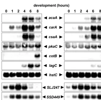
prestar-vation conditions, they did not acquire aggregation compe-tence and thus stayed as a monolayer. The ability of a cell population to respond to and relay cAMP signals can be ana-lyzed at the transcription level by measuring the induction of cAMP pulse-induced genes during aggregation. AX2 and cbfAamcells were spread on phosphate buffer-supported filters,
and total RNA prepared from samples was collected over 14 h. Northern blots were hybridized with DNA probes specific for the cAMP pulse-induced genes carA, acaA, csaA, andpkaC (Fig. 2). Expression of theacaAgene was completely absent in cbfAamcells. The genes carAandcsaA were induced to very
low levels 2 h after starvation of the mutant cells. On Western blots,cbfAamcells did not express any detectable CsaA protein
by 11 h after starvation on filters (data not shown). The mRNA encoding the catalytic subunit of PKA (pkaC) was fairly well expressed in growing and developingcbfAamcells, although the
typical cAMP pulse-induced increase inpkaCexpression seen at 8 h was not observed in the mutant cells (Fig. 2). We also tested for the expression of the postaggregative, cell-type-spe-cific genescotBandlagC. Both markers were well expressed in AX2 cells 8 to 14 h after starvation but were absent in the cbfAamcells under these conditions (Fig. 2).
The Northern blot data suggested that the gene regulatory networks that establish and maintain cAMP relay may not be active incbfAamcells. We used genomewide DNA microarrays
carrying 6,345 targets (19) to characterize the expression ki-netics of developmentally regulated genes incbfAamcells. We
focused on the expression patterns of 118 genes that can be
FIG. 1. Cell density-dependent expression of discoidin I. AX2 and
cbfAamcells were grown in shaken cultures in association with bacteria.
At time points 1 to 4, indicated by the arrows,D. discoideumcells were collected, washed, and analyzed for discoidin I mRNA expression. The indicated samples 1 to 4 corresponded to cell densities of 1.0⫻106, 2.0
⫻106, 4.4⫻106, and 7.9⫻106/ml for AX2 and 1.0⫻106, 2.4⫻106,
3.8⫻106, and 7.7⫻106/ml for strain JH.D. Total RNA was prepared
from the cell samples and analyzed on a Northern blot with a discoidin I␥-specific probe (inset).
on September 8, 2020 by guest
http://ec.asm.org/
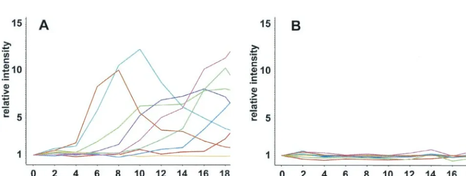
grouped into clusters according to their successive induction at different stages of multicellular development (19) (Fig. 3A). WhencbfAamcells were developed on filters, they did not show
significant induction of any of the developmentally regulated gene clusters (Fig. 3B). Close inspection of the individual ex-pression profiles of the early developmental genes tested on Northern blots revealed that expression of carAwas induced only⬃3-fold incbfAamcells (compared to 38-fold in wild-type
cells) and then stayed constant throughout development at only⬃5% of the wild-type level (not shown). In wild-type cells, csaAexpression increased eightfold 8 h after starvation but was not significantly induced incbfAamcells. The geneacaAwas
not expressed at all incbfAamcells. The only gene from the set
of developmentally regulated genes that was expressed in a manner similar to that in the wild type waspdiA, the phospho-diesterase inhibitor that is repressed by cAMP pulses.
Developmental rescue ofcbfAam cells.The aggregation
de-fect of cbfAamcells most likely results from their inability to
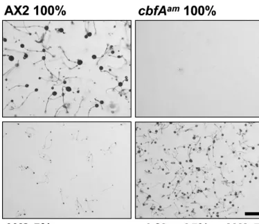
synthesize cAMP at a high rate as a consequence of failing to expressacaA. However, we speculated that they might be able to respond to cells that are making cAMP. We mixed a small number of AX2 cells with cbfAam cells and found that the
cbfAamcells developed significantly better in the presence of
wild-type cells (Fig. 4). Spores isolated from the fruiting bodies were plated clonally on bacterial lawns, and most gave rise to clones with thecbfAamphenotype, thus demonstrating that the
FIG. 2. Expression of developmentally regulated genes in thecbfAammutant JH.D developed on solid support. AX2 andcbfAamcells of strain
JH.D were washed free of medium and spread on black filters at 5⫻107per filter. Cell samples were collected every 2 h. RNA was extracted from
the samples, and 40g of total RNA was loaded in each slot. The origins and preparation of32P-labeled probes are described in Materials and
Methods.
FIG. 3. Microarray analysis of developmentally regulated genes in AX4 andcbfAammutant JH.D developed on solid support. Samples were
collected every 2 h for 18 h and analyzed on chips carrying 6,345 targets. Expression profiles of 118 developmentally regulated genes were extracted and clustered into 9 groups. Each cluster contains⬃10 genes and is color coded. A) wild-type AX4. B)cbfAammutant JH.D.
on September 8, 2020 by guest
http://ec.asm.org/
cbfAamcells had participated in forming the observed fruiting
bodies (data not shown).
To determine ifcbfAam cells developed in response to
ex-tracellular cAMP signals provided by the wild-type cells, we incubated AX2 and mutant cells in buffer and gave them arti-ficial cAMP pulses at 6-min intervals for 4 h, starting 2 h after they were washed, and a final boost of 300M cAMP after 6 h. RNA was collected at 2-h intervals and analyzed on microar-rays. UnlikecbfAamcells developed on filters, those developed
in suspension and given cAMP pulses expressed the early genes, includingacaA(Fig. 5). Accumulation of many of these early gene products was delayed ⬃4 h and was somewhat attenuated compared to that of wild-type cells (Fig. 5). Thus, CbfA appears to play a role in early gene expression but can be partially bypassed if the cells are given exogenous cAMP pulses.
The expression of some critical cAMP pulse-induced genes was analyzed on Northern blots to verify the microarray data. In the wild-type AX2 cells, the genes carA, acaA, andcsaA were strongly induced by exogenous cAMP pulses and reached maximum expression 4 to 6 h after the beginning of starvation (Fig. 6). These genes were also induced in the mutant cells, albeit more slowly (Fig. 6). Expression of pkaC was almost normal in thecbfAamcells under these conditions. Thus, the
time course of gene expression seen on Northern blots con-firmed the DNA microarray data for these genes.
In the DNA microarray experiments, we noticed two genes that were induced incbfAamcells but not in AX4 cells after the
onset of starvation. The geneSLJ247, which encodes CF50, a subunit of counting factor (5), was induced⬃6-fold (SLJ247), whereas expression ofSSD449, an orphan gene, was induced
⬃2-fold (data not shown). We analyzed the expression profiles of these two genes on Northern blots of cAMP-pulsed cells to see if they were in fact overexpressed incbfAamcells compared
to wild-type cells (Fig. 6). It turned out that both genes were in fact underexpressed in growingcbfAamcells compared to AX2
and were strongly induced soon after the beginning of starva-tion, thus giving an apparently stronger induction in the mu-tant than in the wild type. Both genes reached about the same expression levels in both wild-type andcbfAamcells by 2 h after
starvation (Fig. 6). Notably, cAMP-induced repression of
SLJ247was delayed in the mutant strain. Apparently,
under-expression of the gene encoding CF50 in growing cells is un-likely to contribute to the phenotype ofcbfAamcells, sincecf50
null cells form giant streams and fruiting bodies (5). Neverthe-less, the fact that SSD449 and SLJ247 were induced in a cAMP-independent manner within the first 2 h following the removal of nutrients, well before the cells were treated with
FIG. 4. Development of thecbfAammutant in synergy with wild-type cells. Axenically culturedD. discoideumcells were washed and plated on
phosphate-buffered agar. The 100% value corresponds to 108plated cells per 9-cm-diameter petri dish. The pictures were taken 60 h after the cells
were plated. Scale bar: 1 mm.
on September 8, 2020 by guest
http://ec.asm.org/
FIG. 5. Microarray analysis of developmentally regulated genes in AX4 andcbfAammutant JH.D developed in shaken culture with cAMP
pulses. Samples were collected every 2 h for 18 h and analyzed on chips carrying 6,345 targets. Expression profiles of 118 developmentally regulated genes were extracted and clustered into 9 groups. Each cluster contains⬃10 genes and is color coded. A) wild-type AX4. B)cbfAammutant JH.D.
FIG. 6. Northern analysis of developmentally regulated genes in thecbfAammutant JH.D in shaken culture treated with cAMP. Axenically
grown AX2 andcbfAamcells were harvested at 3.0⫻106/ml, washed free of medium, and suspended at 2⫻107/ml in phosphate buffer. The cells
were shaken at 150 rpm at 22°C. Beginning 2 h after the cells were washed, cAMP pulses were applied every 6 min at a 30 nM final concentration, followed by a final boost of 300M cAMP after 6 h. RNA was extracted from cell samples, and 40g of total RNA was loaded in each slot. The origins and preparation of32P-labeled probes are described in Materials and Methods.
on September 8, 2020 by guest
http://ec.asm.org/
cAMP pulse, clearly indicates that cbfAam cells are able to
sense starvation.
We also tested two postaggregation genes,lagCandcotB, for expression in cbfAam cells after cAMP pulsing. As shown in
Fig. 6, cotB expression was absent under the experimental conditions used andlagCexpression was only slightly induced and barely detectable.
When cAMP-treatedcbfAamcells were plated after 8 h on
phosphate-buffered agar plates to allow multicellular develop-ment, they were able to complete development significantly faster and more efficiently than mutant cells that had not been exposed to exogenous cAMP (data not shown). However, the developmental timing ofcbfAamcells was still aberrant, since
fruiting bodies were not formed until 31 h after plating, 6 h later than the formation of wild-type fruiting bodies. More-over, there were fewer fruiting bodies due to inefficient entry of mutant cells into aggregates (data not shown).
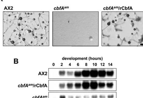
The developmental phenotype ofcbfAamis complemented by recombinant CbfA.To formally prove that the observed devel-opmental defects ofcbfAamcells were exclusively due to CbfA
deficiency, we expressed a nearly full-lengthcbfAcDNA in the
mutant cells under the control of the strong, constitutively activeactin15promoter. Using the CbfA-specific antibody 7F3, we found severalcbfAamtransformants that accumulated
sub-stantial levels of rCbfA (data not shown). These clones all developed normally and gave rise to normal-size fruiting bod-ies (Fig. 7A). Moreover, expression of early developmental genes, such ascsaA, was returned to normal by expression of recombinant CbfA (Fig. 7B).
Constitutive expression of PKA-C restores development of
cbfAam cells. The aggregation defect of the cbfAam mutant
resembled in several respects the phenotype of acaAmutant cells. Wang and Kuspa (41) have shown thatacaAmutant cells can aggregate when they overexpresspkaC, the gene encoding the catalytic subunit of protein kinase A (41). To determine whether the development of cbfAam mutant cells can be
re-stored by expression ofpkaC, we transformedcbfAamcells with
plasmids that allowed the expression of thepkaCcDNA either under the control of the authenticpkaCpromoter or under the constitutively active actin6 promoter. Expression of PKA-C from both plasmids reproducibly overcame the aberrant devel-opment of cbfAamcells (shown in Fig. 8 foract6/pkaC),
sug-FIG. 7. Complementation of thecbfAamphenotype by expression of recombinant CbfA. (A)D. discoideumcells were plated for multicellular
development on phosphate-buffered agar plates.cbfAamcells were transformed with plasmid pDXA-rCbfA (cbfAam/rCbfA). Control AX2 and
mutant cells were transformed with the empty vector pDXA-3H. The pictures were taken 40 h after the cells were plated. Scale bar: 1 mm. (B) Transformants were spread on black filters and allowed to aggregate as described in the legend to Fig. 2. Cell samples were collected every 2 h. RNA was extracted from the samples, and 40g of total RNA was loaded in each slot. Blotted RNAs were hybridized to a radiolabeledcsaA
probe.
on September 8, 2020 by guest
http://ec.asm.org/
gesting that accumulation of constitutive PKA-C can bypass the consequences of the lack of ACA caused by depletion of CbfA.
DISCUSSION
CbfA is a sequence-specific DNA-binding protein that is essential for correct expression of acaAand other genes re-quired to aggregate and develop following starvation and dep-osition ofDictyosteliumcells on a moist support. CbfA contains protein motifs that likely act in DNA binding and transactiva-tion of gene expression. The zinc fingers of CbfA may contrib-ute to DNA binding and/or stabilization of the CbfA structure. The carboxy-terminal domain of CbfA contains an AT hook that is required for binding to the C module of retrotransposon TRE5-A in vitro (18) and that may also mediate binding to AT-rich binding sites in CbfA target gene promoters. The amino-terminal one-third of CbfA contains a jumonji domain that is essential for the regulation of early developmental genes (unpublished data). Hence, we speculate that CbfA is a tran-scription regulator that binds to AT-rich target sites in theD.
discoideumgenome and regulates the transcription of
CbfA-dependent genes.
CbfA seems to be particularly important for the expression of the aggregation-specific adenylyl cyclase ACA. At present, we cannot resolve whether CbfA regulates theacaAgene di-rectly or indidi-rectly. CbfA recognizes two unusual binding sites in the C module of the retrotransposon TRE5-A. These consist of ⬃24-bp oligoadenine sequences surrounded by noncon-served nucleotides. There are similar oligoadenine runs in the 500 bp preceding theacaAgene where CbfA might bind. How-ever, given the generally high A⫹T content of theD. discoi-deum genome, it can be tested experimentally only if CbfA binds to the acaApromoter in vivo. In the absence ofacaA gene expression, the cells cannot generate the extracellular pulses of cAMP that are necessary for chemotactic aggregation and stimulation of adjacent cells. As a result, CbfA-depleted cells fail to aggregate or to accumulate normal levels of early developmental mRNAs. However, if CbfA-depleted cells are developed in mixtures with wild-type cells or are given artificial pulses of cAMP while suspended in buffer, they can express acaA and other early developmental genes and develop into fairly normal fruiting bodies. However, both gene expression and fruiting body formation were delayed incbfAamcells under
these conditions, indicating that the role of CbfA cannot be completely bypassed by artificial cAMP pulses.
CbfA does not appear to be necessary for the prestarvation response, since CbfA-depleted cells express discoidin I in a manner determined by the cell density, as in wild-type cells. In wild-type cells, PSF induces the serine-threonine protein ki-nase YakA, which arrests growth and releases a translation block on pkaC mRNA (35, 36). Our data suggest that the YakA pathway functions normally incbfAamcells, sinceyakA
mutant cells show enhanced growth rates (whereascbfAamcells
grow slowly), and cAMP-pulsedyakAmutant cells express nei-ther carA nor acaA (both of which are induced in cAMP-pulsedcbfAamcells) (Fig. 6). CbfA probably acts downstream
of the YakA pathway and, either directly or indirectly, controls transcription ofacaAand other early genes. When the cells are able to generate and respond to cAMP pulses, a CbfA-inde-pendent transcriptional pathway can take over.
CbfA does not appear to be necessary for cells to sense starvation, since expression of some very early genes increases immediately after mutant cells are washed free of nutrients. The major defects ofcbfAamcells can be attributed to the lack
of ACA, and like those ofacaAnull mutant cells, they can be bypassed by the constitutive PKA activity that results from overexpression of the catalytic subunit encoded by pkaC. It appears that all of the early internal signaling by cAMP is mediated by PKA. WhencbfAamcells are treated with pulses
of cAMP, the MAP kinase ERK2 is transiently activated and inhibits the internal phosphodiesterase so that cAMP gener-ated by the other adenylyl cyclase ACR can accumulate (19). This stimulates PKA activity, which induces acaAand other early developmental genes, allowing thecbfAamcells to escape
their trap.
A Myb-related transcription regulator (Myb2) that is also required for induction ofacaA(28) has been described. Like cbfAamcells, those lackingmybBfail to aggregate but can be
rescued by artificial pulses of cAMP. It is possible that in the absence of high levels of PKA activity, both CbfA and Myb2
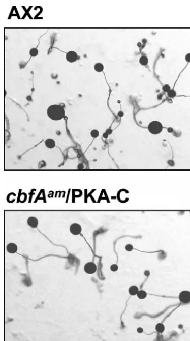
FIG. 8. Complementation of thecbfAamphenotype by expression
of recombinant PKA-C.cbfAamcells were transformed with plasmid
p332 (12), so that expression ofpkaCwas under the control of the
actin6promoter (cbfAam/PKA-C cells). The cells were plated on
phos-phate-buffered agar plates. The pictures were taken 40 h after the cells were plated. Scale bar: 1 mm.
on September 8, 2020 by guest
http://ec.asm.org/
are necessary to induce their target genes. Another gene, amiB, is known to be necessary for aggregation but can be bypassed by cAMP pulsing or ectopic expression of Myb2 (22). It appears to act upstream of CbfA and Myb2, sinceamiBnull mutant cells fail to repress the vegetative gene cprD when starved, suggesting that they cannot sense starvation (22). The AmiB protein contains a domain that is significantly related to MAP kinase phosphatases and may be involved in the regula-tion of YakA, which acts upstream of PKA.
Although pulsing with cAMP partially bypassed the block to developmental gene expression incbfAamcells, expression of
these genes was generally reduced and delayed by 2 to 3 h (Fig. 5 and 6). SincecbfAamcells developed quite efficiently when
mixed with wild-type cells, it may be that wild-type cells pro-vided a secreted factor missing in thecbfAammutant in
addi-tion to cAMP. This factor is unlikely to be CMF, since we found thatcmfAis normally expressed incbfAamcells (data not
presented). However, we cannot presently exclude the possi-bility that a component of the CMF-controlled signaling path-ways is dependent on CbfA, so that thecbfAamcells are unable
to respond properly to CMF.
ACKNOWLEDGMENTS
We are grateful to C. Utner and D. Fuller for expert technical assistance. We thank B. Wetterauer, G. Gerisch, and C. Reymond for gifts of plasmids and antibodies.
This work was supported by grants from the Deutsche Forschungs-gemeinschaft to T.D. and T. W. (WI 1142/2-1 and WI 1142/2-2) and grants from the NIH (GM60447 and GM62350) to W.F.L.
REFERENCES
1. Anjard, C., S. Pinaud, R. R. Kay, and C. D. Reymond.1992. Overexpression of DdPK2 protein kinase causes rapid development and affects the intracel-lular cAMP pathway ofDictyostelium discoideum. Development 115:785– 790.
2. Aubry, L., and R. Firtel.1999. Integration of signaling networks that regulate Dictyosteliumdifferentiation. Annu. Rev. Cell Dev. Biol.15:469–517. 3. Brazill, D. T., D. F. Lindsey, J. D. Bishop, and R. H. Gomer.1998. Cell
density sensing mediated by a G protein-coupled receptor activating phos-pholipase C. J. Biol. Chem.273:8161–8168.
4. Brazill, D. T., R. Gundersen, and R. H. Gomer.1997. A cell-density sensing factor regulates the lifetime of a chemoattractant-induced G␣-GTP confor-mation. FEBS Lett.404:100–104.
5. Brock, D. A., K. Ehrenman, R. Ammann, Y. Tang, and R. H. Gomer.2003. Two components of a secreted cell number-counting factor bind to cells and have opposing effects on cAMP signal transduction inDictyostelium. J. Biol. Chem.278:52262–52272.
6. Bukenberger, M., J. Horn, T. Dingermann, R. P. Dottin, and T. Winckler.
1997. Molecular cloning of a cDNA encoding the nucleosome core histone H3 fromDictyostelium discoideumby genetic screening in yeast. Biochim. Biophys. Acta1352:85–90.
7. Clarke, M., and R. H. Gomer.1995. PSF and CMF, autocrine factors that regulate gene expression during growth and early development of Dictyoste-lium. Experientia51:1124–1134.
8. Clarke, M., S. C. Kayman, and K. Riley.1987. Density-dependent induction of discoidin-I synthesis in exponentially growing cells ofDictyostelium dis-coideum. Differentiation34:79–87.
9. Clarke, M., J. Yang, and S. Kayman.1988. Analysis of the prestarvation response in growing cells ofDictyostelium discoideum. Dev. Genet.9:315– 326.
10. Clissold, P. M., and C. P. Ponting.2001. JmjC: cupin metalloenzyme-like domains in jumonji, hairless and phospholipase A2. Trends Biochem. Sci.
26:7–9.
11. Craig, N. L., R. Craigie, M. Gellert, and A. M. Lambowitz.2002. Mobile DNA II. ASM Press, Washington, D.C.
12. Dammann, H., F. Traincard, C. Anjard, M. X. P. van Bemmelen, C. Rey-mond, and M. Veron.1998. Functional analysis of the catalytic subunit of DictyosteliumPKAin vivo. Mech. Dev.72:149–157.
13. Faure, M., J. Franke, A. L. Hall, G. J. Podgorski, and R. H. Kessin.1990. The cyclic nucleotide phosphodiesterase gene ofDictyostelium discoideum contains 3 promoters specific for growth, aggregation, and late development. Mol. Cell. Biol.10:1921–1930.
14. Firtel, R. A., and A. L. Chapman.1990. Role for cAMP-dependent protein kinase-A in earlyDictyosteliumdevelopment. Genes Dev.4:18–28. 15. Franke, J., M. Faure, L. Wu, A. L. Hall, G. J. Podgorski, and R. H. Kessin.
1991. Cyclic nucleotide phosphodiesterase ofDictyostelium discoideumand its glycoprotein inhibitor—structure and expression of their genes. Dev. Genet.12:104–112.
16. Geier, A., J. Horn, T. Dingermann, and T. Winckler.1996. A nuclear protein factor binds specifically to the 3⬘-regulatory module of the long-interspersed-nuclear-element-likeDictyosteliumrepetitive element. Eur. J. Biochem.241:
70–76.
17. Gomer, R. H.1999. Cell density sensing in a eukaryote. ASM News65:23–29. 18. Horn, J., A. Dietz-Schmidt, I. Zu¨ndorf, J. Garin, T. Dingermann, and T. Winckler. 1999. A Dictyostelium protein binds to distinct oligo(dA)䡠oligo(dT) DNA sequences in the C-module of the retrotranspos-able element DRE. Eur. J. Biochem.265:441–448.
19. Iranfar, N., D. Fuller, and W. F. Loomis.2003. Genome-wide expression analyses of gene regulation during early development ofDictyostelium dis-coideum. Eukaryot. Cell2:664–670.
20. Iranfar, N., D. Fuller, R. Sasik, T. Hwa, M. Laub, and W. F. Loomis.2001. Expression patterns of cell-type-specific genes inDictyostelium. Mol. Biol. Cell12:2590–2600.
21. Jain, R., I. S. Yuen, C. R. Taphouse, and R. H. Gomer.1992. A density-sensing factor controls development inDictyostelium. Genes Dev.6:390–400. 22. Kon, T., H. Adachi, and K. Sutoh.2000.amiB, a novel gene required for the
growth/differentiation transition inDictyostelium. Genes Cells5:43–55. 23. Loomis, W. F.1996. Genetic networks that regulate development in
Dictyo-steliumcells. Microbiol. Rev.60:135.
24. Maeda, M., H. Sakamoto, N. Iranfar, D. Fuller, T. Maruo, S. Ogihara, T. Morio, H. Urushihara, Y. Tanaka, and W. F. Loomis.2003. Changing pat-terns of gene expression inDictyosteliumprestalk cell subtypes recognized by in situhybridization with genes from microarray analyses. Eukaryot. Cell
2:627–637.
25. Mann, S. K. O., J. M. Brown, C. Briscoe, C. Parent, G. Pitt, P. N. Devreotes, and R. A. Firtel.1997. Role of cAMP-dependent protein kinase in control-ling aggregation and postaggregative development inDictyostelium. Dev. Biol.183:208–221.
26. Manstein, D. J., H. P. Schuster, P. Morandini, and D. M. Hunt.1995. Cloning vectors for the production of proteins inDictyostelium discoideum. Gene162:129–134.
27. Morio, T., H. Urushihara, T. Saito, Y. Ugawa, H. Mizuno, M. Yoshida, R. Yoshino, B. N. Mitra, M. Pi, T. Sato, K. Takemoto, H. Yasukawa, J. Wil-liams, M. Maeda, I. Takeuchi, H. Ochiai, and Y. Tanaka.1998. The Dictyo-steliumdevelopmental cDNA project: generation and analysis of expressed sequence tags from the first-finger stage of development. DNA Res.5:335– 340.
28. Otsuka, H., and P. J. M. van Haastert.1998. A novel Myb homolog initiates Dictyostelium development by induction of adenylyl cyclase expression. Genes Dev.12:1738–1748.
29. Parent, C. A., and P. N. Devreotes.1996. Molecular genetics of signal trans-duction inDictyostelium. Annu. Rev. Biochem.65:411–440.
30. Pitt, G. S., N. Milona, J. Borleis, K. C. Lin, R. R. Reed, and P. N. Devreotes.
1992. Structurally distinct and stage-specific adenylyl cyclase genes play dif-ferent roles inDictyosteliumdevelopment. Cell69:305–315.
31. Primpke, G., V. Iassonidou, W. Nellen, and B. Wetterauer.2000. Role of cAMP-dependent protein kinase during growth and early development of Dictyostelium discoideum. Dev. Biol.221:101–111.
32. Rathi, A., and M. Clarke.1992. Expression of early developmental genes in Dictyostelium discoideumis initiated during exponential growth by an auto-crine-dependent mechanism. Mech. Dev.36:173–182.
33. Schulkes, C., and P. Schaap.1995. cAMP-dependent protein kinase activity is essential for preaggregative gene expression inDictyostelium. FEBS Lett.
368:381–384.
34. Shaulsky, G., D. Fuller, and W. F. Loomis.1998. A cAMP-phosphodiester-ase controls PKA-dependent differentiation. Development125:691–699. 35. Souza, G. M., A. M. da Silva, and A. Kuspa.1999. Starvation promotes
Dictyosteliumdevelopment by relieving PufA inhibition of PKA translation through the YakA kinase pathway. Development126:3263–3274. 36. Souza, G. M., S. Lu, and A. Kuspa.1998. YakA, a protein kinase required for
the transition from growth to development inDictyostelium. Development
125:2291–2302.
37. Sussman, M.1987. Cultivation and synchronous morphogenesis of Dictyo-steliumunder controlled experimental conditions. Methods Cell Biol.28:9– 29.
38. Urushihara, H., T. Morio, T. Saito, Y. Kohara, E. Koriki, H. Ochiai, M. Maeda, J. G. Williams, I. Takeuchi, and Y. Tanaka.2004. Analyses of cDNAs from growth and slug stages ofDictyostelium discoideum. Nucleic Acids Res.32:1647–1653.
39. VanDriessche, N., C. Shaw, M. Katoh, T. Morio, R. Sucgang, M. Ibarra, H. Kuwayama, T. Saito, H. Urushihara, M. Maeda, I. Takeuchi, H. Ochiai, W. Eaton, J. Tollett, J. Halter, A. Kuspa, Y. Tanaka, and G. Shaulsky.2002. A transcriptional profile of multicellular development inDictyostelium discoi-deum. Development129:1543–1552.
on September 8, 2020 by guest
http://ec.asm.org/
40. Veron, M., R. Mutzel, M.-L. Lacombe, M.-N. Simon, and V. Wallet.1988. cAMP-dependent protein kinase from Dictyostelium discoideum. Dev. Genet.9:247–258.
41. Wang, B., and A. Kuspa.1997.Dictyosteliumdevelopment in the absence of cAMP. Science277:251–254.
42. Wetterauer, B., G. Jacobsen, P. Morandini, and H. MacWilliams.1993. Mutants ofDictyostelium discoideumwith defects in the regulation of discoi-din I expression. Dev. Biol.159:184–195.
43. Wetterauer, B. W., K. Salger, C. Carballo-Metzner, and H. K. MacWilliams.
1995. Cell-density-dependent repression of discoidin inDictyostelium discoi-deum. Differentiation59:289–297.
44. Winckler, T., C. Trautwein, C. Tschepke, C. Neuha¨user, I. Zu¨ndorf, P. Beck, G. Vogel, and T. Dingermann.2001. Gene function analysis byamberstop codon suppression: CMBF is a nuclear protein that supports growth and development ofDictyosteliumamoebae. J. Mol. Biol.305:703–714. 45. Winckler, T., T. Dingermann, and G. Glo¨ckner.2002.Dictyosteliummobile
elements: strategies to amplify in a compact genome. Cell. Mol. Life Sci.
59:2097–2111.