R E S E A R C H A R T I C L E
Open Access
Serum EBV antibodies and LMP-1 in Polish
patients with oropharyngeal and laryngeal
cancer
Sylwia Fo
ł
tyn
1, Ma
ł
gorzata Strycharz-Dudziak
2*, Bart
ł
omiej Drop
3, Anastazja Boguszewska
1and Ma
ł
gorzata Polz-Dacewicz
1Abstract
Background:The association between Epstein-Barr virus (EBV) and the development of head and neck cancer was reported by many researchers. The aim of the present study was to detect EBV DNA and EBV antibodies in 110 Polish patients with oropharyngeal and laryngeal cancer compared to 40 healthy individuals.
Methods:Frozen tumor tissue fragments were tested using nested PCR assay for EBV DNA detection. Sera from all individuals were investigated using ELISA tests to detect the presence of VCA IgM and IgG, EBNA IgG, EA IgG.
Results:EBV DNA was detected in 52.7% of the patients (25% in controls). EBVCA were detected in 94.5%, EBNA in 96. 4% and EA in 94.5% of patients. The significantly higher level of EA in the patients suggests EBV reactivation. The majority of patients (83%) were infected with wild-type EBV.
Conclusion: Our study showed that this variant seems to be associated with oropharyngeal and laryngeal cancer in the Polish population.
Keywords:EBV antibodies, LMP1, Oropharyngeal cancer, Laryngeal cancer
Background
The Epstein-Barr virus (EBV) is a ubiquitous gammaher-pesvirus that infects more than 90% of the global adult hu-man population [1–3]. For the past two decades, increasing interest has been focused on the EBV-associated cancers including Burkitt’s lymphoma (BL), Hodgkin lymphoma (HL), nasopharyngeal carcinoma and gastric cancer [4–8].
The global burden of mortality from EBV-related can-cers accounts for 1.8% of all cancer deaths in 2010 [9]. The trends indicate that both this burden and life-expectancy in the world population will increase. About 92% of all EBV-associated cancer deaths are caused by nasopharyngeal cancer and gastric cancer. After primary infection, EBV establishes latent infection in B lympho-cytes with periodic reactivation and viral transmission in the oropharyngeal epithelium. Chronic viral infection is
an important risk factor, particularly for tongue cancer and oropharyngeal cancer [1, 7, 8, 10–14]. The associ-ation between the latent EBV infection and the develop-ment of head and neck cancer (HNC) was reported by several investigators [11, 12, 14, 15].
The present study analysed the serum level of EBV antibodies (VCA IgM and IgG, EBNA IgG, EA IgG) and determined LMP1 variant in patients with oropharyngeal and laryngeal cancers in the Polish population.
Methods Patients
The present study comprised of a group of 110 patients with a diagnosed cancer of the pharynx or larynx who were hospitalized in Otolaryngology Division of the Hos-pital in Radom, Poland. The results were compared to the control group, involving 40 persons hospitalized at the Otolaryngology Ward due to diseases other than cancer. There were no statistically significant differences between the patients and the control group (age, sex, to-bacco and alcohol consumption (Table 1)). The
socio-* Correspondence:malgorzata.strycharz-dudziak@umlub.pl
2Chair and Department of Conservative Dentistry with Endodontics, Medical
University of Lublin, Lublin, Poland
Full list of author information is available at the end of the article
demographic and clinicopathological characteristics of the study group are shown in Table 2 in relation to EBV DNA. The research material consisted of the sera and frozen tumor tissue fragments. This research was ap-proved by the Ethics Committee and is in accordance with the GCP regulations (no. KE-0254/133/2013).
DNA extraction from fresh frozen tumour tissue
Fragments of the fresh frozen tumour tissue (20 mg), both from the patients with OSCC and from the control subjects (biopsies), were cut and homogenized in a man-ual homogenizer Omni TH/Omni International/Kenne-sewa/Georgia/USA. DNA was extracted using a protocol as described in the DNeasy Tissue Kit Handbook (Qia-genGmBH, Hilden, Germany). Purified DNA was quantified by spectrophoto-metery (Epoch Microplate Spectrophotom-eter, BioTek Instruments Inc., Vinooski, Vermont, USA). The isolates were kept at −20 °C until the test was con-ducted. To verify the quality of the obtained DNA (presence of inhibitors of Polymerase Chain Reaction), aβ-globinassay was performed.
Detection of viruses
EBV DNA detection: All PCR reactions were carried out in the final volume of 25 μl using HotStartTaq DNA Polymerase (Qiagen, Germany). Concentrations of PCR reaction components were prepared as follows: 2.0 mM MgCl2, 0.2 mM dNTPs, 0.5μM of each forward and
re-verse primers and 0.5 U of HotStartTaq polymerase. During each run the samples were tested together with one negative (nuclease-free water) and positive control (EBV-positive cell line, Namalwa, ATCC-CRL-1432).
Amplification of EBNA-2 gene
The nested PCR was carried out for amplification of EBNA-2. The sequence of primers used for PCR was as follows: outer pair 5′ –TTT CAC CAA TAC ATG ACC
C–3′, 5′ –TGG CAA AGT GCT GAG AGC AA–3′
and inner pair 5′ –CAA TAC ATG AAC CRG AGT CC
–3′, 5′ – AAG TGC TGA GAG CAA GGC MC– 3′.
2μl of extracted DNA was subjected to the PCR mixture with the concentration as described above. The first-round amplification consisted of the activation of poly-merase 95 °C for 15 min, 35 cycles of 94 °C for 1 min, 55 °C for 1 min, 72 °C for 2 min and the final extension at 72 °C for 5 min. The second-round amplification was performed with 1μl of first round PCR product in 30 cy-cles with an annealing temperature at 60 °C. The Table 1Epidemiological characteristics of patients and control
group
Patients (n= 110)
Controls (n= 40)
p
n % n %
Sex female 8 7.3 2 5.0 >0.05
male 102 92.7 38 95.0
Age <49 12 10.9 6 15.0 >0.05
50–59 46 41.8 16 40.0
>60 52 47.3 18 45.0
Place of residence urban 72 65.5 28 70.0 >0.05
rural 38 34.5 12 30.0
Smoking yes 94 85.5 36 90.0 >0.05
no 16 14.5 4 10.0
Alcohol abuse yes 50 45.5 14 35.0 >0.05
no 60 54.5 13 65.0
EBV DNA positive 58 52.7 10 25.0 <0.001
negative 52 47.3 30 75.0
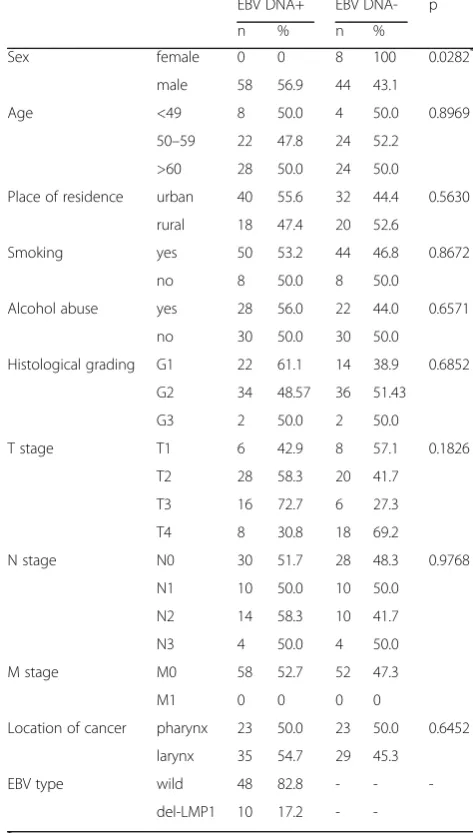
Table 2Epidemiological and clinical characteristics of patients in relation to EBV DNA
EBV DNA+ EBV DNA- p
n % n %
Sex female 0 0 8 100 0.0282* male 58 56.9 44 43.1
Age <49 8 50.0 4 50.0 0.8969
50–59 22 47.8 24 52.2
>60 28 50.0 24 50.0
Place of residence urban 40 55.6 32 44.4 0.5630
rural 18 47.4 20 52.6
Smoking yes 50 53.2 44 46.8 0.8672
no 8 50.0 8 50.0
Alcohol abuse yes 28 56.0 22 44.0 0.6571
no 30 50.0 30 50.0
Histological grading G1 22 61.1 14 38.9 0.6852
G2 34 48.57 36 51.43
G3 2 50.0 2 50.0
T stage T1 6 42.9 8 57.1 0.1826
T2 28 58.3 20 41.7
T3 16 72.7 6 27.3
T4 8 30.8 18 69.2
N stage N0 30 51.7 28 48.3 0.9768
N1 10 50.0 10 50.0
N2 14 58.3 10 41.7
N3 4 50.0 4 50.0
M stage M0 58 52.7 52 47.3
M1 0 0 0 0
Location of cancer pharynx 23 50.0 23 50.0 0.6452
larynx 35 54.7 29 45.3
EBV type wild 48 82.8 - -
-del-LMP1 10 17.2 -
-*
amplicons 368 bp, 473 bp in length (depending on the EBV type EBV-1 and EBV-2, respectively) were separated on 2% agarose gel and purified using a Gel-Out kit (A&A Biotechnology, Poland) for further analysis. Puri-fied PCR products were sent to Genomed Warsaw com-pany for sequencing.
Genotyping of LMP1
PCR screening for the EBV LMP1 subtype based on exon 3, defined as wild-type (wtLMP1) or del-LMP1, was done using specific primers: forward 5′-AGC GAC TCT GCT GGA AAT GAT- 3′; revers 5′-TGA TTA GCT AAG GCA TTC CCA- 3′. Concentrations of PCR reaction components were prepared as follows: 2.0 mM MgCl2, 0.2 mM dNTPs, 0.5 μM of each forward
and revers primers and 0.5 U Hot Start DNA polymer-ase and 5 μl of extracted DNA. The reaction mixture (25 μl) was incubated at 95 °C for 15 min., followed by 45 cycles at 94 °C for 1 min., 57 °C for 1 min., 72 °C for 1 min., a final extension at 72 °C for 10 mins. The PCR products were analyzed by gel electrophoresis in a 3% agarose gel and visualized under UV light.
Serological tests
To detect antibody levels serological tests were used with ELISA method. Designed antibodies: anti-VCA IgM (Nova-Lisa Epstein-Barr Virus VCA IgM/Nova Tec Immunodiag-nostica GmbH/Germany/catalog number: EBVM0150), anti-VCA IgG (NovaLisa Epstein-Barr Virus VCA IgG/ Nova Tec Immunodiagnostica GmbH/Germany/catalog number: EBVG0150), and anti-EBNA IgG (NovaLisa Epstein-Barr Virus EBNA IgG/Nova Tec Immunodiagnos-tica GmbH/Germany/catalog number: EBVG0580), anti-bodies anti-EA IgG (ELISA-VIDITEST anti-EA (D) EBV IgG/Vidia/Czech Republic/catalog number: ODZ-006). All tests were performed according to the manufacturer’s instructions.
The NovaTec Epstein-Barr Virus (EBV) IgG-ELISA is intended for the qualitative determination of IgG class antibodies against Epstein-Barr virus. Samples are con-sidered positive if the absorbance value is higher than 10% over the cut-off. The level of antibodies is expressed as NovaTec-Units = NTU.
ELISA-VIDITEST anti-EA is semiquantitative test. Samples with absorbances higher than 110% of the cut-off value are considered positive.
Statistical analysis
Descriptive statistics were used to characterize patient baseline characteristics. The Mann-Whitney U-test and Kruskal-Wallis test were used to compare antibody levels. Pearson’s chi-square test was used to investigate the relationship between EBNA2 subtypes and LMP1
subtype and clinical and demographical parameters. Statistical significance was defined asp< 0.05.
Results
EBV DNA was detected in 52.70% of the patients with pharyngeal and laryngeal cancer and in 25.0% of con-trols (p< 0.001). In all patients with EBV DNA type 1 of the virus was detected. Epidemiological and demo-graphical characteristics of the patients and controls are shown in Table 1.
The prevalence of EBV in patients group was signifi-cantly higher in males than in females (p< 0.05). No sta-tistically significant differences were found between EBV infection and other demographical features, smoking and alcohol consumption (Table 2). The majority of pa-tients (82.8%) were infected with wild type of EBV.
All the patients and controls were EBVCA IgM anti-bodies negative. IgG EBVCA antianti-bodies were detected in 94.5% of patients, EBNA in 96.4% (Table 3). A statisti-cally significant difference was observed in the preva-lence of IgG EA antibodies in patients and controls (94.5% vs 22.5%). High level of EA was stated in 70% of cancer cases and in 0% of controls (p< 0.05). The level of IgG EBVCA and IgG EBNA antibodies was higher in the patients than in control group; however, the differ-ence was not statistically significant.
Patients with an advanced stage of tumour development (T3-T4 stage) had a significantly higher level of EBNA antibodies than patients with T1-T2 stage (p< 0.05), while the level of EBVCA antibodies was the highest in patients with N1 stage. The level of EBVCA antibodies decreased with more advanced nodal stage (p< 0.05) (Table 4).
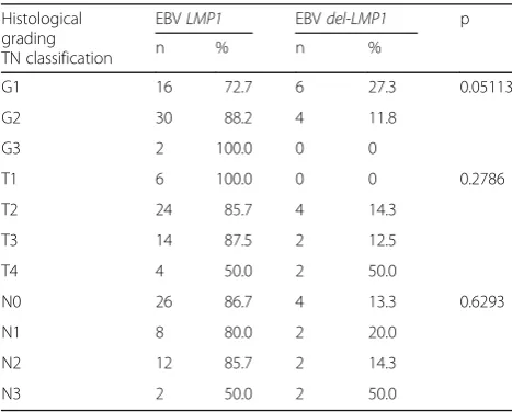
In patients infected with the wild-type of EBV, the level of anti-EBVCA was significantly higher than in cases with EBV type with deletion (p< 0.05). There was no relationship between EBV type on the basis of the
Table 3EBVCA, EBNA and EA level in serum of patients and control group
Antibodies level (IgG) Patients Controls p
n % n %
EBVCA negative 6 5.5 0 0 >0.05
low 24 21.8 14 35.0
high 80 72.7 26 65.0
EBNA negative 4 3.6 0 0 >0.05
low 23 20.9 12 30.0
high 83 75.5 28 70.0
EA negative 6 5.5 31 77.5 <0.05*
low 27 24.5 9 22.5
high 77 70.0 0 0
*
sequence in LMP1 gene, histological grading, and TN stage (Table 5).
Discussion
Two major types of EBV (EBV-1 and EBV-2) have been identified (a classification based on the EBNA2 gene se-quence) and according to the researchers they differ in geographic distribution [6, 16]. According to some inves-tigators, these genetic differences in EBV DNA sequence may be responsible for varied interactions with host cells, immunological response and cancer transform-ation, but the role of specific EBV types in the etiology of different cancers is not fully understood [17, 18]. EBV type 1 has a greater potential to transform B lympho-cytes than EBV type 2 and is more common in Western and Asian countries, while EBV-2 is more frequently de-tected in Africa [16]. The results of our present and pre-vious studies are consistent with the existing reports as type 1 EBV was detected in all examined patients [19]. Similar results were presented by Neves et al. [18], who demonstrated that 96.3% of the examined Portuguese in-dividuals carried EBV type 1.
As the disease progresses, EBV is activated into the rep-licative stage, in which major viral proteins are expressed [20, 21]. Patients with nasopharyngeal carcinoma show an elevated level of antibodies to several EBV antigens, in-cluding the viral capsid antigen (VCA), early antigen (EA) and EB nuclear antigen (EBNA) [22–25]. Traditional as-says of EBV antibodies have been very useful in clinical diagnosis of nasopharyngeal cancer (NPC) [26].
Our study revealed that more than half of the patients were EBV positive but also the level of IgG EBV anti-bodies was higher in patients than in controls and it in-creased with the tumour development stage. Moreover, the difference in EBV DNA prevalence between patients and controls was statistically significant.
Serological testing was performed both in the patients and in the controls to clarify whether there is a relation-ship between pharyngeal and laryngeal cancer develop-ment and past EBV infection. Various studies revealed that IgG EBV antibodies were detected in a greater part of the examined patients and their level was higher in the patients with pharyngeal and laryngeal cancers than in controls [24, 27, 28]. Other researchers report IgG EBVCA antibodies detection in more than 60% of pa-tients with laryngeal and pharyngeal cancers [29].
Our study did not reveal any significant difference be-tween the presence and the level of EBV antibodies and demographical features of the patients, cancer location and histological grading of malignant lesions. However, patients with an advanced stage of tumour development (T3-T4 stage) had a significantly higher level of EBNA antibodies than patients with T1-T2 stage, while the level of EBVCA antibodies was the highest in patients with N1 stage. The more advanced nodal stage was, the more the level of these antibodies decreased.
The level of EA antibodies among our patients was higher than in control group. Our results are similar to other researchers’findings and may indicate a reactiva-tion of latent EBV infecreactiva-tion, because a high titer of EA antibodies can be seen in cases of reactivation of latent infection [25].
Statistically significant differences were observed by Chen et al. [30] in the scores/levels of six markers (EVB-IgG, VCA-(EVB-IgG, EA-D p43-(EVB-IgG, EA-(EVB-IgG, EA-IgG + EBNA1-IgG, and EA-D p45-IgG) between NPC patients and healthy subjects. The lytic cycle of EBV is important and plays more active roles in oncogenesis [31].
Many authors show that EBV DNA load and IgA anti-bodies are more effective and useful in the clinical diagno-sis and screening of NPC [32–34]. In our study only IgG antibodies were analysed. Immunoglobulins against viral proteins, including EA-IgG, VCA-IgA, and Rta-IgG, may be used as molecular biomarkers for predicting the prog-nosis of nasopharyngeal cancer. According to Tay et al. [24], EBV DNA load correlated with EA IgA serology Table 5Relationship between EBV type according to the LMP1
gene sequence and histological grading, TN stage
Histological grading TN classification
EBVLMP1 EBVdel-LMP1 p
n % n %
G1 16 72.7 6 27.3 0.05113
G2 30 88.2 4 11.8
G3 2 100.0 0 0
T1 6 100.0 0 0 0.2786
T2 24 85.7 4 14.3
T3 14 87.5 2 12.5
T4 4 50.0 2 50.0
N0 26 86.7 4 13.3 0.6293
N1 8 80.0 2 20.0
N2 12 85.7 2 14.3
N3 2 50.0 2 50.0
Table 4Association between EBVCA, EBNA, EA antibodies level in patients serum and selected features (pvalue)
Histological grading TN classification
EBVCA IgG EBNA IgG EA IgG
Histological grade G 0.9139 0.2222 0.5587
T stage 0.6359 0.0174a 0.1759 N stage 0.0126a 0.5920 0.4201 EBV type
LMP1vsdel-LMP1
0.0379a 0.4479 0.3622 Location of cancer 0.8875 0.9363 0.9485
a
titers may be useful in detecting early NPC in screening studies.
EBV is transmitted via oral route and primary infec-tion establishes a lifelong virus latent infecinfec-tion. The es-tablishment of latent EBV infection in premalignant nasopharyngeal epithelial cells and the expression of latent viral genes are crucial features of NPC [14, 35].
Latent infection was divided into different subgroups due to specific viral proteins expression [12, 31, 34]. Nasopharyngeal carcinoma can display both latency type I and II EBV infections [36]. During type I latency EBNA1, EBER1 and 2, BamHI-A rightward transcripts (BART) are expressed, but type II latency can also ex-press latent membrane protein 1 (LMP1). The oncogenic role ofLMP1 is well established. In nasopharyngeal car-cinoma LMP1 expression is associated with TNM stage and lymph node metastasis [37]. The EBV variant with a 30 bp deletion ((amino acids 346–355) includes part of C terminal activating region 2) isolated from an NPC tumor had a greater transforming activity than the refer-enceLMP1 [38]. The 30 bp deletion variant (del-LMP1) was first detected in EBV isolated from cell lines derived from NPC patients from Southern China [39]. Molecular studies demonstrated that a higher frequency of naso-pharyngeal cancer detected in Asian population contains a variant of EBV LMP1 gene with a 30-bp deletion (del-LMP1) [16, 34].
According to some researchers, EBVdel-LMP1plays a key role in nasopharyngeal cancer development and might be detected at higher frequencies in NPC patients than in the general population. Other investigators, how-ever, suggest that del-LMP1 is only a geographic vari-ation – it is more common in the Chinese population but not involved in the pathogenesis of NPC, as no asso-ciation was found between the del-LMP1 and NPC. Hadhri et al. [40] found thatdel-LMP1 variant was sig-nificantly more frequent in NPC (71.42%) than in con-trol biopsies (52%) in Tunisia. Tiwawech et al. [16] also reported that a significant association between the
del-LMP1 variant and NPC susceptibility was found in the Thai. Moreover, the frequency ofdel-LMP1in NPC pa-tients was associated with the clinical stage of NPC [39].
Our study demonstrated that in the Polish population with oropharyngeal and laryngeal cancer wild-type LMP1 was more frequent (83%). A limitation of our re-search is, however, small size of del-LMP1group, which makes statistical data comparing relationship between EBV type on the basis of the sequence in LMP1 gene and histological grading or TN stage not sufficiently strong. Neves et al. [18] demonstrated that EBV-2 and
wt-LMP1 were associated with NPC in the Portuguese population. Their research performed in a similar ethnic group–Portuguese individuals–revealed no predomin-ance of a specificLPM1variant as not only both variants
but also co-infection was common in this population. However, contrary to the Chinese population these au-thors found that the majority of NPC patients were wt-LMP1, which pointed to a differential geographic associ-ation of EBV-strains with NPC development.
Although the association between EBV infection and head and neck cancer was reported in various studies, the mechanism of malignancy development is still not clear. Understanding the role of the EBV latent genes expressed in pharyngeal and laryngeal cancers is crucial in determining the role of viral infection in the develop-ment and progression of cancer in this area.
Conclusions
Our results reveal that EBV DNA and a high level of antibodies, particularly EA, are most frequent and the wild type EBV is predominant in Polish patients with both pharyngeal and laryngeal carcinoma. However, fur-ther studies are necessary to clarify the role of Epstein-Barr virus in cancer development because genetic and epigenetic changes occur after EBV infection.
Abbreviations
BL:Burkitt’s lymphoma; del-LMP1: deletion variant of latent membrane protein 1; EA: Early antigen; EBNA: Epstein-Barr nuclear antigen; EBV: Epstein-Barr virus; EBVCA: Epstein-Barr viral capsid antigen; HL: Hodgkin lymphoma; HNC: Head and neck cancer; LMP1: Latent membrane protein 1; NPC: Nasopharyngeal cancer; TN: Tumour, node; VCA: Viral capsid antigen; wt-LMP1: wild type latent membrane protein 1
Acknowledgements Not applicable.
Funding
This study was supported by a Research Grant from the Medical University of Lublin, Lublin, Poland (DS 233).
Availability of data and materials
All data generated or analysed during this study are included in this published article.
Authors’contributions
SF: Conceived the study, its design, data and clinical samples collection. MS-D: data analysis, manuscript preparation. BD: Statistical and data analysis. AB: carried out serological and molecular identification. MP-D: conceived the study, data analysis, coordination and help in drafting the manuscript. All authors read and approved the final manuscript.
Competing interests
The authors declare that they have no competing interests.
Consent for publication Not applicable.
Ethics approval and consent to participate
This research was approved by the Ethics Committee and is in accordance with the GCP regulations (no. KE-0254/133/2013).
All participants provided written informed consent to participate in this study according to forms required by the Local Ethics Committee.
Publisher’s Note
Author details
1Department of Virology, Medical University of Lublin, Lublin, Poland.2Chair
and Department of Conservative Dentistry with Endodontics, Medical University of Lublin, Lublin, Poland.3Chair and Department of Public Health, Medical University of Lublin, Lublin, Poland.
Received: 8 March 2017 Accepted: 19 May 2017
References
1. Hillbertz NS, Hirsch JM, Jalouli J, Jalouli MM, Sand L. Viral and molecular aspects of oral cancer. Anticancer Res. 2012;32(10):4201–12.
2. Young LS, Rickinson AB. Epstein-Barr virus: 40 years on. Nat Rev Cancer. 2004;4(10):757–68.
3. Macsween KF, Higgins CD, McAulay KA, Williams H, Harrison N, Swerdlow AJ, et al. Infectious mononucleosis in university students in the United Kingdom: evaluation of the clinical features and consequences of the disease. Clin Infect Dis. 2010;50(5):699–706.
4. Tsang CM, Tsao SW. The role of Epstein-Barr virus infection in the pathogenesis of nasopharyngeal carcinoma. Virol Sin. 2015;30(2):107–21. 5. Raab-Traub N. Nasopharyngeal carcinoma: an evolving role for the
Epstein-Barr virus. Curr Top Microbiol Immunol. 2015;390:339–63.
6. Deng Z, Uehara T, Maeda H, Hasegawa M, Matayoshi S, Kiyuna A, et al. Epstein-Barr virus and human papillomavirus infections and genotype distribution in head and neck cancers. PLoS One. 2014;9(11), e113702. 7. Jalouli J, Jalouli MM, Sapkota D, Ibrahim SO, Larsson PA, Sand L. Human
Papilloma virus, Herpes Simplex virus and Epstein Barr virus in oral squamous cell carcinoma from eight different countries. Anticancer Res. 2012;32:571–80.
8. Alibek K, Kakpenova A, Baiken Y. Role of infectious agents in the carcinogenesis of brain and head and neck cancers. Infect Agent Cancer. 2013;8(1):7. 9. Khan G, Hashim MJ. Global burden of deaths from Epstein-Barr virus
attributable malignancies 1990–2010. Infect Agent Cancer. 2014;9(1):38. 10. Gandini S, Negri E, Boffetta P, La Vecchia C, Boyle P. Mouthwash and oral
cancer risk quantitative meta-analysis of epidemiologic studies. Ann Agric Environ Med. 2012;19(2):173–80.
11. Sand L, Jalouli J. Viruses and oral cancer. Is there a link? Microbes Infect. 2014;16(5):371–8.
12. Bornkamm GW. Epstein-Barr virus and the pathogenesis of Burkitt’s lymphoma: more questions than answers. Int J Cancer. 2009;124(8):1745–55. 13. Shield KD, Ferlay J, Jemal A, Sankaranarayanan R, Chaturvedi AK, Bray F, et
al. The global incidence of lip, oral cavity, and pharyngeal cancers by subsite in 2012. CA Cancer J Clin. 2017;67(1):51–64.
14. Young LS, Dawson CW. Epstein-Barr virus and nasopharyngeal carcinoma. Chin J Cancer. 2014;33(12):581–90.
15. Kis A, Fehér E, Gáll T, Tar I, Boda R, Tóth ED, et al. Epstein-Barr virus prevalence in oral squamous cell cancer and in potentially malignant oral disorders in an eastern Hungarian population. Eur J OralSci. 2009;117(5):536–40.
16. Tiwawech D, Srivatanakul P, Karalak A, Ishida T. Association between EBNA2 and LMP1 subtypes of Epstein-Barr virus and nasopharyngeal carcinoma in Thais. J Clin Virol. 2008;42(1):1–6.
17. IARC monographs on the evaluation of carcinogenic risks to humans. A review of human carcinogens. Biological agents. Lyon: World Health Organization; 2012. p. 255.
18. Neves M, Marinho-Dias J, Ribeiro J, Esteves M, Maltez E, Baldaque I, et al. Characterization of Epstein-Barr virus strains and LMP1-deletion variants in Portugal. J Med Virol. 2015;87(8):1382–8.
19. Polz D, PodsiadłoŁ, Stec A, Polz-Dacewicz M. Prevalence of EBV genotypes in Polish, Taiwanese and Arabic healthy students and association between genotypes and 30-bp deletion in the LMP-1 gene phylogenetic analysis. Pol J Microbiol. 2014;63(1):105–9.
20. Makielski KR, Lee D, Lorenz LD, Nawandar DM, Chiu YF, Kenney SC, et al. Human papillomavirus promotes Epstein-Barr virus maintenance and lytic reactivation in immortalized oral keratinocytes. Virology. 2016;495:52–62. 21. Ji MF, Huang QH, Yu X, Liu Z, Li X, Zhang LF, et al. Evaluation of plasma Epstein-Barr virus DNA load to distinguish nasopharyngeal carcinoma patients from healthy high-risk populations in Southern China. Cancer. 2014; 120(9):1353–60.
22. Abdulamir AS, Hafidh RR, Abu Bakar F, Abbas K. Novel Epstein-Barr virus immunoglobulin G-based approach for the specific detection of nasopharyngeal carcinoma. Am J Otolaryngol. 2010;31(6):410–7.
23. Linde A. Diagnosis of Epstein-Barr virus-related diseases. Scand J Infect Dis Suppl. 1996;100:83–8.
24. Tay JK, Chan SH, Lim CM, Siow CH, Goh HL, Loh KS. The role of Epstein-Barr Virus DNA load and serology as screening tools for nasopharyngeal carcinoma. Otolaryngol Head Neck Surg. 2016;155(2):274–80.
25. De Paschale M, Clerici P. Serological diagnosis of Epstein-Barr virus infection: problems and solutions. World J Virol. 2012;1(1):31–43.
26. Chang KP, Hsu CL, Chang YL, Tsang NM, Chen CK, Lee TJ, et al. Complementary serum test of antibodies to Epstein-Barr virus nuclear antigen-1 and early antigen: a possible alternative for primary screening of nasopharyngeal carcinoma. Oral Oncol. 2008;44(8):784–92.
27. Chien YC, Chen JY, Liu MY, Yang HI, Hsu MM, Chen CJ, et al. Serologic markers of Epstein-Barr virus infection and nasopharyngeal carcinoma in Taiwanese men. N Engl J Med. 2001;345(26):1877–82.
28. Coghill AE, Hsu WL, Pfeiffer RM, Juwana H, Yu KJ, Lou PJ, et al. Epstein-Barr virus serology as a potential screening marker for nasopharyngeal carcinoma among high-risk individuals from multiplex families in Taiwan. Cancer Epidemiol Biomarkers Prev. 2014;23(7):1213–9.
29. Roy A, Dey S, Chatterjee R. Prevalence of serum IgG and IgM antibodies against Epstein-Barr virus capsid antigen in Indian patients with respiratory tract carcinomas. Neoplasma. 1994;41(1):29–33.
30. Chen H, Luo YL, Zhang L, Tian LZ, Feng ZT, Liu WL. EA-D p45-IgG as a potential biomarker for nasopharyngeal carcinoma diagnosis. Asian Pac J Cancer Prev. 2013;14(12):7433–8.
31. Murata T. Regulation of Epstein–Barr virus reactivation from latency. Microbiol Immunol. 2014;58(6):307–17.
32. Li Y, Wang K, Yin SK, Zheng HL, Min DL. Expression of Epstein-Barr virus antibodies EA-IgG, Rta-IgG, and VCA-IgA in nasopharyngeal carcinoma and their use in a combined diagnostic assay. Genet Mol Res. 2016;15(1). 33. Peng YH, Xu YW, Huang LS, Zhai TT, Dai LH, Qiu SQ, et al. Autoantibody
signatures combined with Epstein-Barr virus capsid antigen-IgA as a biomarker panel for the detection of nasopharyngeal carcinoma. Cancer Prev Res (Phila). 2015;8(8):729–36.
34. Kikuchi K, Noguchi Y, de Rivera MW, Hoshino M, Sakashita H, Yamada T, et al. Detection of Epstein-Barr virus genome and latent infection gene expression in normal epithelia, epithelial dysplasia, and squamous cell carcinoma of the oral cavity. Tumour Biol. 2016;37(3):3389–404.
35. Kang MS, Kieff E. Epstein-Barr virus latent genes. Exp Mol Med. 2015;47:e131. 36. Frappier L. Role of EBNA1 in NPC tumourigenesis. Semin Cancer Biol. 2012;
22(2):154–61.
37. Wang A, Zhang W, Jin M, Zhang J, Li S, Tong F, Zhou Y. Differential expression of EBV proteins LMP1 and BHFR1 in EBV-associated gastric and nasopharyngeal cancer tissues. Mol Med Rep. 2016;13(5):4151–8.
38. Tzellos S, Farrell PJ. Epstein-Barr virus sequence variation–biology and disease. Pathogens. 2012;1(2):156–74.
39. Ai J, Xie Z, Liu C, Huang Z, Xu J. Analysis of EBNA-1 and LMP-1 variants in diseases associated with EBV infection in Chinese children. Virol J. 2012;9:13. 40. Hadhri-Guiga B, Khabir AM, Mokdad-Gargouri R, Ghorbel AM, Drira M, Daoud J, Frikha M, et al. Various 30 and 69 bp deletion variants of the Epstein-Barr virus LMP1 may arise by homologous recombination in nasopharyngeal carcinoma of Tunisian patients. Virus Res. 2006;115(1):24–30.
• We accept pre-submission inquiries
• Our selector tool helps you to find the most relevant journal
• We provide round the clock customer support
• Convenient online submission
• Thorough peer review
• Inclusion in PubMed and all major indexing services • Maximum visibility for your research
Submit your manuscript at www.biomedcentral.com/submit