R E S E A R C H
Open Access
Frequent detection of parental consanguinity in
children with developmental disorders by a
combined CGH and SNP microarray
Yao-Shan Fan
1*, Xiaomei Ouyang
1, Jinghong Peng
1, Stephanie Sacharow
2, Mustafa Tekin
2, Deborah Barbouth
2,
Olaf Bodamer
2, Roman Yusupov
3, Christina Navarrete
4, Ana H Heller
4and Sérgio DJ Pena
5Abstract
Background:Genomic microarrays have been used as the first-tier cytogenetic diagnostic test for patients with developmental delay/intellectual disability, autism spectrum disorders and/or multiple congenital anomalies. The use of SNP arrays has revealed regions of homozygosity in the genome which can lead to identification of uniparental disomy and parental consanguinity in addition to copy number variations. Consanguinity is associated with an increased risk of birth defects and autosomal recessive disorders. However, the frequency of parental consanguinity in children with developmental disabilities is unknown, and consanguineous couples may not be identified during doctor’s visit or genetic counseling without microarray.
Results:We studied 607 proband pediatric patients referred for developmental disorders using a 4 × 180 K array containing both CGH and SNP probes. Using 720, 360, 180, and 90 Mb as the expected sizes of homozygosity for an estimated coefficient of inbreeding (F) 1/4, 1/8, 1/16, 1/32, parental consanguinity was detected in 21cases (3.46%). Conclusion:Parental consanguinity is not uncommon in children with developmental problems in our study population, and can be identified by use of a combined CGH and SNP chromosome microarray. Identification of parental consanguinity in such cases can be important for further diagnostic testing.
Keywords:Chromosome microarray, Consanguinity, Developmental disabilities
Background
Studies of the human genome using genomic arrays (also called chromosomal microarrays) in the last decade have revolutionized clinical cytogenetics and changed the standard of care in medical genetics [1-4]. The resolution of the studies has evolved from the megabase level of the bacterial artificial chromo-some (BAC) arrays during the early 2000s to the kilo-base or exon level of the current oligonucleotide arrays. The traditional microarrays were designed for detection of gene copy number variations (CNVs) by comparative genomic hybridization (CGH), and were popularly called CGH arrays. The second type of ar-rays containing a high density of probes for single
nucleotide polymorphisms (SNPs), so called SNP array, reveals both CNVs and homozygosity in the genome. The third type of genomic arrays uses a combination of probes designed to detect CNVs and probes to geno-type SNPs. The use of genomic microarrays has signifi-cantly improved accuracy and diagnostic yield in comparison with the conventional karyotype. Recently, genomic microarrays have been used as the first-tier cytogenetic diagnostic test for patients with develop-mental delay/intellectual disability, autism spectrum disorders and/or multiple congenital anomalies [2-4]. Many studies have shown the advantages of gen-omic arrays in detecting pathogenic CNVs. Our la-boratory has performed CGH array studies on about 2500 patients with developmental disorders since 2005, and reported a 14% detection rate for patho-genic CNVs [5,6].
* Correspondence:[email protected]
1Department of Pathology and Mailman Center for Child Development, Room 7050, University of Miami Miller School of Medicine, 1601 NW 12th Avenue, Miami, FL 33136, USA
Full list of author information is available at the end of the article
The use of SNP arrays has revealed regions of homo-zygosity (ROH) in the genome which can lead to identi-fication of uniparental disomy (UPD) and parental consanguinity in addition to CNVs [7,8]. It is well known that consanguinity is associated with an in-creased risk of congenital anomalies and autosomal re-cessive diseases [9-12]. The recently published Geneva International Consanguinity Workshop report dis-cussed the health impact of consanguinity and high-lighted the importance of evidence-based counseling recommendations for consanguineous marriage and for undertaking genomic and social research in defining the various influences and outcomes of consanguinity [12]. However, the frequency of parental consanguinity in children with developmental disabilities is unknown, and consanguineous couples may not be identified dur-ing doctor’s visit or genetic counseling without micro-array study as our data have shown. Here we report our study results on patients referred for developmental problems using a combined CGH/SNP array with a focus on detection of parental consanguinity.
Results
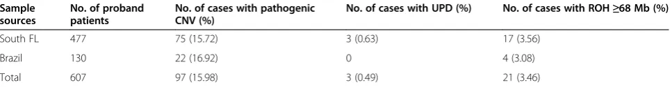
A total of 607 proband pediatric patients were tested over a one year period, including 130 from Brazil and 477 from south Florida. Pathogenic CNVs were detected in 97 cases (15.98%). SNP genotyping suggested presence of UPD in three cases (0.49%), including two UPD20 and a UPD14. Our results suggested presence of parental consanguinity in 21 cases (3.46%) including 4 (3.07%) from Brazil and 17 (3.56%) referred from hospitals in south Florida (Table 1).
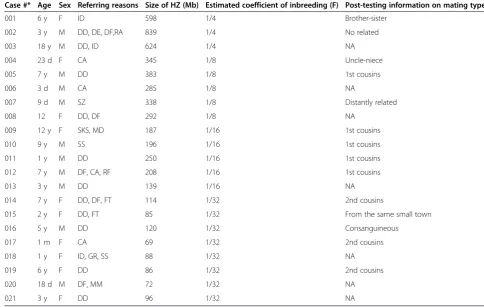
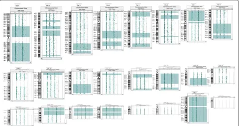
ROHs larger than 5 Mb were observed in 86 cases (data details not provided in this paper) including 21 with results suggesting presence of parental consan-guinity. Among the 21 patients (Table 2), three had an average of 686 Mb of ROH with an estimated coeffi-cient of inbreeding (F) ¼ (Figure 1); five had an aver-age 329 Mb of ROH with an estimated F 1/8; five had an average of 196 Mb of ROH with an estimated F 1/16; and eight had an average 91 Mb of ROH with an estimated F 1/32. Post-testing information on parental relationship was available in 14 of the 21 cases. A par-ental relationship of 1st cousins was confirmed in all the 4 cases with an estimated F1/16. Among the five
cases with an estimated F 1/32 suggestive of a mating type of half first cousins or first cousins once removed, three were said having parents as 2nd cousins; One couple was consanguineous but relationship was not specified; In the remaining case, parents said they are not related but from the same small city. In the three cases with F1/8 suggestive of a 2nd degree relation-ship, one was an uncle-niece mating; one said to be first cousins; and the other was said to be distant rela-tives. Among the two children with F1/4, one was a product of a brother-sister mating, and the other’s parents said they were not related.
Discussion
In addition to pathogenic CNVs and UPDs, approxi-mately 3.5% of children with intellectual and/or devel-opmental disabilities in our study populations showed greater than 68 Mb of ROH, suggesting presence of parental consanguinity. Post-testing surveys confirmed the 1st cousin relationships for all the cases with an estimated F 1/16 and presence of consanguinity in 4 out of 5 cases with an estimated F1/32 although the information from medical records was incomplete or inconsistent with the suggested mating type. In our practice, we report results suggesting presence of par-ental consanguinity but not specify the biological rela-tionship of the parents as recommended by the ACMG guidelines [13], because SNP array analysis is not designed to be a paternity test or to assign a spe-cific relationship between the parents of the proband patient. Also, the observed size of ROH may not pre-cisely predict the true biological relationship due to variables such as recombination during meiosis, mul-tiple loops of consanguinity or mulmul-tiple generations of breeding within a relatively closed community. For the patients showing an estimated F ¼ or 1/8, we have in-cluded the percentage of homozygosity in our clinical report and stated that the result may suggest a first-or second-degree parental relationship first-or incestuous mating.
Incestuous parental relationships identified by SNP-based microarrays have been reported previously. One was a 3-year-old boy with multiple medical conditions showing 668 Mb of genomic homozygosity which was consistent with the patient being conceived as the
Table 1 Summary of array study results
Sample sources
No. of proband patients
No. of cases with pathogenic CNV (%)
No. of cases with UPD (%) No. of cases with ROH≥68 Mb (%)
South FL 477 75 (15.72) 3 (0.63) 17 (3.56)
Brazil 130 22 (16.92) 0 4 (3.08)
product of a mating between first-degree relatives [14]. In a report on a SNP data, ROH of greater than 20% of the genome were identified in two of 5000 samples an-alyzed in a cytogenetics laboratory in Australia [8]. Our study has shown that a level of ROH greater than 20% correlates with an estimated coefficient of in-breeding F1/4 which is suggestive of a first degree rela-tionship such as parent–child or brother-sister mating, as exemplified in case 001. However, an incestuous re-lationship, which represents a complicated social and legal issue, may not be readily identified during a doc-tor’s visit or genetic counseling, as shown in our post-testing survey. Genetic counseling for consanguinity can be complicated particularly when incestuous mat-ing is uncovered by use of microarrays.
The frequency of consanguineous mating may vary significantly in different geographic regions or in dif-ferent ethnic populations [12,15-17]. Our study pa-tients were referred from hospitals in South Florida (mainly Miami-Dade and Broward counties) and Brazil. Hispanic or Latino Americans account for 65% of the population in Miami-Dade and 25% of the population
in Broward (2010 census data). The Hispanic or Latino Americans in Florida originate mainly from Cuba (32%) and Puerto Rico (28%) and Mexico (9%). It was esti-mated in 2008 that over one million of Brazilians live in USA and about 300,000 of them live in Florida. First cousin marriage is legal in Florida. The rate of consan-guineous marriage in Miami-Dade and Broward coun-ties is unknown but is likely to be higher than the generally estimated average (<1%) in USA. The rate of consanguineous mating in Brazil was estimated as 1.6% with a heterogeneous geographic distribution and half of the consanguineous marriages have a coef-ficient of inbreeding F 1/16 or higher [18]. There was a consensus that consanguineous marriages are asso-ciated with an increased risk of congenital mal-formations and autosomal recessive disease [12]. The estimated excess risks of morbidity and precocious mortality for the children with F1/4, 1/8, 1/16 and 1/32 are about 40%, 20%, 10%, 5%, respectively [9,10]. A recent review suggests that the risk for congenital heart disease is increased in consanguineous mar-riages [19].
Table 2 The cases with parental consanguinity
Case #* Age Sex Referring reasons Size of HZ (Mb) Estimated coefficient of inbreeding (F) Post-testing information on mating type
001 6 y F ID 598 1/4 Brother-sister
002 3 y M DD, DE, DF,RA 839 1/4 No related
003 18 y M DD, ID 624 1/4 NA
004 23 d F CA 345 1/8 Uncle-niece
005 7 y M DD 383 1/8 1st cousins
006 3 d M CA 285 1/8 NA
007 9 d M SZ 338 1/8 Distantly related
008 12 F DD, DF 292 1/8 NA
009 12 y F SKS, MD 187 1/16 1st cousins
010 9 y M SS 196 1/16 1st cousins
011 1 y M DD 250 1/16 1st cousins
012 7 y M DF, CA, RF 208 1/16 1st cousins
013 3 y M DD 139 1/16 NA
014 7 y F DD, DF, FT 114 1/32 2nd cousins
015 2 y F DD, FT 85 1/32 From the same small town
016 5 y M DD 120 1/32 Consanguineous
017 1 m F CA 69 1/32 2nd cousins
018 1 y F ID, GR, SS 88 1/32 NA
019 6 y F DD 86 1/32 2nd cousins
020 18 d M DF, MM 72 1/32 NA
021 3 y F DD 96 1/32 NA
Different from previous clinical studies on the fam-ilies with a history of consanguineous mating, we detected parental consanguinity in about 3.5% of chil-dren referred for developmental problems. Most of these children were referred for congenital anomalies or multiple malformations, developmental delay or in-tellectual disability, dysmorphic features, failure to thrive/growth retardation or short stature. The finding of parental consanguinity in these cases is highly sug-gestive of an underlying recessive cause. For example, following the array study, case 012 was diagnosed with Schimke immune-osseous dysplasia, an autosomal-recessive pleiotropic disorder caused by loss of
func-tion mutafunc-tions in SMARCAL1 leading to
spondyl-oepiphyseal dysplasia, renal dysfunction and T-cell immunodeficiency [20,21]. A diagnosis of autosomal recessive spinal muscular atrophy has been suspected in case 009. In case 001, the 6 year-old girl initially re-ferred for mental problems had a history of proximal renal tubular acidosis (RTA) at 18 months of age, basal ganglia and corneal calcifications secondary to RTA. Further diagnostic work-up has revealed increased ex-cretion of Krebs cycle intermediate and a tentative diagnosis of 2-ketoglutarate dehydrogenase deficiency, a very rare metabolic disorder [22-24]. Involvement of multiple recessive mutant genes or a complex genetic mechanism is possible in some patients, particularly in
the children as products of incestuous mating. Con-sanguinity may not be noted in many cases without microarray studies and identification of the biological relationship of the parents in such cases is clinically important for additional diagnostic testing. Further studies of phenotype and possible correlating meta-bolic deficiency, as well as sequencing of the ROH re-gions would determine the genetic mechanisms of the developmental problems in these children.
Methods
Patients
Patients, all pediatric but one, included in this report were referred for developmental delay or intellectual disability, autism spectrum disorders, and/or congeni-tal anomalies from November 1, 2011 to March 5, 2013. Approximately 80% of the patient samples origi-nated in the Genetics Clinic at the University Miami and other hospitals in south Florida. However, many of the patients were of Latin American or Caribbean descent or referred from Latin American countries. The remaining 20% of patient samples were received from Brazil.
Microarray studies
used in our laboratory as a clinical test for over a year. The array contains 180,000 oligonucleotide probes, in-cluding approximately 120,000 CGH probes and 60,000 SNP probes with a resolution of 5 ~ 10 Mb for ROH de-tection. Array studies were performed using a protocol recommended by Agilent. Briefly, DNA was prepared from a blood sample and purified using the QIAamp DNA Blood Midi Kit (Qiagen) or DNeasy Blood & Tissue Kit (Qiagen) when a sample was less than 0.5 ml. DNA was quantified, digested and then labeled with Cy5 (testing DNA) and Cy3 (reference DNA) and hybridized to the microarray at 65°C for 21 ~ 24 hours. The microarray was washed and analyzed using an Agilent C microarray scan-ner and CytoGenomics Edition 2.0.6.0 software.
Criteria for reporting
We used the same criteria for detection and interpret-ation of CNVs as previously reported [5,6]. A size of 20 Mb of interstitial ROH or 10 Mb of telomeric ROH was used as the threshold for UPD when ROH was ob-served on a single chromosome [7,8]. For detection of parental consanguinity, we calculated the theoretic size of ROH for coefficient of inbreeding (F) 1/4, 1/8, 1/16, and 1/32 respectively using the total 2,881 Mb of auto-somal genome (GRCh37/hg19) and its variable range using the middle line between theoretic average sizes (Table 3). Based on this calculation, a result was reported suggesting presence of parental consanguinity when ROHs were observed on multiple chromosomes with a total size≥68 Mb which correlates with an mated F 1/32. However, we did not specify the esti-mated coefficient of inbreeding and the likelihood of biologic relationship of the parents in our laboratory report. ROHs on multiple chromosomes with a total size of 10 ~ 67 Mb or ROHs on a single chromosome which do not meet the criteria for UPD were reported as identical by decent (shared parental ancestry). We reported the individual ROH with a size ≥5 Mb either on a single chromosome or on multiple chromosomes to clinicians (data not shown) because rarely an ROH may contain recessive mutations responsible for the clinical phenotype. We have reported and interpreted ROH using above described criteria starting in late
2011 and our practice appears to be consistent with the ACMG standards and guidelines recently pub-lished [13].
Retrospective survey on parental consanguinity
Referring clinicians were contacted to obtain informa-tion on the parent’s family relationship of the children for whom laboratory results suggested presence of parental consanguinity. The clinician provided infor-mation based on either the notes of clinical geneticists or family history in medical records. In some cases, we obtained information on patient diagnosis after microarray study.
This report is a summary of clinical cases tested in a CLIA certified and Florida State licensed clinical la-boratory with patients identification removed. This is not an experimental research using human material and therefore no IRB approval is necessary.
Consent
Written informed consent was obtained from the pa-tient’s guardian/parent/next of kin for the publication of this report and any accompanying images.
Abbreviations
ACMG:American college of medical genetics; BAC: Bacterial artificial chromosome; CGH: Comparative genomic hybridization; CNV: Copy number variation; F: Coefficient of inbreeding; SNP: Single nucleotide polymorphisms; ROH: Regions of homozygosity; UPD: Uniparental disomy.
Competing interests
The authors declare that they have no competing interests.
Authors’contributions
YSF analyzed the laboratory data, drafted and finalized the manuscript. XO and JP performed microarray analysis and prepared the laboratory data. SS, MT, DB, OB, RY, CN, AHH, SDJP clinically examined the patients and collected clinical data. All authors read and approved the final manuscript.
Author details
1Department of Pathology and Mailman Center for Child Development, Room 7050, University of Miami Miller School of Medicine, 1601 NW 12th Avenue, Miami, FL 33136, USA.2Department of Human Genetics, University of Miami Miller School of Medicine, Miami, FL, USA.3Memorial Regional Hospitals, Miami, FL, USA.4Jackson Memorial Hospital, Hollywood, FL, USA. 5
Núcleo de Genética Médica, Belo Horizonte, Belo Horizonte, MG, Brazil.
Received: 15 July 2013 Accepted: 28 August 2013 Published: 20 September 2013
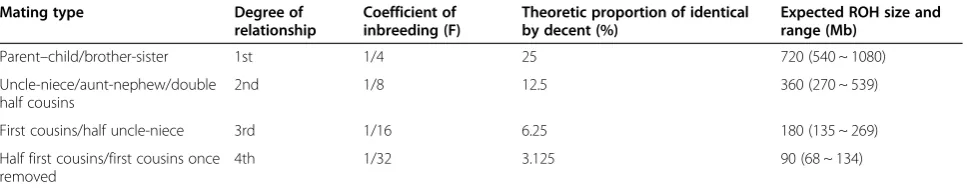
Table 3 Theoretic size of ROH for coefficient of inbreeding (F) expected from the total 2881 Mb autosomal genome (GRCh37/hg19)
Mating type Degree of
relationship
Coefficient of inbreeding (F)
Theoretic proportion of identical by decent (%)
Expected ROH size and range (Mb)
Parent–child/brother-sister 1st 1/4 25 720 (540 ~ 1080)
Uncle-niece/aunt-nephew/double half cousins
2nd 1/8 12.5 360 (270 ~ 539)
First cousins/half uncle-niece 3rd 1/16 6.25 180 (135 ~ 269)
Half first cousins/first cousins once removed
References
1. Lee C, Iafrate AJ, Brothman AR:Copy number variations and clinical cytogenetic diagnosis of constitutional disorders.
Nat Genet2007,39(Suppl 7):S48–S54.
2. Miller DT, Adam MP, Aradhya S, Biesecker LG, Brothman AR, Carter NP, Church DM, Crolla JA, Eichler EE, Epstein CJ, Faucett WA, Feuk L, Friedman JM, Hamosh A, Jackson L, Kaminsky EB, Kok K, Krantz ID, Kuhn RM, Lee C, Ostell JM, Rosenberg C, Scherer SW, Spinner NB, Stavropoulos DJ, Tepperberg JH, Thorland EC, Vermeesch JR, Waggoner DJ, Watson MS,et al:
Consensus statement: chromosomal microarray is a first-tier clinical diagnostic test for individuals with developmental disabilities or congenital anomalies.Am J Hum Genet2010,86:749–764.
3. Manning M, Hudgins L:Professional practice and guidelines committee. Array-based technology and recommendations for utilization in medical genetics practice for detection of chromosomal abnormalities. Genet Med2010,12:742–545.
4. Kaminsky EB, Kaul V, Paschall J, Church DM, Bunke B, Kunig D, Moreno-De -Luca D, Moreno-De-Luca A, Mulle JG, Warren ST, Richard G, Compton JG, Fuller AE, Gliem TJ, Huang S, Collinson MN, Beal SJ, Ackley T, Pickering DL, Golden DM, Aston E, Whitby H, Shetty S, Rossi MR, Rudd MK, South ST, Brothman AR, Sanger WG, Iyer RK, Crolla JA,et al:An evidence-based approach to establish the functional and clinical significance of copy number variants in intellectual and developmental disabilities.Genet Med
2011,13:777–784.
5. Fan YS, Jayakar P, Zhu H, Barbouth D, Sacharow S, Morales A, Carver V, Benke P, Mundy P, Elsas LJ:Detection of pathogenic gene copy number variations in patients with mental retardation by genomewide oligonucleotide array comparative genomic hybridization.Hum Mutat
2007,28:1124–1132.
6. Xiang B, Zhu H, Shen Y, Miller DT, Lu K, Hu X, Andersson HC, Narumanchi TM, Wang Y, Martinez JE, Wu BL, Li P, Li MM, Chen TJ, Fan YS: Genome-wide oligonucleotide array comparative genomic hybridization for etiological diagnosis of mental retardation: a multicenter experience of 1499 clinical cases.J Mol Diagn2010,12:204–212.
7. Papenhausen P, Schwartz S, Risheg H, Keitges E, Gadi I, Burnside RD, Jaswaney V, Pappas J, Pasion R, Friedman K, Tepperberg J:UPD detection using homozygosity profiling with a SNP genotyping microarray. Am J Med Genet A2011,155A:757–768.
8. Bruno DL, White SM, Ganesamoorthy D, Burgess T, Butler K, Corrie S, Francis D, Hills L, Prabhakara K, Ngo C, Norris F, Oertel R, Pertile MD, Stark Z, Amor DJ, Slater HR:Pathogenic aberrations revealed exclusively by single nucleotide polymorphism (SNP) genotyping data in 5000 samples tested by molecular karyotyping.J Med Genet2011,48:831–839.
9. Freire-Maia N:Effects of consanguineous marriages on morbidity and precocious mortality: genetic counseling.Am J Med Genet1984,18:401–406. 10. Baird PA, McGillivray B:Children of incest.J Pediatr1982,101:854–857. 11. Bundey S, Alam H:A five-year prospective study of the health of children
in different ethnic groups, with particular reference to the effect of inbreeding.Eur J Hum Genet1993,1:206–219.
12. Hamamy H, Antonarakis SE, Cavalli-Sforza LL, Temtamy S, Romeo G, Kate LP, Bennett RL, Shaw A, Megarbane A, Van Duijn C, Bathija H, Fokstuen S, Engel E, Zlotogora J, Dermitzakis E, Bottani A, Dahoun S, Morris MA, Arsenault S, Aglan MS, Ajaz M, Alkalamchi A, Alnaqeb D, Alwasiyah MK, Anwer N, Awwad R, Bonnefin M, Corry P, Gwanmesia L, Karbani GA,et al:
Consanguineous marriages, pearls and perils: Geneva international consanguinity workshop report.Genet Med2011,13:841–847. 13. Rehder CW, David KL, Hirsch B, Toriello HV, Wilson CM, Kearney HM:
American college of medical genetics and genomics: standards and guidelines for documenting suspected consanguinity as an incidental finding of genomic testing.Gene Med2013,15:150–152.
14. Schaaf CP, Scott DA, Wiszniewska J, Beaudet AL:Identification of incestuous parental relationships by SNP-based DNA microarrays. Lancet2011,377:555–556.
15. Bener A, El Ayoubi HR, Chouchane L, Ali AI, Al-Kubaisi A, Al-Sulaiti H, Teebi AS:Impact of consanguinity on cancer in a highly endogamous population.Asian Pac J Cancer Prev2009,10:35–40.
16. Shami SA, Qaisar R, Bittles AH:Consanguinity and adult morbidity in Pakistan.Lancet1991,338:954–955.
17. Simpson JL, Martin AO, Elias S, Sarto GE, Dunn JK:Cancers of the breast and female genital system: search for recessive genetic factors through analysis of human isolate.Am J Obstet Gynecol1981,141:629–636.
18. Liascovich R, Rittler M, Castilla EE:Consanguinity in South America: demographic aspects.Hum Hered2001,51:27–34.
19. Shieh JT, Bittles AH, Hudgins L:Consanguinity and the risk of congenital heart disease.Am J Med Genet A2012,158A:1236–1241.
20. Boerkoel CF, Takashima H, John J, Yan J, Stankiewicz P, Rosenbarker L, André JL, Bogdanovic R, Burguet A, Cockfield S, Cordeiro I, Fründ S, Illies F, Joseph M, Kaitila I, Lama G, Loirat C, McLeod DR, Milford DV, Petty EM, Rodrigo F, Saraiva JM, Schmidt B, Smith GC, Spranger J, Stein A, Thiele H, Tizard J, Weksberg R, Lupski JR,et al:Mutant chromatin remodeling protein SMARCAL1 causes schimke immuno-osseous dysplasia. Nat Genet2002,30:215–220.
21. Bansbach CE, Boerkoel CF, Cortez D:SMARCAL1 and replication stress: an explanation for SIOD?Nucleus2010,1:245–248.
22. Bonnefont JP, Chretien D, Rustin P, Robinson B, Vassault A, Aupetit J, Charpentier C, Rabier D, Saudubray JM, Munnich A:Alpha-ketoglutarate dehydrogenase deficiency presenting as congenital lactic acidosis. J Pediatr1992,121:255–258.
23. Guffon N, Lopez-Mediavilla C, Dumoulin R, Mousson B, Godinot C, Carrier H, Collombet JM, Divry P, Mathieu M, Guibaud P:2-Ketoglutarate
dehydrogenase deficiency, a rare cause of primary hyperlactataemia: report of a new case.J Inherit Metab Dis1993,16:821–830.
24. Dunckelmann RJ, Ebinger F, Schulze A, Wanders RJ, Rating D, Mayatepek E:2-Ketoglutarate dehydrogenase deficiency with intermittent 2-ketoglutaric aciduria.Neuropediatrics2000,31:35–38.
doi:10.1186/1755-8166-6-38
Cite this article as:Fanet al.:Frequent detection of parental
consanguinity in children with developmental disorders by a combined CGH and SNP microarray.Molecular Cytogenetics20136:38.
Submit your next manuscript to BioMed Central and take full advantage of:
• Convenient online submission
• Thorough peer review
• No space constraints or color figure charges
• Immediate publication on acceptance
• Inclusion in PubMed, CAS, Scopus and Google Scholar
• Research which is freely available for redistribution