Open Access
Review
The Peter Pan paradigm
J Craig Cohen* and Janet E Larson
Address: The Brady Laboratory, Section of Neonatology, Department of Pediatrics, Stony Brook University Medical Center, Stony Brook, NY 11794, USA
Email: J Craig Cohen* - [email protected]; Janet E Larson - [email protected] * Corresponding author
Abstract
Genetic and environmental agents that disrupt organogenesis are numerous and well described. Less well established, however, is the role of delay in the developmental processes that yield functionally immature tissues at birth. Evidence is mounting that organs do not continue to develop postnatally in the context of these organogenesis insults, condemning the patient to utilize under-developed tissues for adult processes. These poorly differentiated organs may appear histologically normal at birth but with age may deteriorate revealing progressive or adult-onset pathology. The genetic and molecular underpinning of the proposed paradigm reveals the need for a comprehensive systems biology approach to evaluate the role of maternal-fetal environment on organogenesis.
You may delay, but time will not
Benjamin Franklin
USA Founding Father
Background
The fragility of the developmental process is well known with estimates as high as 70% of conceptions lost in early pregnancy [1]. Genetic defects are known to produce numerous changes in both metabolic and morphologic characteristics of the fetus [2]. The choreography of genes necessary for successful fetal development is amply dem-onstrated by embryonic lethal [3] and other phenotypes [4-8] in knockout mouse models. Thus, genetic defects introduced in the developmental cascade contribute to structural malformations. But organogenesis requires the genetic choreography to occur not just in sequence but also during a sensitive time period. What happens when the genes regulating the developmental time clock [9,10] are mutated or delayed by epigenetic factors? Can the organ continue to develop outside the fetal environment
and if not, what are the consequences of retaining fetal characteristics in an adult?
The best model for addressing these questions related to organogenesis and timing is the premature infant. Mod-ern neonatology has extended viability to 23 weeks in the 40 week human gestation, but what are the consequences of completing less than 60% of gestation on the human fetus? The obvious immediate consequences seen in any neonatal unit include poor lung function, intestinal defi-ciencies, immune system defidefi-ciencies, and poor renal function. These immediate problems combined with ther-apies used to combat them can lead to significant morbid-ity and mortalmorbid-ity in the premature infant.
Published: 8 January 2008
Theoretical Biology and Medical Modelling 2008, 5:1 doi:10.1186/1742-4682-5-1
Received: 6 June 2007 Accepted: 8 January 2008
This article is available from: http://www.tbiomed.com/content/5/1/1
© 2008 Cohen and Larson; licensee BioMed Central Ltd.
Infants surviving the immediate organ deficiencies and therapies of premature birth, however, do not grow into healthy adult. Rather, numerous epidemiological and clinical observations have shown that prematurity is asso-ciated with many adult onset diseases (Table 1). The epi-demiologic basis for late or adult-onset diseases, following either premature birth or low birth weight, includes poor pre-natal maternal nutrition and smoking during pregnancy both of which are reviewed previously [11,12]. But these epidemiologic correlations do no pro-vide a theoretical, molecular basis for explaining late onset of diseases related to delays in organogenesis.
Conceptualizing the effect of small developmental distur-bances on solid organs such as the lung, liver, intestines and kidneys can be difficult because morphological differ-ences may be minor. If one considers an organ with func-tional characteristics that are morphologic, however, then the concept of developmental disruption becomes obvi-ous. A good example would be the hand. Numerous examples of impaired development through either genetic defects or blood flow interruption result in obvious deformities including webbing, polydactyl and complete and partial absences of digits. Visualization of changes in solid organ development is less obvious but the effects on function have the same impact as a hand without a thumb.
As an example, lung growth and development (Fig. 1A), like the hand, is precisely timed throughout gestation [13]. In humans functional respiratory structures are com-plete by birth although alveolarization continues until an adult number of alveoli are reached at 2–3 years of age; however, in the mouse and rat functional alveolarization is completed after birth by day of life (DOL) 5. The embry-onic human lung can be identified at 28 days with trachea and bronchi expanding from the lung bud. During the pseudoglandular stage further branching occurs and the airways are lined with multipotent stem cells. Branches of terminal bronchioles are formed and respiratory bronchi-oles begin as out pockets. These pockets of epithelial cells expand during the saccular and alveolar stages to form ter-minal alveoli which are necessary for gas exchange.
Nor-mal lung organogenesis is a carefully programmed process that can be disrupted and result in abnormal lung struc-ture and function.
Three types of disruptions can occur in lung organogene-sis (Fig. 1B–D). The most obvious are genetic mutations that completely block lung growth (Fig. 1B) resulting in a non-functional lung and fetal or neonatal demise [14-16]. The less obvious yet equally important disruptions to nor-mal lung function are those that impair or impede specific events in organogenesis. A genetic mutation or environ-mental insult to the fetus through the mother can result in either a premature (Fig. 1C) or prolonged (Fig. 1D) tran-sition to a later stage of development. In the former case, structures, which are normally propagated, are lost along with their function. In the latter case, slower growth results in late development of important structures and functions prior to normal birth. In both cases, the histo-logic structure of the lung may grossly appear normal; however, the actual functional equivalency of the cells may be significantly altered.
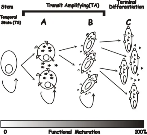
Two questions arise from these models. First, can discon-tinuity between stages in a developmental cascade alter the cell type or their function? During each stage of orga-nogenesis, stem cells are progressing through stages of transit amplifying (TA) cells in which the functional char-acteristics of each generation is maturing to that of a ter-minally differentiated cell type (Fig. 2). During organogenesis the environment progresses through differ-ent temporal states (TS) in which the TA cells produce products necessary for differentiation that are not neces-sary needed for final functionality. As an example, in TS-A (Fig. 2) may produce products that are required for devel-opment of specialized structures. Proteins produced in TS-B, however, may induce rapid cell growth and increased tissue complexity. If this process is abbreviated as pro-posed in Fig. 1C, then the terminal differentiation state can not be reached and downstream functional character-istics of the cell will be missing.
Secondly, when there is a delay in progression (Fig. 1D), does normal organogenesis continue postnatally? Because the fetal environment plays a significant role in differenti-ation, preterm birth arrests organ development with retention of fetal characteristics. Thus, all or part of the organ would remain in a fetal temporal state (e.g. TS-B; Fig. 2), significantly different from that of the terminally differentiated tissue (TS-C, Fig. 2).
Delay and disruption of organogenesis could result in two different phenotypes because the immature tissue would be missing different functional gene sets. Distinguishing these two would be dependent upon a developmental gene expression knowledge base. However, it is possible
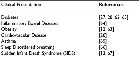
Table 1: Late-onset clinical presentations associated with premature birth.
Clinical Presentation References
Diabetes [27, 28, 62, 63]
Inflammatory Bowel Diseases [64]
Obesity [12, 63]
Cardiovascular Disease [28]
Asthma [65]
to get similar morphologic phenotypes but different phys-iology. As illustrated in Figure 3, if one takes a case in which a protein is required for triggering the normal final stage of complex organ architecture (Fig. 3A), disrupting
the stage that precedes this protein could result in its pre-mature activation without sufficient support structure (Fig. 3B), resulting in a hypoplastic structure. Likewise, prolonging the preceding stage by delaying activation of these proteins could lead to some gain in complexity in the supporting structure. However, similar decreased structural complexity of this final stage would result (Fig.
Architecture and developmental programming
Figure 3
Architecture and developmental programming. Normal architecture depends on building layers that are based on both quantity and timing of components with an overall structural size limitation (Panel A). Premature activation of subsequent stages disrupts primary base structure requiring downstream compo-nents to fill a space larger than normally required resulting in less complexity (Panel B). Likewise, prolonging production of the upstream components increases their complexity at the expense of downstream structures (Panel C), resulting in a hypoplastic structure similar to that seen in disrupted growth of upstream components.
Lung development and the effects of developmental disruption
Figure 1
Lung development and the effects of developmental disruption. A-Normal lung differentiation; B-Disrupted growth; C-Prema-ture transition resulting in abbreviated pseudoglandular stage; D-Delayed transition leading to incomplete canalicular and saccular/alveolar stages.
Stem cell differentiation during organogenesis
Figure 2
3C) due to a decrease in the time interval between activa-tion of terminal differentiaactiva-tion and birth. Morphologi-cally both tissues would appear hypoplasia of the terminal structure; because the balance among structural components is altered the physiology could be quite dif-ferent. Thus, distinctions between delayed and disrupted organogenesis will be dependent upon biochemical mak-ers of differentiation, structural characteristics of the tis-sue, and perhaps most importantly the physiologic functions of the modified organ.
There are numerous examples of failed differentiation due to specific gene defects leading to deficient cell types. In the lung, mutations in lamellar body formation lead to structural changes leading to chronic inflammatory dis-eases [17-19]. Mutation of Arnt, a gene required for hepatic vascularization, results in a persistent embryonic liver phenotype that demonstrate decreased survival and changes in fat and carbohydrate metabolism after birth [20]. Bapx1 is required for separation of the pancreas and spleen into distinct organs [21]. In each of these single gene defects, slow or inhibited organogenesis results in a suboptimal functioning organ and a subsequent pathol-ogy.
The physical environment of the fetus can also impact the dynamics of organogenesis. One of the more common prototypes is congenital diaphragmatic hernias which result in hypoplastic lung disease. Surgical blocking of the trachea [22] or in utero cystic fibrosis transmembrane con-ductance regulator (CFTR) gene therapy [23] to promote stretch induced lung differentiation can reverse the effect, but postnatally this condition has a poor prognosis with high morbidity and mortality. Likewise, amniotic fluid blockage to the intestines alters villus formation and func-tion [24].
Probably the least understood and most subtle effectors of developmental delay are fetal environmental insults. Maternal smoking, alcohol consumption, diabetes, and diehylstilbesterol use have all been linked to significant long term consequences on the fetus [11,19,25-28].
Discussion
What are diseases of organogenesis that result from devel-opmental delay and not disruption called? In premature infants terms like fetal and immature are used to describe their gestational correct, underdeveloped organs. We pro-pose that the temporal state of many tissues can become fixed at birth, resulting in organs that are functionally immature. Thus, we use the term Peter Pan Paradigm for diseases that occur due to developmental delay or disrup-tion resulting in a seemingly histological, normal organ with functional immaturity. Developmental delay pro-duces tissues that at birth are in a gestational abnormal
temporal state, either fetal (e.g. TS-B; Fig. 2) or hybrid (e.g. TS-B/C). Thus, like the fictional namesake, these affected organs never fully mature and remain at a nexus between the fetal and mature temporal states.
Changes in the organs trapped in either a complete or par-tial fetal temporal state can be varied. As illustrated in Fig. 2 receptors and secreted products may be different. In addition, functions such as regulated secretion could be affected. This functional immaturity results in progressive incapacitation of the organ with associated late-onset dis-ease manifestations. Possible syndromes included in this category would be any progressive or late-onset disease that exhibited multi-factorial genetics. Obviously, these diseases would present with very heterogeneous pheno-types because the more severe the delay, the earlier the average temporal state of the tissue.
The prototypical disease in the Peter Pan paradigm is Cystic Fibrosis (CF). The natural history of this disease includes progressive multi-system failure. There is a lack of phenotype genotype correlation suggesting the role of multiple genes or environment [29,30]. CF is a recessive disorder yet the human phenotype can occur in the absence of homozygous mutations in the CFTR gene [31] and the heterozygous knockout mice have a unique lung phenotype [32]. Expression during embryonic lung growth is associated with genes required for normal devel-opment [3,10,33]. CFTR expression occurs at high levels in the developing lung [34] and other tissues [35]. Fur-thermore, transient in utero replacement to increase or decrease expression of CFTR in animal models alters the lung and intestinal CF phenotype [23,36-43]. Thus, there are multiple independent lines of evidence suggesting a role of CFTR in normal organogenesis of the lung, intes-tines, and other organs.
As previously described regarding progression of develop-ment from one stage to another (Fig. 1), the recent finding that CFTR influences stretch-induced differentiation [37] further delineates CF as a Peter Pan disease. Stretch induced differentiation is essential for normal lung devel-opment [49-52]. Amniotic fluid flow is also essential for normal gut differentiation [24]. Thus, the major organs involved in the CF phenotype are all dependent upon amniotic fluid flow and mechanochemical regulated dif-ferentiation [51,53]. In the total absence of CFTR this flow would be dependent upon other, less effective, mecha-nisms to drive development such as rho kinase [54] and fetal breathing [55]. In addition, environmental exposure of the mother to nicotine or other agents could affect the final phenotype by affecting muscle contractions neces-sary for stretch induction [25,56,57]. Thus, the lack of phenotype-genotype correlation and a common mechan-ochemical origin would account for the variable effects seen in multiple organ systems.
The intestine may be one of the more susceptible organs for Peter Pan diseases. As discussed earlier inflammatory bowel diseases are linked epidemiologically with prema-turity (Table 1). Thus, delay in intestinal development could alter cell maturation and the metabolic balance of the intestines. One of the most common Peter Pan dis-eases may prove to be obesity and diabetes. The ability to maintain metabolic balance depends on developmental and transcriptional control systems of the intestinal epi-thelium [58,59]. Thus, interruption or delay leading to under differentiation of intestinal epithelium would have a significant impact on metabolic activity. In what may be considered an early prelude to Peter Pan Paradigm, a rela-tionship between metabolic balance and fetal develop-ment was originally proposed in the "Thrifty Phenotype Hypothesis"[60].
Lung diseases such as asthma, allergies, and sudden infant death syndrome (SIDS) may be related to delay in devel-opment. All of these conditions are associated with pre-maturity (Table 1) and from Fig. 1, one can readily visualize how slowing progress in differentiation of suc-ceeding structures would have a cascading effect on lung function. The central role of stretch-induced differentia-tion to lung development [37,51,53,54] strongly suggests that deficiencies in amniotic fluid volume, depression of fetal breathing [55], or inhibition of smooth muscle con-tractions [49,52] with agents such as nicotine [57] could depress and delay differentiation of the bronchioles lead-ing to under differentiated cell types and airways. In fact this is the case as previous studies showed persistence of fetal gene expression and functional immature phenotype in allergic asthma [61].
Competing interests
The author(s) declare that they have no competing inter-ests.
Authors' contributions
JCC and JEL developed this concept jointly.
Acknowledgements
JCC and JEK are supported by the Brady Russell Foundation.
References
1. Jauniaux E, Burton GJ: Pathophysiology of histological changes in early pregnancy loss. Placenta 2005, 26:114-123.
2. Jones KL: Smiths Recognizable Patterns of Human Malformation Philade-phia, PA: W.B. Saunders Company; 1988.
3. Copp AJ: Death before birth: clues from gene knockouts and mutations. Trends Genet 1995, 11:87-93.
4. Guilbault C, Saeed Z, Downey GP, Radzioch D: Cystic Fibrosis Mouse Models. Am J Respir Cell Mol Biol 2006.
5. Bhangoo A, Anhalt H, Ten S, King SR: Phenotypic variations in lipoid congenital adrenal hyperplasia. Pediatr Endocrinol Rev 2006, 3:258-271.
6. Dunger DB, Petry CJ, Ong KK: Genetic variations and normal fetal growth. Horm Res 2006, 65(Suppl 3):34-40.
7. Reecy JM, Spurlock DM, Stahl CH: Gene expression profiling: insights into skeletal muscle growth and development. J Anim Sci 2006, 84(Suppl):E150-154.
8. Adelman CA, Petrini JH, Attwooll CL: Modeling disease in the mouse: lessons from DNA damage response and cell cycle control genes. J Cell Biochem 2006, 97:459-473.
9. Edwards RG: Regulatory systems in early mammalian devel-opment, with especial reference to polarity and totipotency. Ital J Anat Embryol 2001, 106:85-100.
10. Pourquie O: Clocks regulating developmental processes. Curr Opin Neurobiol 1998, 8:665-670.
11. Jaddoe VW, Witteman JC: Hypotheses on the fetal origins of adult diseases: contributions of epidemiological studies. Eur J Epidemiol 2006, 21:91-102.
12. Gluckman PD, Hanson MA, Pinal C: The developmental origins of adult disease. Maternal and Child Nutrition 2005, 1:130-141. 13. Hislop AA: Airway and blood vessel interaction during lung
development. J Anat 2002, 201:325-334.
14. Vergnes L, Peterfy M, Bergo MO, Young SG, Reue K: Lamin B1 is required for mouse development and nuclear integrity. Proc Natl Acad Sci USA 2004, 101:10428-10433.
15. Kikkawa Y, Miner JH: Molecular dissection of laminin alpha 5 in vivo reveals separable domain-specific roles in embryonic development and kidney function. Dev Biol 2006, 296:265-277. 16. Muglia LJ: Genetic analysis of fetal development and
parturi-tion control in the mouse. Pediatr Res 2000, 47:437-443. 17. Guttentag SH, Akhtar A, Tao JQ, Atochina E, Rusiniak ME, Swank RT,
Bates SR: Defective surfactant secretion in a mouse model of Hermansky-Pudlak syndrome. Am J Respir Cell Mol Biol 2005, 33:14-21.
18. Lyerla TA, Rusiniak ME, Borchers M, Jahreis G, Tan J, Ohtake P, Novak EK, Swank RT: Aberrant lung structure, composition, and function in a murine model of Hermansky-Pudlak syn-drome. Am J Physiol Lung Cell Mol Physiol 2003, 285:L643-653. 19. Yin Y, Ma L: Development of the mammalian female
repro-ductive tract. J Biochem (Tokyo) 2005, 137:677-683.
20. AYim SH, Shah Y, Tomita S, Morris HD, Gavrilova O, Lambert G, Ward JM, Gonzalez FJ: Disruption of the Arnt gene in endothe-lial cells causes hepatic vascular defects and partial embry-onic lethality in mice. Hepatology 2006, 44:550-560.
21. Asayesh A, Sharpe J, Watson RP, Hecksher-Sorensen J, Hastie ND, Hill RE, Ahlgren U: Spleen versus pancreas: strict control of organ interrelationship revealed by analyses of Bapx1-/-mice. Genes Dev 2006, 20:2208-2213.
23. Larson JE, Cohen JC: Improvement of pulmonary hypoplasia associated with congenital diaphragmatic hernia by in utero CFTR gene therapy. Am J Physiol Lung Cell Mol Physiol 2006, 291:L4-10.
24. Cellini C, Xu J, Buchmiller TL: Effect of esophageal ligation on small intestinal development in normal and growth-retarded fetal rabbits. J Pediatr Gastroenterol Nutr 2006, 43:291-298. 25. Schuller HM, Jull BA, Sheppard BJ, Plummer HK: Interaction of
tobacco-specific toxicants with the neuronal alpha(7) nico-tinic acetylcholine receptor and its associated mitogenic sig-nal transduction pathway: potential role in lung carcinogenesis and pediatric lung disorders. Eur J Pharmacol 2000, 393:265-277.
26. Landau LI: Paediatric basis of adult lung disease. Paediatric Res-piratory Reviews 2006, 7:S251-S254.
27. Mericq V: Prematurity and insulin sensitivity. Horm Res 2006, 65(Suppl 3):131-136.
28. Osmond C, Barker DJ: Fetal, infant, and childhood growth are predictors of coronary heart disease, diabetes, and hyper-tension in adult men and women. Environ Health Perspect 2000, 108(Suppl 3):545-553.
29. Haston CK, McKerlie C, Newbigging S, Corey M, Rozmahel R, Tsui LC: Detection of modifier loci influencing the lung phenotype of cystic fibrosis knockout mice. Mamm Genome 2002, 13:605-613.
30. Cutting GR: Modifier genetics: cystic fibrosis. Annu Rev Genomics Hum Genet 2005, 6:237-260.
31. Groman JD, Meyer ME, Wilmott RW, Zeitlin PL, Cutting GR: Vari-ant cystic fibrosis phenotypes in the absence of CFTR muta-tions. N Engl J Med 2002, 347:401-407.
32. Cohen JC, Lundblad LK, Bates JH, Levitzky M, Larson JE: The "Gold-ilocks Effect" in Cystic Fibrosis: identification of a lung phe-notype in the cftr knockout and heterozygous mouse. BMC Genet 2004, 5:21.
33. Kaplan F: Molecular determinants of fetal lung organogenesis. Mol Genet Metab 2000, 71:321-341.
34. Broackes-Carter FC, Mouchel N, Gill D, Hyde S, Bassett J, Harris A: Temporal regulation of CFTR expression during ovine lung development: implications for CF gene therapy. Hum Mol Genet 2002, 11:125-131.
35. McGrath SA, Basu A, Zeitlin PL: Cystic fibrosis gene and protein expression during fetal lung development. Am J Respir Cell Mol Biol 1993, 8:201-208.
36. Bueler H: Adeno-associated viral vectors for gene transfer and gene therapy. Biol Chem 1999, 380:613-622.
37. Cohen JC, Larson JE: Cystic fibrosis transmembrane conduct-ance regulator (CFTR) dependent cytoskeletal tension dur-ing lung organogenesis. Dev Dyn 2006, 235:2736-2748. 38. Cohen JC, Larson JE: Pathophysiologic consequences following
inhibition of a CFTR-dependent developmental cascade in the lung. BMC Dev Biol 2005, 5:2.
39. Cohen JC, Morrow SL, Cork RJ, Delcarpio JB, Larson JE: Molecular pathophysiology of cystic fibrosis based on the rescued knockout mouse model. Mol Genet Metab 1998, 64:108-118. 40. Larson JE, Delcarpio JB, Farberman MM, Morrow SL, Cohen JC:
CFTR modulates lung secretory cell proliferation and differ-entiation. Am J Physiol Lung Cell Mol Physiol 2000, 279:L333-341. 41. Larson JE, Morrow SL, Delcarpio JB, Bohm RP, Ratterree MS,
Blan-chard JL, Cohen JC: Gene transfer into the fetal primate: evi-dence for the secretion of transgene product. Mol Ther 2000, 2:631-639.
42. Larson JE, Morrow SL, Happel L, Sharp JF, Cohen JC: Reversal of cystic fibrosis phenotype in mice by gene therapy in utero. Lancet 1997, 349:619-620.
43. Morrow SL, Larson JE, Nelson S, Sekhon HS, Ren T, Cohen JC: Mod-ification of development by the CFTR gene in utero. Mol Genet Metab 1998, 65:203-212.
44. Galietta LJ, Folli C, Caci E, Pedemonte N, Taddei A, Ravazzolo R, Zegarra-Moran O: Effect of inflammatory stimuli on airway ion transport. Proc Am Thorac Soc 2004, 1:62-65.
45. Girod S, Zahm JM, Plotkowski C, Beck G, Puchelle E: Role of the physiochemical properties of mucus in the protection of the respiratory epithelium. Eur Respir J 1992, 5:477-487.
46. Mackie AD, Thornton SJ, Edenborough FP: Cystic fibrosis-related diabetes. Diabet Med 2003, 20:425-436.
47. Brennan AL, Geddes DM, Gyi KM, Baker EH: Clinical importance of cystic fibrosis-related diabetes. J Cyst Fibros 2004, 3:209-222. 48. Flora KD, Benner KG: Liver disease in cystic fibrosis. Clin Liver
Dis 1998, 2:51-61.
49. Featherstone NC, Jesudason EC, Connell MG, Fernig DG, Wray S, Losty PD, Burdyga TV: Spontaneous propagating calcium waves underpin airway peristalsis in embryonic rat lung. Am J Respir Cell Mol Biol 2005, 33:153-160.
50. Jernigan NL, Drummond HA: Vascular ENaC proteins are required for renal myogenic constriction. Am J Physiol Renal Physiol 2005, 289:F891-901.
51. Ingber DE: Cellular mechanotransduction: putting all the pieces together again. Faseb J 2006, 20:811-827.
52. Featherstone NC, Connell MG, Fernig DG, Wray S, Burdyga TV, Losty PD, Jesudason EC: Airway smooth muscle dysfunction precedes teratogenic congenital diaphragmatic hernia and may contribute to hypoplastic lung morphogenesis. Am J Respir Cell Mol Biol 2006, 35:571-578.
53. Ingber DE: Mechanical control of tissue growth: Function fol-lows form. Proc Natl Acad Sci USA 2005, 102:11571-11572. 54. Moore KA, Polte T, Huang S, Shi B, Alsberg E, Sunday ME, Ingber DE:
Control of basement membrane remodeling and epithelial branching morphogenesis in embryonic lung by Rho and cytoskeletal tension. Dev Dyn 2005, 232:268-281.
55. Inanlou MR, Baguma-Nibasheka M, Kablar B: The role of fetal breathing-like movements in lung organogenesis. Histol His-topathol 2005, 20:1261-1266.
56. Sekhon HS, Keller JA, Proskocil BJ, Martin EL, Spindel ER: Maternal nicotine exposure upregulates collagen gene expression in fetal monkey lung. Association with alpha7 nicotinic acetyl-choline receptors. Am J Respir Cell Mol Biol 2002, 26:31-41. 57. Jesudason EC, Smith NP, Connell MG, Spiller DG, White MR, Fernig
DG, Losty PD: Peristalsis of airway smooth muscle is develop-mentally regulated and uncoupled from hypoplastic lung growth. Am J Physiol Lung Cell Mol Physiol 2006, 291:L559-565. 58. Schmitz G, Langmann T: Metabolic learning in the intestine:
adaptation to nutrition and luminal factors. Horm Metab Res 2006, 38:452-454.
59. Heimerl S, Moehle C, Zahn A, Boettcher A, Stremmel W, Langmann T, Schmitz G: Alterations in intestinal fatty acid metabolism in inflammatory bowel disease. Biochim Biophys Acta 2006, 1762:341-350.
60. Hales CN, Barker DJ: Type 2 (non-insulin-dependent) diabetes mellitus: the thrifty phenotype hypothesis. Diabetologia 1992, 35:595-601.
61. Bousquet J, Jacot W, Yssel H, Vignola AM, Humbert M: Epigenetic inheritance of fetal genes in allergic asthma. Allergy 2004, 59:138-147.
62. Hales CN: Non-insulin-dependent diabetes mellitus. Br Med Bull 1997, 53:109-122.
63. Hofman PL, Cutfield WS: Insulin sensitivity in people born pre-term, with low or very low birth weight and small for gesta-tional age. J Endocrinol Invest 2006, 29:2-8.
64. Mikulecky M, Cierna I: Seasonality of births and childhood inflammatory bowel disease. Wien Klin Wochenschr 2005, 117:554-557.
65. Jaakkola JJ, Ahmed P, Ieromnimon A, Goepfert P, Laiou E, Quansah R, Jaakkola MS: Preterm delivery and asthma: a systematic review and meta-analysis. J Allergy Clin Immunol 2006, 118:823-830.
66. Rosen CL, Larkin EK, Kirchner HL, Emancipator JL, Bivins SF, Surovec SA, Martin RJ, Redline S: Prevalence and risk factors for sleep-disordered breathing in 8- to 11-year-old children: associa-tion with race and prematurity. J Pediatr 2003, 142:383-389. 67. Hoppenbrouwers T, Hodgman JE, Rybine D, Fabrikant G, Corwin M,