R E S E A R C H
Open Access
Compartmentalization in PVC
super-phylum: evolution and impact
Sandrine Pinos
1,2*, Pierre Pontarotti
2, Didier Raoult
1, Jean Pierre Baudoin
1and Isabelle Pagnier
1Abstract
Background:The PVC super-phylum gathers bacteria from seven phyla (Planctomycetes, Verrucomicrobiae, Chlamydiae, Lentisphaera, Poribacteria, OP3, WWE2) presenting different lifestyles, cell plans and environments.
Planctomycesand severalVerrucomicrobiaeexhibit a complex cell plan, with an intracytoplasmic membrane inducing the compartmentalization of the cytoplasm into two regions (pirellulosome and paryphoplasm). The evolution and function of this cell plan is still subject to debate. In this work, we hypothesized that it could play a role in protection of the bacterial DNA, especially against Horizontal Genes Transfers (HGT). Therefore, 64 bacterial genomes belonging to seven different phyla (whose four PVC phyla) were studied. We reconstructed the evolution of the cell plan as precisely as possible, thanks to information obtained by bibliographic study and electronic microscopy. We used a strategy based on comparative phylogenomic in order to determine the part occupied by the horizontal transfers for each studied genomes.
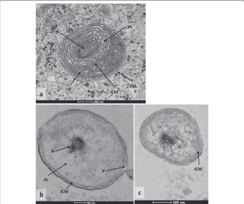
Results:Our results show that the bacteriaSimkania negevensis(Chlamydiae) andCoraliomargarita akajimensis (Verrucomicrobiae), whose cell plan were unknown before, are compartmentalized, as we can see on the
micrographies. This is one of the first indication of the presence of an intracytoplasmic membrane in aChlamydiae. The proportion of HGT does not seems to be related to the cell plan of bacteria, suggesting that
compartmentalization does not induce a protection of bacterial DNA against HGT. Conversely, lifestyle of bacteria seems to impact the ability of bacteria to exchange genes.
Conclusions:Our study allows a best reconstruction of the evolution of intracytoplasmic membrane, but this structure seems to have no impact on HGT occurrences.
Reviewers:This article was reviewed by Mircea Podar and Olivier Tenaillon.
Keywords:Evolution, Bacteria, PVC-super phylum, Microscopy, Compartmentalization
Background
The PVC super-phylum gathers seven bacterial phyla (Planctomycetes, Verrucomicrobiae, Chlamydiae, Lenti-sphaerae, Poribacteria, OP3, WWE2) [1–4], and comports 37 species of bacteria whose genome was entirely se-quenced. The monophyly of this super phylum have been discussed a lot in the last years, due to the difficulty to ob-tain a consensual phylogeny [5–14]. Recently, it seems that a global consensus was reached, that include all these bacteria in a same super-phylum [2, 5]. This idea is
confirmed by the discovery in 2012 and 2014 of a molecu-lar signature conserved in all PVC bacteria [1, 15]. The phylogenetic relations between PVC and other bacteria are still a controversial subject [5, 13]. PVC bacteria are present in an important variety of environments: water, soils, vertebrates, amoeba, insects…[5, 16–21].
The bacteria of this super-phylum show different in-teresting characteristics [11, 22]. Some of these
fea-tures can be found in other bacteria: some
Planctomycetes are implicated in carbon and nitrogen cycles [23, 24] or synthesize special sterols [25], many Chlamydiae are pathogens of mammals [26, 27], some Chlamydiae and Verrucomicrobiae are symbionts [19]. But PVC present also genetic and cellular features
unusual among bacteria: compartmentalization in
* Correspondence:[email protected] 1
Aix Marseille Université, URMITE, UM63, CNRS 7278, IRD 198, INSERM 1095, 27 Bd Jean Moulin, 13385 Marseille Cedex 5, France
2Aix Marseille Université, CNRS, Centrale Marseille, I2M UMR 7373, Evolution
Biologique et Modélisation, 13385 Marseille, Cedex 5, France
Verrucomicrobiae and Planctomycetes [28, 29], ab-sence of tubulin like protein FtZ in Planctomycetes and Chlamydiae [30, 31], crateiform surface and
bud-ding reproduction [32] in Planctomycetes. Among
these characteristics, we were specifically interested in the compartmentalization of bacteria. This feature is characterized by a specific cell plan, and concerns all the Planctomycetes [29, 33, 34], some Verrucomicro-biae [28], one Lentisphaera and one Poribacteria [35]. An intracytoplasmic membrane (ICM) separates the cytoplasm of bacteria into two distinct compartments: the pirellulosome inside (containing the DNA [36]), and the paryphoplasm outside (the size and shape of these two compartments varied a lot among PVC bacteria). This ICM is a lipid bilayer in contact with proteins [29, 32, 33] presenting structural similarities with some proteins from eukaryotic membranes, such as clathrins [37, 38]. Some compartmentalized bacteria also present a more complex cell plan, with the presence of an-other compartment, like the anamoxosome inCandidatus Kuenenia Stuttgartiensis[32, 39, 40], or a double internal membrane with ribosomes [41] in the same compartment that DNA [36] in Gemmata obscuriglobus [42]. Compartmentalization in PVC bacteria is still debatable and three opinions are defended actually : 1- As some fea-tures of PVC bacteria are current in Eukaryota[15], this observation allows some people to assume that the compartmentalization of PVC is the precursor of Eukaryotic nuclei, but this idea is not very popular [34, 43–48]. 2- Another proposition is that the compart-ment is structurally and functionally similar to a nucleus but that these two structures appeared independently [49]. 3- Some people identify the membrane as an invagination of a Gram negative external membrane [50].
Considering the reality of the existence of a
compartmentalization in PVC bacteria, it would be in-teresting to determine the function of this specific cell plan; indeed, no previous studies had been able to deter-mine it. Here we assumed that this membrane could be a protection against Horizontal Gene Transfers (HGT). This hypothesis was based on observations of the impact of different membranes already studied : the role of the internal membrane in Eukaryotes, or the influence of host membrane on genomes from intracellular bacteria (which limits the contact between foreign elements and DNA). We used a phylogenomic strategy of HGT detec-tion to reconstruct the history of HGT during the evolu-tion of PVC bacteria, before and after appearance and disappearance of compartmentalization.
Results
Lifestyle of species and evolution of cell plan
PVC bacteria present two different cell plan, compart-mentalized by an intra cytoplasmic membrane (ICM)
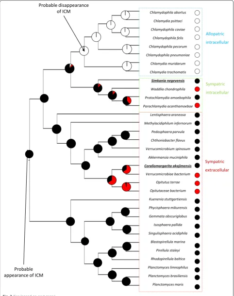
or non compartmentalized. They also present two dif-ferent lifestyles: allopatric lifestyle, for bacteria isolated from other microorganisms (obligate intracellular teria living in non amoeba cells) and sympatric, for bac-teria in contact with other microorganisms. The studies already published allowed determining cell plan of 26 PVC bacteria: PVC super phylum presented 18 com-partmentalized species distributed in three phyla (Verrucomicrobiae, Planctomycetes and Lentisphaerae) and eight non compartmentalized bacteria in one phyla (Chlamydiae). These bacteria were used in our study, but we also selected seven bacteria with an unknown cell plan, distributed in two phyla (Verrucomicrobiae and Chlamydiae). These data allowed the reconstruc-tion of cell plan evolureconstruc-tion with a good reliability, how-ever it was still difficult to conclude about ancestral state at different nodes of the tree, especially in Chla-mydiae phylum (their common ancestor presented a probability of 80.2 % to be compartmentalized). It was interesting to add new information in the dataset, in order to improve this reconstruction.
The electron microscopic pictures obtained for the thin sections of Simkania negevensis (Fig. 1a) and Coraliomargarita akajimensis (Fig. 1b, c), revealed the presence of a potential intracytoplasmic membrane in these two bacteria. In both cases, cells contain structures identified as a pirellulosome and a paryphoplasm, sepa-rated by the intracytoplasmic membrane. The nucleoid is contained within the pirellulosome. For S.negevensis, we noticed some differences compared to the other compartmentalized species: we identified the membrane of the phagocytic vacuole surrounding the Chlamydiae, related to the phagocytosis of S.negevensis by the amoeba. An important presence of internal membranes is also detected. C.akajimensis presents a compartmen-talized cell plan with an intracytoplasmic membrane close to the external membrane.
Relation between cell plan and genomes contents
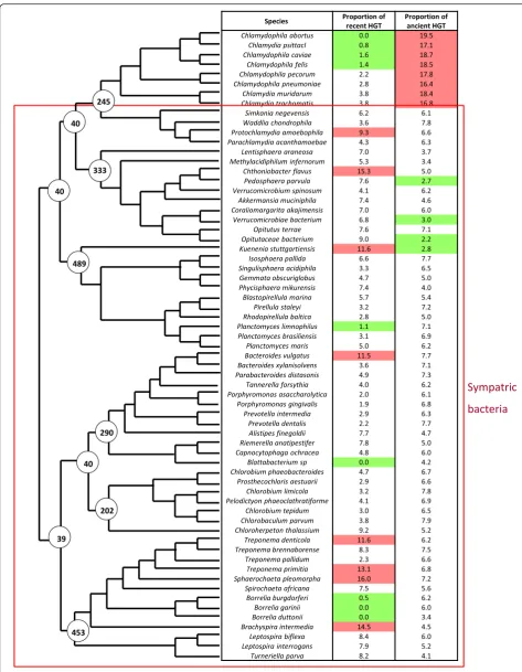
HGT were detected by phylogenomic methods, the position of HGT in species tree allows to differentiate HGT occurred only in modern species (specific HGT) and HGT occurred in their ancestors (non specific HGT). We identified 27275.0 proteins acquired by HGT (13.2 % of all proteins in proteomes studied). The proteomes of sympatric compartmentalized bacteria present 13040.0 HGT with 7259.0 specific HGT (6.2 % of proteomes) and 5781.0 non specific (4.9 % of pro-teomes). The proteomes of sympatric non compart-mentalized bacteria contain 12686.0 HGT, whom 5466.0 specific (6.7 % of proteomes) and 7220.0 non
specific (8.9 % of proteomes). Allopatric non
9.8*10-1 / 5.1*10-1 respectively) (Fig. 3). The quantities of HGT were compared between the ancestors of stud-ied bacteria, according to their most probable cell plan. This comparison did not allow to detect a significant differences between ancestral bacteria depending on cell plan (ANOVA test, p-value = 6.2*10-1). These ob-servations suggest that intracytoplasmic membrane has no impact on HGT frequency.
The compartmentalization of bacteria could disturb only some particular mechanisms of HGT processes, leading to a low impact on HGT global proportions. The study of mobilome revealed that no element present proportions varying significantly according cell plan (Additional file 1). The lifestyle of bacteria seems to clearly influence the variations of some mobilome el-ements; indeed, the intracellular bacteria present more abundant proportions of transposases than extracellular bacteria (Kruskal test :p-value = 2.0*10-2). Conjugation genes are significantly over represented in the genomes of Intracellular allopatric bacteria, and underrepre-sented in extracellular compartmentalized bacteria (Kruskal test :p-value = 3.0*10-2).
Discussion
Lifestyle of species and evolution of cell plan
The results of electronic microscopy allowed to defined S.negevensis and C.akajimensis as compartmentalized bacteria. TheChlamydiaeis characterized by an import-ant presence of internal membranes. This observation is probably related to the folds of the intracytoplasmic membrane, as it is visible for the G.obscuriglobus in the micrographies and the three dimensional reconstruction published by Santarella-Mellwig et al in 2010 [38] and 2013 [37] respectively. Conversely, the cell plan observed is different from that identified in the cryotomographies realized by Pilhofer et al in 2014 [51]. The differences could be related to the variations in methodology used and in the growth stage observed. The possible compartmentalization of S.negevensis is very interesting because the cell plan is unknown in a large majority of sympatric Chlamydiae. C.akajimensis presents a more classical compartmentalized cell plan, as it was already observed in some PVC bacteria by Santarella-Mellwig in 2013 [37] and Kuo-Chang Lee and al in 2009 [28]. These micrographies allowed an improving in reconstruction of cell plan evolution among PVC bacteria, especially in Chlamydiae. They permitted to analyze the impact of ICM in the phyla of intracellular sympatric Chlamydiae,
with two bacteria presenting a known cell plan and the probable presence of compartmentalization in their last common ancestor (with a probability of 71 %, against 57 % without the information about S.negevensis). The micrography of C.akajimensis completes the studies already realized on theVerrucomicrobiae and reinforces the probability of compartmentalization in the last
com-mon ancestor of Verrucomicrobiae (probability of
compartmentalization = 83 %, against 79 % without the cell plan of C.akajimensis). These observations allowed to conclude with fewer uncertainties our study concern-ing the HGT in compartmentalized bacteria.
Cell plan evolution reconstruction indicated that compartmentalization appeared at the root of PVC super-phylum and disappeared once, in the group of allopatric intracellular bacteria. This disappearance seems to occurred simultaneously with bacteria conver-sion to intracellular allopatric lifestyle. Intracellular allo-patric bacteria are isolated from other bacteria, so their genomes are protected against HGT. Conversely, extra-cellular and intraextra-cellular sympatric bacteria are more ex-posed to exchanges because they are in contact with many microorganisms. An hypothesis could be that the intracytoplasmic membrane plays a role in protection of genomes against HGT, as host membrane of intracellular bacteria or eukaryotic membrane [52]. This intracyto-plasmic membrane became useless in allopatric bacteria, due to the absence of HGT, leading to its disappearance. However, this hypothesis needs to be confirmed by data concerning genomes evolution in compartmentalized and non compartmentalized bacteria.
Relation between cell plan and genomes contents
The analysis of HGT in the different groups of bacteria highlighted the role of lifestyle on HGT process and the absence of impact of compartmentalization. The import-ant decrease of HGT proportions observed inChlamydiae after their conversion to the intracellular allopatric lifestyle is probably due to the physical isolation from other micro-organisms, which prevents the opportunities of HGT [52]. This agrees with previous studies showing that the pre-dominant evolutionary process in intracellular bacteria is genome reduction leading to small genome sizes [53, 54] with the bacteria from amoeba as the exception [55, 56]. The similarity of HGT proportions between compartmen-talized and non compartmencompartmen-talized bacteria suggests that the presence on an intracytoplasmic membrane has no impact on HGT process. We could imagine that this (See figure on previous page.)
absence of difference is due to a problem of our strategy but three observations demonstrate the efficiency of our method:Chlamydiaepresent an important rate of ancient HGT with eukaryotes, significantly higher than other bac-teria (ANOVA testp-value = 3.3 × 10⁻⁶). This observation is consistent with the existing literature dedicated to transfers between plants and Chlamydiae [57, 58] that supports a role ofChlamydiaein chloroplast endosymbi-osis; The HGT proportions detected in two Planctomy-cetes (Planctomyces maris and Candidatus Kuenenia stuttgartiensis) correspond to these observed by Kamneva et al in 2012 [5]; Spirochaetes seem to exchanges more with Firmicutes as it was already detected in previous studies [59, 60].
A similar HGT proportion in compartmentalized and non-compartmentalized bacteria does not neces-sary contradict the hypothesis of protection against HGT by compartmentalization. Indeed, the intracyto-plasmic membrane of PVC bacteria can represents a barrier to HGT by disturbing only some transfers mechanisms mediated by mobilome elements. This perturbation could lead or not to a decrease of HGT level, depending if the deficiency of some mechanisms are offset by the others. The absence of difference in mobilome elements quantities and proportions be-tween the different groups of bacteria indicated that the compartmentalization has no impact, not only on HGT proportions, but also on elements implicated in HGT process. The lifestyle seems to have an influence on two mobilomes elements, proportions of transpo-sases and conjugation genes. The high proportion of transposases in intracellular allopatric bacteria could be related to the importance of transposable elements in genomes of intracellular bacteria [61, 62]. However, conjugation genes present other uses in bacteria than conjugation [63, 64]. So we can assume that the abundance of conjugation genes in bacteria has prob-ably no influence on the HGT.
The function of compartmentalization in PVC bacteria remains unknown and the causes of intracytoplasmic membrane disappearance in intracellular isolated bac-teria is still unresolved.
Conclusions
The different pictures obtained for Verrucomicrobiae and Chlamydiaeallow a best definition of ancestral and modern states of compartmentalization and so, permit to reconstruct intracytoplasmic membrane evolution in
PVC bacteria. But this compartmentalization seems have no significant impact on HGT quantity, HGT proportion or partners of transfers.
Methods
Bacteria selection and genomes recovery
We selected bacteria from 4 phyla of PVC super-phylum:Planctomycetes, Verrucomicrobia, Lentisphaerae, and Chlamydiae. Bacteria from three phylogenetically close phyla were chosen as negative controls, based on a reference tree [5]: Bacteroidetes, Spirochaetes and Chlorobi. We retrieved the proteomes of all bacteria selected in genomes NCBI database (Additional file 2).
Lifestyle and cell plan determination
The lifestyle of selected bacteria is determined by a bib-liographic study of each bacterium. Two types of life-styles are known for bacteria, according the possibility of exchanges with other microorganisms: allopatric or sym-patric bacteria [65, 66]. Symsym-patric bacteria are bacteria living in community with other microorganisms, they exchange easily genes by lateral transfer through their interactions. Allopatric bacteria are living in cells and are not in contact with other microorganisms, more spe-cialized, with a reduced genome size and less genetic ex-changes. They are obligate intracellular bacteria, living in non amoeba cells.
Cell plans of bacteria are determined thanks to trans-mission electron microscopy pictures already available in bibliography [28, 29, 34, 35] and microscopic observations of the bacteria whose cell plan is unknown. First, the spe-cies Simkania negevensis (DSM27360T, type strain) was grown within a culture of its amoebal host A. castellanii. At H6, and H16 post-infection, cultures were centrifuged at 2000 rpm/min during 10 min, and the pellets were fixed for electron microscopy in a fixative solution (2.5 % glutar-aldehyde in 0.1 M sodium cacodylate buffer). The second species,Coraliomargarita akajimensis(DSM 45221T, type strain) was cultivated directly into Bacto Marine Broth medium (Difco). Growth occurred after 6 days of cultiva-tion, and when the bacterial suspension reached the expo-nential growth phase, it was centrifuged at 5500 rpm/min during 30 min, and fixed for electron microscopy in the same fixative solution. An aliquot of each culture was kept for DNA extraction, followed by a standard 16S rRNA PCR and sequencing, in order to confirm the bacterial identification. For both bacteria, Electron microscopic observations were realized in different steps: for (See figure on previous page.)
embedding, cells were fixed for 1 h with glutaraldehyde 2.5 % in 0.1 M sodium cacodylate buffer and washed three times. Cells were post-fixed for 1 h with 1 % OsO4 diluted in 0.2 M Potassium hexa-cyanoferrate (III) / 0.1 M sodium cacodylate solution. After four 5 min washes with distilled water, cells were gradually dehydrated with ethanol by successive 10 min baths in 30, 50, 70, 96, 100 and 100 % ethanol. Substitution was achieved by successively placing the cells in 25, 50 and 75 % Epon solutions for 15 min. Cells were placed for 1 h in 100 % Epon solution and in fresh Epon 100 % over-night under vacuum at room-temperature. Polymerization took place with cells in fresh 100 % Epon for 24 h at 60°. Ultrathin 70 nm sections were cut with a UC7 ultramicrotome (Leica) and placed on HR25 300 Mesh Copper/Rhodium grids (TAAB, UK). Electron micrographs were obtained on a Tecnai G20 TEM operated at 200 keV equipped with a 4096 × 4096 pixels resolution Eagle camera (FEI)).
Phylogeny and cell plan evolution reconstruction
We reconstructed the species tree of PVC bacteria and Bacteroidetes-Chlorobi-Spirochaetes thanks to 12 markers common to the 65 species (three ribosomal proteins (16S, 23S, 30S), two elongation factors (Tu and Ts), two DNA polymerase subunits, CTP synthetase, and four tRNA ligases). The selected sequences were concatenated and Mega5 [67] software is used to perform the phylogeny (alignment by Muscle algorithm, manual removal of non-conserved positions, tree building with NJ and ML methods with 150 bootstraps, comparison of phylogenies and selection of a consensus (Additional file 3)).
Thanks to information about compartmentalization in bacteria, we reconstructed the ancestral state of cell plan with parsimony method (Mesquite software [68] and Phytools package on R software [69]. Phytools allowed the reconstruction of ancestral sates of discrete character by a method of Maximum likelihood). The tree obtained allowed us to date the different events occurred during evolution of the phyla studied (appearance and dis-appearance of compartmentalization, conversion to intracellular allopatric or sympatric lifestyle).
Mobilome study
We studied 9 elements directly or indirectly implicated in the horizontal transfers: phages (complete or incom-plete), conjugation genes and plasmids, these three ele-ments are involved in entrance of foreign sequences in bacteria; Transposases, integrases and CRISP (candidate or confirmed), regulating positively or negatively the se-quences integration in recipient genome; and tRNA used for sequences translation. Different databases were used as RepBase [70] and CRISPfinder [71] to detect the mobilome elements (the complete list of databases used is presented in the Additional file 4). For each element
we counted the quantity in each species; if this quantity is related to the genome size (CRISPs, transposases, inte-grases and conjugation genes), we calculated the propor-tion of these elements in the genomes. We used the same statistical tests as those used for HGT analysis, in order to determine the elements overrepresented in the different classes of bacteria.
HGT detection
OrthoMCL [72] was used for construction of orthologous groups. Genes absent to all orthologous groups are either acquired by HGT or generated de novo (ORFans). Blast against nr database allowed the identification genes pre-senting no identifiable homologous genes (e-value < 10-4 and coverage > 50 %), considered as ORFans.
Phylopattern [73] pipeline allowed the detection of gen-etic events in four steps: comparison between species tree and each orthologous groups; detection of missing species in orthologous groups; reconstruction of ancestral state for each proteins, based on the pattern of presence/ab-sence of sequences in modern species; identification of two types of genetic events: gains and losses. Gains could be HGT, de novo genes, or artifacts. We focused on gene gains and Blast [74] each sequences against nr database (NCBI). HGT candidates were identified thanks to filtering on Blast results: For each blast results, sequences derived from phylum where gene gain was detected were removed (for example, if gain was identified inAkkermansia muci-niphila (Verrucomicrobiae), all sequences in Blast results derived fromVerrucomicrobiaewere removed). Sequences with e-value > 10-5, coverage < 60 % or identities < 30 % were also removed. Then, species of the first ten sequences remaining in Blast result were identified. If species identified do not belong to one of the sister phyla studied, gene gain is probably an HGT (for example if gain was identified inAkkermansia muciniphila, gain is consid-ered as HGT only if the first ten hits do not belong to the phyla Planctomycetes or Chlamydiae). The position on gains in species tree allows to differentiate HGT occurred only in modern species (specific HGT) and HGT occurred in the ancestor (non specific HGT). We calculated the quantity of HGT and the percentage of proteins present in each species, due to transfers. For each HGT detected (recent or ancient), we determined with which organism the transfers were realized.
distribution and ANOVA test for data with a normal dis-tribution). Tests of Nemenuyi (data not normally distrib-uted) or Tukey (data normally distribdistrib-uted) were performed to obtain a comparison of each pairs of clas-ses. We realized also these statistical analysis with bac-terial groups based on the phyla, to determine if classes based on phyla present the same results that classes based on cell plan. If it had been the case, it would have been impossible to determine if differences observed were related to cell plan or to phylogenetic relations.
Reviewers’comments
Reviewer summary
Reviewer 1 (Mircea Podar)
“In their study, Pinos et al tested their hypothesis that cellular compartmentalization, observed in some repre-sentatives of the PVC super phylum, is correlated to the frequency of horizontal gene transfer. The authors performed ultrastructural analyses of a representative from the Chlamydia and one from Verrucomicrobiae, which were shown to be compartmentalized. That information was combined with results from literature on other PVC species and was correlated with phylogenomic analyses on HGT frequencies, based on completed genomes. The result indicates that
compartmentalization is in fact not significantly
impacting HGT, which has a stronger correlation with the organismal life-style.
The study is interesting and original, even though the conclusion did not support the original hypothesis. It does provide a foundation for additional tests and fo-cused study of related organisms/genomes, which may increase the signal to link compartmentalization to more subtle evolutionary genomic effects.”
Reviewer 2 (Olivier Tenaillon)
“In the manuscript entitled “Compartmentalization in PVC super-phylum: evolution and impact”, Sandrine Pinos and colleagues use both microscopy and a phylo-genetic approach to study Compartmentalization and its potential impact on horizontal gene transfer.
The idea is interesting, but the paper is not very clear in its present form. The usage of microscopy is not in-troduced properly, statistics and methods are over-simplified and impossible to reproduce. I would consider therefore that the paper needs very substantial rewriting and the phylogenetic analysis should include uncertainty in the phylogeny.”
Reviewer recommendations to authors
Reviewer 1 (Mircea Podar): The manuscript needs ex-tensive work in correcting the many language errors (spelling, syntax, grammar). It also does not provide
sufficient detail to evaluate the power of the phyloge-nomic analyses that were performed. While I like the concept of this study and the overall approach, the manuscript requires a complete overhaul.
Reviewer 2 (Olivier Tenaillon): How robust is the tree? Before being able to conclude on the ancestry of a trait, the robustness of the phylogeny has to be tested. In my experience a 16S phylogeny is not very robust and much more loci should be involved in the reconstruc-tion, bootstrap values should be presented. Character re-construction should also take into account the tree uncertainty. This is critical for both figures and both character mapping procedures.
Reviewer 2 (Olivier Tenaillon): line 219: “If species identified do not belong to one of the close phyla stud-ied, gene gain is probably an HGT” how close is close, the threshold use should be clearly defined?”
Author’s response: The different comments and ques-tions concerning our phylogenomic strategy indicate an important problem in our presentation of this method and its results. In order to correct these problems, we ex-panded the methodology part of phylogenomic strategy, by adding indications and some examples (we detailed some points, as what we want to say by“If species identi-fied do not belong to one of the close phyla studied, gene gain is probably an HGT”, in this case,“close phyla” re-fers to species belonging to the sister phylum). We also added, in the Additional file 3, phylogenies of PVC bac-teria and Bacteroidetes-Spirochaetes-Chlorobi, with the indication of bootstrap values. These phylogenies are more robust than those of the first version, due to the use of ten supplementary markers, for alignment and phylo-genetic reconstruction. We want to underline that some results obtained by our phylogenomic method are conver-gent with results already observed in previous studies (line 155–161), this is an indication of the relative effi-ciency of our method.
The improvement of the phylogeny allowed a better re-construction of cell plan evolution in PVC bacteria. How-ever, In order to take into account the problem of phylogenetic uncertainties, we used another method of ancestral reconstruction that not only considers these un-certainties, but also provides the probabilities of each state of cell plan at each nodes of the tree. The results of this analysis were included in the Figure 2, they led to a better vision of PVC bacteria evolution and highlighted the importance of microscopic data.
Reviewer 2 (Olivier Tenaillon): Introduction
-The link between horizontal gene transfer and
and the reconstruction of ancestral states”. What is meant by this sentence is not clear to me. I think developing more on the subject would improve the introduction.
Author’s response: This sentence is difficult to explain without the knowledge about the compartmentalization and the lifestyle of PVC bacteria, however, it could be long to detail all these information in introduction. Conse-quently, we decided to remove this sentence from the intro-duction, we presented and explained it in the results part. This sentence resumed the idea that if Chlamydiae are the only non compartmentalized PVC bacteria sequenced and also the only intracellular bacteria of this super-phylum, if the compartmentalization protects genomes against HGT, its lost in the intracel-lular species could be related to the isolation of these bacteria that induces a low proportion of HGT, and leads to the uselessness of compartmentalization. So, the bibliographic knowledge about the presence or absence of compartmentalization in species suggests the hypothesis of a role of the ICM in protection against HGT.
Reviewer 2 (Olivier Tenaillon): Results - The result section starts with the description of microscopic struc-tures. It is not introduced in anyway. The two first para-graphs should be inverted: first mention that the compartmentalization is unknown in some species, then present the evidence for the ones lacking information.
Discussion - again no clear explanation of the interest of the microscopy.
Author’s response: As the result of the comments of re-viewers, we decided to reorganize the results of microscopic analyzes to improve our presentation. We insisted on the importance of these data in our study by adding explana-tions of their use in ancestral reconstruction (71–80) and their role in the study of the relation between HGT and compartmentalization (97–99). We realized a more de-tailed ancestral state reconstruction, with a more complex method (analysis performed thanks to R software, with phylotools package, by a maximum likelihood method) that provided the probabilities of each cell plan states at each nodes of the tree. These analyzes highlighted the im-portance to add our data to the study, indeed our supple-ment of information allowed to improve significantly the probability of compartmentalization for several nodes of tree.
Reviewer 2 (Olivier Tenaillon): Results - The term allopatric and sympatric should clearly be defined in the present context and not in the method. Being an evolutionary biologist rather than a microbiologist, I do not support the use of these terms that refer to speciation and not to accessibility of foreign DNA,
but as the author have previously used them… It
should nevertheless be explicitly defined, with the present definition it is not clear who has access to other microbes.
Author’s response: Indeed the terms allopatry and sympatry needed to be clearly defined, we added the complete definition in methodology part and we also put a short definition in the results part.
Reviewer 2 (Olivier Tenaillon): The mobilome ana-lysis are not described enough to be reproducible.
Author’s response: We improved the paragraphs dedi-cated to the mobilome analysis by adding explanations about our strategy (elements selected and calculation of proportions). We completed these explanations by a table of the databases used to retrieve the mobilome elements, available in the Additional file 4.
Reviewer 2 (Olivier Tenaillon):“line 206“Genes ab-sent to all orthologous groups are either acquired by HT or generated de novo”: this sentence is not clear. Some-times HT is used someSome-times HGT.
Author’s response:The term HT indicates an horizon-tal transfer of sequence, this sequence could contain one or more genes (average of genes implicated by transfer is 1.5). The term HGT refers to a transfer of one gene. As the use of these two terms complicated our manuscript, we decided to use only the term of HGT, and we adapted our data in consequence.
Reviewer 2 (Olivier Tenaillon): line 206 NR database: the author should say what it is and use either nr or NR. Author’s response:Indeed we used the nr database, we corrected this mistakes in manuscript.
Reviewer 2 (Olivier Tenaillon): Figure - species names should be written in full and in italic with no underscore. Line 70–75 Statistics should be given to compare“sympatric”groups.
Author’s response: The figures were corrected and we added the statistics for sympatric bacteria comparison in the paragraph.
Additional files
Additional file 1:Mobilome distribution in the different bacterial groups according lifestyle and cell plan. The different graphics allow a better sight of the different levels for each mobilome feature. Red stars show significant differences between groups. (PDF 175 kb)
Additional file 2:Characteristics of studied species. This table presents the lifestyles, genomes and proteomes features of each species studied. (PDF 125 kb)
Additional file 3:Phylogeny of PVC bacteria and phylogeny of
Bacteroidetes-Chlorobi-Spirochaetes. The phylogenies of studied bacteria were realized with Maximum likelihood method, thanks to Mega6 software, the bootstraps values are indicated at each node. (PDF 234 kb)
Additional file 4:Databases used for the mobilome study. The mobilome elements studied are indicated in the first column, the second column contains the corresponding database and the third column presents the url of websites for these databases. (PDF 177 kb)
Acknowledgements
assistance in statistical analyzes and Pauline Fournerie for her implication in electronic microscopy. We also thank the Xegen company for the assistance in HGT detection by using of Phylopattern software. This work was supported by the Assistance Publique - Hopitaux de Marseille (Marseille Public University Hospital System).
Funding
This work was supported by the Assistance Publique - Hopitaux de Marseille (Marseille Public University Hospital System). The funders had no role in study design, data collection and interpretation, or the decision to submit the work.
Availability of data and materials
The dataset supporting the conclusions of this article is included within the article and its Additional files 1, 2, 3 and 4.
Authors’contribution
PS carried out the design of the study, the strategy elaboration and the collect of data, performed the statistical analysis and interpretation of results and drafted the manuscript. PP participated in strategy elaboration, data interpretation and revised the manuscript. RD conceived the study, participated in its design and coordination and revised the manuscript. BJP coordinated and realized the microscopic observations and helped in data interpretation. PI coordinated and participated in microscopic observations, helped in data interpretation and revised the manuscript. All authors read and approved the manuscript.
Competing interests
The authors declare that they have no competing interests.
Consent for publication
Not applicable.
Ethics approval and consent to participate
Not applicable.
Received: 13 May 2016 Accepted: 2 August 2016
References
1. Gupta RS, Bhandari V, Naushad HS. Molecular signatures for the PVC clade (planctomycetes, verrucomicrobia, chlamydiae, and lentisphaerae) of bacteria provide insights into their evolutionary relationships. Front Microbiol. 2012;3:327.
2. Wagner M, Horn M. The planctomycetes, verrucomicrobia, chlamydiae and sister phyla comprise a superphylum with biotechnological and medical relevance. Curr Opin Biotechnol. 2006;17(3):241–9.
3. Cho JC, Vergin KL, Morris RM, Giovannoni SJ. Lentisphaera araneosa gen. nov., sp. nov, a transparent exopolymer producing marine bacterium, and the description of a novel bacterial phylum, lentisphaerae. Environ Microbiol. 2004;6(6):611–21.
4. Siegl A, Kamke J, Hochmuth T, Piel J, Richter M, Liang C,…Hentschel U. Single-cell genomics reveals the lifestyle of Poribacteria, a candidate phylum symbiotically associated with marine sponges. ISME J. 2011;5(1):61–70. 5. Kamneva OK, Knight SJ, Liberles DA, Ward NL. Analysis of genome content
evolution in PVC bacterial super-phylum: assessment of candidate genes associated with cellular organization and lifestyle. Genome Biol Evol. 2012;4(12):1375–90.
6. Yarza P, Richter M, Peplies J, Euzeby J, Amann R, Schleifer KH,… Rosselló-Móra R. The All-Species Living Tree project: A 16S rRNA-based phylogenetic tree of all sequenced type strains. Syst. Appl. Microbiol. 2008;31(4):241–250. 7. Griffiths E, Gupta RS. Phylogeny and shared conserved inserts in proteins
provide evidence that verrucomicrobia are the closest known free-living relatives of chlamydiae. Microbiology. 2007;153(8):2648–54.
8. Brochier C, Philippe H. Phylogeny: a non-hyperthermophilic ancestor for bacteria. Nature. 2002;417(6886):244.
9. Wu D, Hugenholtz P, Mavromatis K, Pukall R, Dalin E, Ivanova NN,…Eisen JA. A phylogeny-driven genomic encyclopaedia of Bacteria and Archaea. Nat. 2009;462(7276):1056–1060.
10. Jenkins C, Fuerst JA. Phylogenetic analysis of evolutionary relationships of the planctomycete division of the domain bacteria based on amino acid sequences of elongation factor Tu. J Mol Evol. 2001;52(5):405–18. 11. Pilhofer M, Rappl K, Eckl C, Bauer AP, Ludwig W, Schleifer KH, Petroni G.
Characterization and evolution of cell division and cell wall synthesis genes in the bacterial phyla verrucomicrobia, lentisphaerae, chlamydiae, and planctomycetes and phylogenetic comparison with rRNA genes. J Bacteriol. 2008;190(9):3192–202.
12. Gupta RS, Griffiths E. Critical issues in bacterial phylogeny. Theor Popul Biol. 2002;61(4):423–34.
13. Jun SR, Sims GE, Wu GA, Kim SH. Whole-proteome phylogeny of prokaryotes by feature frequency profiles: an alignment-free method with optimal feature resolution. Proc Natl Acad Sci. 2010;107(1):133–8. 14. Ciccarelli FD, Doerks T, Von Mering C, Creevey CJ, Snel B, Bork P.
Toward automatic reconstruction of a highly resolved tree of life. Science. 2006;311(5765):1283–7.
15. Lagkouvardos I, Jehl MA, Rattei T, Horn M. Signature protein of the PVC superphylum. Appl Environ Microbiol. 2014;80(2):440–5.
16. Buckley DH, Huangyutitham V, Nelson TA, Rumberger A, Thies JE. Diversity of planctomycetes in soil in relation to soil history and environmental heterogeneity. Appl Environ Microbiol. 2006;72(7):4522–31.
17. Wang J, Jenkins C, Webb RI, Fuerst JA. Isolation of gemmata-like and Isosphaera-like planctomycete bacteria from soil and freshwater. Appl Environ Microbiol. 2002;68(1):417–22.
18. Sangwan P, Kovac S, Davis KE, Sait M, Janssen PH. Detection and cultivation of soil verrucomicrobia. Appl Environ Microbiol. 2005;71(12):8402–10.
19. Horn M. Chlamydiae as symbionts in eukaryotes. Annu Rev Microbiol. 2008;62:113–31.
20. Zwart G, Crump BC, Kamst-van Agterveld MP, Hagen F, Han SK. Typical freshwater bacteria: an analysis of available 16S rRNA gene sequences from plankton of lakes and rivers. Aquat Microb Ecol. 2002;28(2):141–55. 21. Bergmann GT, Bates ST, Eilers KG, Lauber CL, Caporaso JG, Walters WA,…
Fierer N. The under-recognized dominance of Verrucomicrobia in soil bacterial communities. Soil Biol. Biochem. 2011;43(7):1450–1455.
22. Ward N, Staley JT, Fuerst JA, Giovannoni S, Schlesner H, Stackebrandt E. The order planctomycetales, including the genera planctomyces, pirellula, gemmata and Isosphaera and the candidatus genera brocadia, kuenenia and scalindua. New York: Springer; 2006. p. 757–93.
23. Shu Q, Jiao N. Different planctomycetes diversity patterns in latitudinal surface seawater of the open sea and in sediment. J Microbiol. 2008;46(2):154–9.
24. Strous M, Pelletier E, Mangenot S, Rattei T, Lehner A, Taylor MW,…Barbe V. Deciphering the evolution and metabolism of an anammox bacterium from a community genome.Nature. 2006;440(7085):790–794.
25. Pearson A, Budin M, Brocks JJ. Phylogenetic and biochemical evidence for sterol synthesis in the bacterium gemmata obscuriglobus. Proc Natl Acad Sci. 2003;100(26):15352–7.
26. Bébéar C, De Barbeyrac B. Genital chlamydia trachomatis infections. Clin Microbiol Infect. 2009;15(1):4–10.
27. Corsaro D, Greub G. Pathogenic potential of novel chlamydiae and diagnostic approaches to infections due to these obligate intracellular bacteria. Clin Microbiol Rev. 2006;19(2):283–97.
28. Lee KC, Webb RI, Janssen PH, Sangwan P, Romeo T, Staley JT, Fuerst JA. Phylum verrucomicrobia representatives share a compartmentalized cell plan with members of bacterial phylum planctomycetes. BMC Microbiol. 2009;9(1):5. 29. Lindsay MR, Webb RI, Strous M, Jetten MS, Butler MK, Forde RJ, Fuerst
JA. Cell compartmentalisation in planctomycetes: novel types of structural organisation for the bacterial cell. Arch Microbiol. 2001;175(6):413–29.
30. Chopra I, Storey C, Falla TJ, Pearce JH. Antibiotics, peptidoglycan synthesis and genomics: the chlamydial anomaly revisited. Microbiology. 1998;144:2673–8.
31. McCoy AJ, Maurelli AT. Building the invisible wall: updating the chlamydial peptidoglycan anomaly. Trends Microbiol. 2006;14:70–7.
32. Lage OM, Bondoso J, Lobo-da-Cunha A. Insights into the Ultrastructural morphology of novel planctomycetes. Antonie Van Leeuwenhoek. 2013;104(4):467–76.
34. Fuerst JA, Sagulenko E. Beyond the bacterium: planctomycetes challenge our concepts of microbial structure and function. Nat Rev Microbiol. 2011;9:403–13.
35. Fieseler L, Horn M, Wagner M, Hentschel U. Discovery of the novel candidate phylum“poribacteria”in marine sponges. Appl Environ Microbiol. 2004;70(6):3724–32.
36. Fuerst JA, Webb RI. Membrane-bounded nucleoid in the eubacterium gemmatata obscuriglobus. Proc Natl Acad Sci. 1991;88(18):8184–8. 37. Santarella-Mellwig R, Pruggnaller S, Roos N, Mattaj IW, Devos DP.
Three-dimensional reconstruction of bacteria with a complex endomembrane system. PLoS Biol. 2013;11(5):e1001565.
38. Santarella-Mellwig R, Franke J, Jaedicke A, Gorjanacz M, Bauer U, Budd A, Devos DP. The compartmentalized bacteria of the planctomycetes-verrucomicrobia-chlamydiae superphylum have membrane coat-like proteins. PLoS Biol. 2010;8(1):e1000281.
39. Van de Graaf AA, Mulder A, de Bruijn P, Jetten MS, Robertson LA, Kuenen JG. Anaerobic oxidation of ammonium is a biologically mediated process. Appl Environ Microbiol. 1995;61(4):1246–51.
40. Strous M, Fuerst JA, Kramer EH, Logemann S, Muyzer G, van de Pas-Schoonen KT,…Jetten MS. Missing lithotroph identified as new planctomycete. Nat. 1999;400(6743):446–449.
41. Fuerst JA. Intracellular compartmentation in planctomycetes. Annu Rev Microbiol. 2005;59:299–328.
42. Lonhienne TG, Sagulenko E, Webb RI, Lee KC, Franke J, Devos DP,…Fuerst JA. Endocytosis-like protein uptake in the bacterium Gemmata
obscuriglobus. Proc. Natl. Acad. Sci. 2010;107(29):12883–12888. 43. Devos DP, Reynaud EG. Evolution. Intermediate steps. Science.
2010;330:1187–8.
44. McInerney JO, Martin WF, Koonin EV, Allen JF, Galperin MY, Lane N,… Embley TM. Planctomycetes and eukaryotes: a case of analogy not homology. Bioessays. 2011;33(11):810–817.
45. Fuchsman CA, Rocap G. Whole-genome reciprocal BLAST analysis reveals that planctomycetes do not share an unusually large number of genes with eukarya and archaea. Appl Environ Microbiol. 2006;72(10):6841–4. 46. Budd A, Devos DP. Evaluating the evolutionary origins of unexpected
character distributions within the bacterial planctomycetes-verrucomicrobia-chlamydiae superphylum. Front Microbiol. 2012;3:401.
47. Forterre P. A new fusion hypothesis for the origin of eukarya: better than previous ones, but probably also wrong. Res Microbiol. 2011;162(1):77–91. 48. Fuerst JA, Sagulenko E. Keys to eukaryality: planctomycetes and ancestral
evolution of cellular complexity. Front Microbiol. 2012;3(167):b94. 49. Fuerst JA. The PVC superphylum: exceptions to the bacterial definition?
Antonie Van Leeuwenhoek. 2013;104(4):451–66.
50. Devos DP. PVC bacteria: variation of, but not exception to, the gram-negative cell plan. Trends Microbiol. 2014;22(1):14–20.
51. Pilhofer M, Aistleitner K, Ladinsky MS, König L, Horn M, Jensen GJ. Architecture and host interface of environmental chlamydiae revealed by electron cryotomography. Environ Microbiol. 2014;16(2):417–29.
52. Hirt RP, Alsmark C, Embley TM. Lateral gene transfers and the origins of the eukaryote proteome: a view from microbial parasites. Curr Opin Microbiol. 2015;23:155–62.
53. Merhej V, Royer-Carenzi M, Pontarotti P, Raoult D. Massive comparative genomic analysis reveals convergent evolution of specialized bacteria. Biol Direct. 2009;4(1):13.
54. Wolf YI, Koonin EV. Genome reduction as the dominant mode of evolution. Bioessays. 2013;35(9):829–37.
55. Molmeret M, Horn M, Wagner M, Santic M, Kwaik YA. Amoebae as training grounds for intracellular bacterial pathogens. Appl Environ Microbiol. 2005;71(1):20–8.
56. Moliner C, Fournier PE, Raoult D. Genome analysis of microorganisms living in amoebae reveals a melting pot of evolution. FEMS Microbiol Rev. 2010;34(3):281–94.
57. Subtil A, Collingro A, Horn M. Tracing the primordial chlamydiae: extinct parasites of plants? Trends Plant Sci. 2014;19(1):36–43.
58. Facchinelli F, Colleoni C, Ball SG, Weber AP. Chlamydia, cyanobiont, or host: who was on top in the ménage à trois? Trends Plant Sci. 2013;18(12):673–9. 59. Viswanathan VK. Spirochaetes and their twisted ways. Gut Microbes.
2012;3(5):399–400.
60. Bellgard MI, Wanchanthuek P, La T, Ryan K, Moolhuijzen P, Albertyn Z. Genome sequence of the pathogenic intestinal spirochete brachyspira
hyodysenteriae reveals adaptations to its lifestyle in the porcine large intestine. PLoS One. 2009;4:e4641.
61. Plague GR, Dunbar HE, Tran PL, Moran NA. Extensive proliferation of transposable elements in heritable bacterial symbionts. J Bacteriol. 2008;190(2):777–9.
62. Bordenstein SR, Reznikoff WS. Mobile DNA in obligate intracellular bacteria. Nat Rev Microbiol. 2005;3(9):688–99.
63. Cascales E, Christie PJ. The versatile bacterial type IV secretion systems. Nat Rev Microbiol. 2003;1(2):137–49.
64. Christie PJ, Atmakuri K, Krishnamoorthy V, Jakubowski S, Cascales E. Biogenesis, architecture, and function of bacterial type IV secretion systems. Annu Rev Microbiol. 2005;59:451–85.
65. Georgiades K. Genomics of epidemic pathogens. Clin Microbiol Infect. 2012;18(3):213–7.
66. Merhej V, Notredame C, Royer-Carenzi M, Pontarotti P, Raoult D. The rhizome of life: the sympatric rickettsia felis paradigm demonstrates the random transfer of DNA sequences. Mol Biol Evol. 2011;28(11):3213. 67. Tamura K, Peterson D, Peterson N, Stecher G, Nei M, Kumar S. MEGA5:
molecular evolutionary genetics analysis using maximum likelihood, evolutionary distance, and maximum parsimony methods. Mol Biol Evol. 2011;28:2731–9.
68. MESQUITE : http://mesquiteproject.org/ consulted in June and July 2016. Accessed June 2015.
69. Revell LJ. phytools: an R package for phylogenetic comparative biology (and other things). Methods Ecol Evol. 2012;3(2):217–23.
70. Repbase: http://www.girinst.org/repbase/ consulted in march 2015. Accessed March 2015.
71. CRISPfinder: http://crispr.i2bc.paris-saclay.fr/ consulted in February 2015. Accessed February 2015.
72. Fischer S, Brunk BP, Chen F, Gao X, Harb OS, Iodice JB, Shanmugam D, Roos DS, Stoeckert CJ. Using OrthoMCL to assign proteins to OrthoMCL-DB groups or to cluster proteomes into New ortholog groups. Current Protocols in Bioinformatics. 2011;35(6):12. 1–6.12.19.
73. Gouret P, Thompson JD, Pontarotti P. PhyloPattern: regular expressions to identify complex patterns in phylogenetic trees. BMC Bioinf. 2009;10(1):298. 74. Altschul SF, Gish W, Miller W, Myers EW, Lipman DJ. Basic local alignment
search tool. J Mol Biol. 1990;215(3):403–10.
• We accept pre-submission inquiries
• Our selector tool helps you to find the most relevant journal
• We provide round the clock customer support
• Convenient online submission
• Thorough peer review
• Inclusion in PubMed and all major indexing services
• Maximum visibility for your research
Submit your manuscript at www.biomedcentral.com/submit